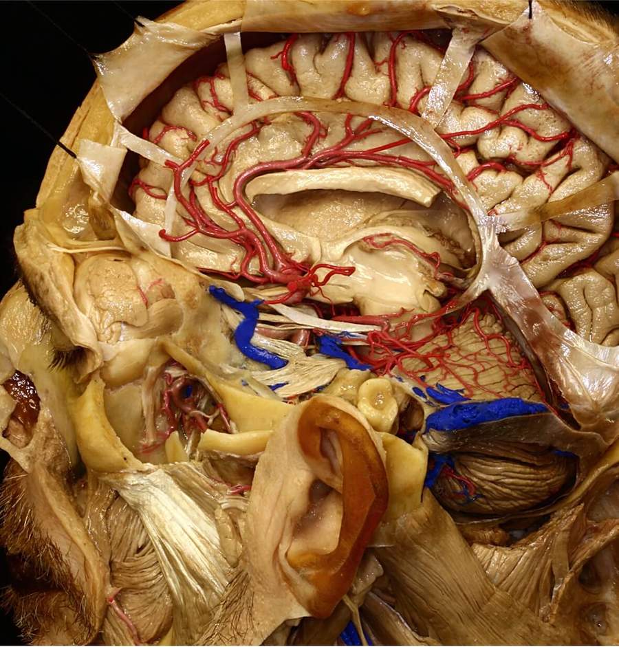
波動率。 視頻

前斜位切除術和視神經減壓術

最後更新:2021年4月7日

摘要

背景幾種涉及視神經管或視神經管內結構的疾病可引起視力損害。已經開發了幾種技術來減壓視神經。

客觀的:描述微創硬膜外前臥位切除術(MiniEx)視神經減壓術,詳細介紹其手術解剖、臨床病例,並建立概念證明。

方法:解剖解剖在屍體頭部進行,以顯示外科解剖,並逐步顯示MiniEx入路。此外,我們還應用這些手術理念對6例臨床視神經減壓。

結果: MiniEx入路允許使用無鑽技術進行硬膜外前斜位切除術和近270°視神經減壓。在MiniEx入路中,皮膚切口、顳肌剝離和開顱術更小,視神經、前臥突和上眶裂的暴露程度與標準技術相同。所有接受該技術手術的患者視力均有改善。

結論MiniEx入路是硬膜外前斜位切除術和視神經減壓的傳統入路的一個很好的選擇。它可以作為更複雜的外科手術的一部分,也可以作為單一的外科手術。

廣告

阿特拉斯選擇雙極鉗(2022年2月上市)

為你的每一個外科手術設計的

大腦和脊柱手術的五種針尖尺寸

無與倫比的不粘和低調的特點

了解更多並在網上購物

介紹

一些涉及視神經管或視神經管內容物的疾病可引起眼窩病,從而導致視力損害。1、2額眶創傷(由視神經管骨折或神經內挫傷、血管痙攣、壞死或水腫引起)、顱內腫瘤(鞍結節腦膜瘤或蝶骨腦膜瘤和視神經膠質瘤)、繼發病變(粘液囊腫、鼻旁眶竇腫瘤)、纖維或骨發育不良、炎症性假瘤、Graves眼眶病或血管病變(頸動脈眼動脈瘤)是可能壓迫視神經的最常見疾病。3 - 5許多報告顯示,早期視神經管段減壓可改善視力。5-13

在許多手術場景中(如血管和顱底手術),微創開顱術已日益成為傳統開顱術的流行選擇。與傳統技術相比,它們有幾個優點。各種微創技術減壓視神經最近已被描述。然而,一些報告隻包括實驗研究,沒有相關的臨床應用,其他報告描述了內鏡輔助或包括需要硬膜內手術的技術。14-22

經鎖眼入路的硬膜外視神經減壓術可提供滿意的視神經減壓術。在這篇文章中,我們提出了一種通過微創入路的硬膜外視神經減壓技術,作為Dolenc先前描述的方法的修正。1、2

方法

采用微創硬膜外前斜位切除術(MiniEx)和翼側小開顱入路視神經減壓,在顯微鏡下(×4 - ×25)對4例福爾馬林固定屍體頭部進行手術。動脈灌注紅色矽膠靜脈灌注藍色矽膠。另取2例標本和2個幹顱骨,分析視神經與視神經管、前臥突、海綿竇、眶上裂的解剖關係。為避免測量誤差,用卡尺測量骨瓣的大小和麵積。

此外,6例臨床病例采用硬膜外前臥位切除術入路與MiniEx進行視神經減壓。本研究分析了這些病例的手術結果,包括視覺和神經疾病。

結果

骨的關係

視神經管位於眶尖。它的邊界是內側的蝶骨體,上方的蝶骨小翼,外側的前臥突和視神經支柱的內側麵,以及下方的上表麵視神經支柱和蝶骨體的鄰近部分(圖1)。視神經管向後向內側,在垂直軸上呈橢圓形。23

圖1。前床突、眶上裂和視神經管的骨關係。(一)高級視圖。前突從蝶骨小翼的內側端向後突出。前凸突的前附著從凸突基部向內側延伸至平麵,形成視神經管的頂部。前突的後附著物,視神經支柱,從前突的下內側延伸到蝶骨體,形成視神經管的下壁。另一個小突起,中床突,位於頸動脈溝的內側,位於前床突尖端的水平,向上和側向突出。(B)左側頸動脈斜突孔的上位視圖。一個骨橋從前突尖延伸到中突尖,在動脈周圍形成一個幾乎完整的骨環,稱為頸動脈斜突孔。(C)左側視神經管、視神經支柱和右側眶上裂斜後視圖。 The optic canal has an oval shape and is formed superiorly by the anterior attachment of the anterior clinoid, inferiorly by the optic strut, laterally by the medial side of the anterior clinoid process and the optic strut, and medially by the sphenoid body. The superior orbital fissure has a triangular shape and is formed superiorly by the lesser wing of the sphenoid bone, medially by the optic strut and the sphenoid body, and inferiorly and laterally by the greater sphenoid wing. The maxillary strut is the bridge of bone separating the superior orbital fissure from the foramen rotundum. (D) Intraorbital view of the optic canal and superior orbital fissure. The optic strut separates the optic canal and superior orbital fissure and forms the floor of the optic canal and the superomedial part of the roof of the superior orbital fissure. Ant., anterior; Car., carotid; Caroticoclin., caroticoclinoid; Clin., clinoid; Fiss., fissure; For., foramen; Gr., greater; Inf., inferior; Less., lesser; Max., maxillary; Mid., middle; Orb., orbital; Post., posterior; Proc., process; Rotund., rotundum; Sphen., sphenoid; Sulc., sulcus; Sup., superior. (Images courtesy of AL Rhoton, Jr.)

圖1。前床突、眶上裂和視神經管的骨關係。(一)高級視圖。前突從蝶骨小翼的內側端向後突出。前凸突的前附著從凸突基部向內側延伸至平麵,形成視神經管的頂部。前突的後附著物,視神經支柱,從前突的下內側延伸到蝶骨體,形成視神經管的下壁。另一個小突起,中床突,位於頸動脈溝的內側,位於前床突尖端的水平,向上和側向突出。(B)左側頸動脈斜突孔的上位視圖。一個骨橋從前突尖延伸到中突尖,在動脈周圍形成一個幾乎完整的骨環,稱為頸動脈斜突孔。(C)左側視神經管、視神經支柱和右側眶上裂斜後視圖。 The optic canal has an oval shape and is formed superiorly by the anterior attachment of the anterior clinoid, inferiorly by the optic strut, laterally by the medial side of the anterior clinoid process and the optic strut, and medially by the sphenoid body. The superior orbital fissure has a triangular shape and is formed superiorly by the lesser wing of the sphenoid bone, medially by the optic strut and the sphenoid body, and inferiorly and laterally by the greater sphenoid wing. The maxillary strut is the bridge of bone separating the superior orbital fissure from the foramen rotundum. (D) Intraorbital view of the optic canal and superior orbital fissure. The optic strut separates the optic canal and superior orbital fissure and forms the floor of the optic canal and the superomedial part of the roof of the superior orbital fissure. Ant., anterior; Car., carotid; Caroticoclin., caroticoclinoid; Clin., clinoid; Fiss., fissure; For., foramen; Gr., greater; Inf., inferior; Less., lesser; Max., maxillary; Mid., middle; Orb., orbital; Post., posterior; Proc., process; Rotund., rotundum; Sphen., sphenoid; Sulc., sulcus; Sup., superior. (Images courtesy of AL Rhoton, Jr.)

前突是一種骨的突出,從蝶骨小翼的內側端向後延伸到海綿竇頂的前部。從上看,它呈三角形,底部位於腹側,尖端位於後方(圖1A-C)。24日,25

前臥突基部有1個外側和2個內側附著點。側麵,基底附著在蝶骨小翼上,蝶骨小翼位於眶上裂的上方。內側,基底通過2根連接:前根從前床突基部向內側延伸至蝶骨體,形成視神經管頂,後根稱為視支,從視神經下方向內側延伸至蝶骨體,形成視神經管底(圖1C)。

視神經支柱的上表麵形成視神經管的底部,下表麵形成眶上裂頂的內側部分和海綿竇頂的前部(圖1D)。前凸突的外層由密集的皮質骨和內部的鬆質骨組成。它可能包含靜脈通道,也可能是充氣的,包括通過視神經支柱與蝶竇相通的空氣細胞。如果術後蝶竇被打開,特別是硬膜內前臥位切除術或硬膜外前臥位切除術後硬腦膜被打開,術後可能發生腦脊液泄漏。5、10 - 13

硬腦膜的關係

覆蓋前床突上表麵的硬腦膜繼續向內側形成環繞頸內動脈的遠端硬腦膜環的外側部分。這個硬腦膜與覆蓋蝶骨小翼上表麵的硬腦膜橫向相連。在內側,它延伸到視神經下方,包括視神經支柱的上表麵,形成遠端硬腦膜環的前部。硬腦膜從這裏向內和向後延伸覆蓋頸動脈溝的上部並從上環的內側延伸。前側,頸前突上表麵硬腦膜覆蓋小翼前根,並附著於蝶平麵後緣(圖2)。頸內動脈的頸前突段位於硬腦膜近端和遠端環之間(圖2C和3F)。

圖2。視神經管、眶上裂和海綿竇的硬腦膜、動脈和神經關係。(A)海綿竇上外側視圖。海綿竇從眶上裂延伸到岩尖。岩上竇穿過梅克爾洞口上方,與海綿竇的後部相連。覆蓋外側壁的硬腦膜已被移除,三叉神經節暴露在外。動眼神經、滑車神經和眼神經向前延伸,彙合於眶上裂。眼神經向下收縮露出外展神經。(B)海綿竇和鞍區頂部的上方視圖。前突被硬腦膜覆蓋,硬腦膜向外側延伸包括蝶骨小翼的上側麵。 The falciform ligament, the dural fold extending above the optic nerve proximal to the entrance of the nerve into the bony optic canal, extends from the base of the anterior clinoid to the tuberculum. The carotid artery exits the cavernous sinus on the medial side of the anterior clinoid process. The oculomotor nerve enters the narrow oculomotor cistern in the posterior part of the roof of the cavernous sinus referred as the oculomotor triangle. (C) Lateral view of the cavernous sinus. The anterior clinoid process has been removed, and the dural roof of the oculomotor triangle has been removed to expose the clinoid segment of the internal carotid artery in the clinoidal triangle and the posterior bend of the intracavernous carotid below the oculomotor triangle. The dura that covers the superior aspect of the clinoid continues medially around the carotid artery and forms the distal ring. The trigeminal nerve and the petrolingual ligament, extending from the petrous apex to the lingual process of the sphenoid bone, have been partially removed to expose the entrance of the petrous carotid into the cavernous sinus. The cavernous segment of the artery turns abruptly forward to course along the carotid sulcus and lateral part of the body of the sphenoid. It passes forward in a horizontal direction and terminates by moving upward along the medial side to the distal ring. The abducens nerve passes lateral to the internal carotid artery and medial to the ophthalmic nerve in the lower part of the cavernous sinus. (D–F) The relationship of the meningoperiorbital band and anterior clinoid process. (D) A right pterional craniotomy. The junction of the dura and periorbital forms the meningoperiorbital band at the lateral margin of the superior orbital fissure. (E) The anterior clinoid process has been exposed. After dividing the meningoperiorbital band, the dura of the middle fossa has to be peeled away from the anterior part of the cavernous sinus to show the anterior clinoid process. (F) Lateral exposure of the superior orbital fissure, anterior clinoid process, and cavernous sinus. The lateral edge of the superior orbital fissure (red arrow) is located anterolateral to the anterior clinoid process. After the meningoperiorbital band is divided, the dura has to be peeled posterior to the level of the interrupted vertical line to expose the anterior clinoid process for clinoidectomy. A., artery; Ant., anterior; Bas., basilar; Car., carotid; Cav., cavernous; Clin., clinoid; CN, cranial nerve; Dist., distal; Falc., falciform; Fiss., fissure; Front., frontal; Gang., ganglion; Interclin., interclinoidal; Lig., ligament; Men., meningo; Mid., middle; Oculom., oculomotor; Ophth., ophthalmic; Orb., orbital; P.C.A., posterior cerebral artery; Pet., petrosal, petrous; Petroclin., petroclinoidal; Petroling., petrolingual; Petrosphen., petrosphenoidal; Post., posterior; S.C.A., superior cerebellar artery; Seg., segment; Sup., superior; Temp., temporal; Tent., tentorial; Triang., triangle. (Images courtesy of AL Rhoton, Jr.)

圖2。視神經管、眶上裂和海綿竇的硬腦膜、動脈和神經關係。(A)海綿竇上外側視圖。海綿竇從眶上裂延伸到岩尖。岩上竇穿過梅克爾洞口上方,與海綿竇的後部相連。覆蓋外側壁的硬腦膜已被移除,三叉神經節暴露在外。動眼神經、滑車神經和眼神經向前延伸,彙合於眶上裂。眼神經向下收縮露出外展神經。(B)海綿竇和鞍區頂部的上方視圖。前突被硬腦膜覆蓋,硬腦膜向外側延伸包括蝶骨小翼的上側麵。 The falciform ligament, the dural fold extending above the optic nerve proximal to the entrance of the nerve into the bony optic canal, extends from the base of the anterior clinoid to the tuberculum. The carotid artery exits the cavernous sinus on the medial side of the anterior clinoid process. The oculomotor nerve enters the narrow oculomotor cistern in the posterior part of the roof of the cavernous sinus referred as the oculomotor triangle. (C) Lateral view of the cavernous sinus. The anterior clinoid process has been removed, and the dural roof of the oculomotor triangle has been removed to expose the clinoid segment of the internal carotid artery in the clinoidal triangle and the posterior bend of the intracavernous carotid below the oculomotor triangle. The dura that covers the superior aspect of the clinoid continues medially around the carotid artery and forms the distal ring. The trigeminal nerve and the petrolingual ligament, extending from the petrous apex to the lingual process of the sphenoid bone, have been partially removed to expose the entrance of the petrous carotid into the cavernous sinus. The cavernous segment of the artery turns abruptly forward to course along the carotid sulcus and lateral part of the body of the sphenoid. It passes forward in a horizontal direction and terminates by moving upward along the medial side to the distal ring. The abducens nerve passes lateral to the internal carotid artery and medial to the ophthalmic nerve in the lower part of the cavernous sinus. (D–F) The relationship of the meningoperiorbital band and anterior clinoid process. (D) A right pterional craniotomy. The junction of the dura and periorbital forms the meningoperiorbital band at the lateral margin of the superior orbital fissure. (E) The anterior clinoid process has been exposed. After dividing the meningoperiorbital band, the dura of the middle fossa has to be peeled away from the anterior part of the cavernous sinus to show the anterior clinoid process. (F) Lateral exposure of the superior orbital fissure, anterior clinoid process, and cavernous sinus. The lateral edge of the superior orbital fissure (red arrow) is located anterolateral to the anterior clinoid process. After the meningoperiorbital band is divided, the dura has to be peeled posterior to the level of the interrupted vertical line to expose the anterior clinoid process for clinoidectomy. A., artery; Ant., anterior; Bas., basilar; Car., carotid; Cav., cavernous; Clin., clinoid; CN, cranial nerve; Dist., distal; Falc., falciform; Fiss., fissure; Front., frontal; Gang., ganglion; Interclin., interclinoidal; Lig., ligament; Men., meningo; Mid., middle; Oculom., oculomotor; Ophth., ophthalmic; Orb., orbital; P.C.A., posterior cerebral artery; Pet., petrosal, petrous; Petroclin., petroclinoidal; Petroling., petrolingual; Petrosphen., petrosphenoidal; Post., posterior; S.C.A., superior cerebellar artery; Seg., segment; Sup., superior; Temp., temporal; Tent., tentorial; Triang., triangle. (Images courtesy of AL Rhoton, Jr.)

圖3。經翼點入路逐步左前臥位移除和視神經減壓術的手術視圖。(A)插圖(左上)顯示頭部的位置和頭皮切口的位置。用盔下剝離術反映頭皮,暴露額骨和顳肌上部和筋膜。麵神經在顴弓上方的顳淺筋膜外表麵。顳肌筋膜淺層在筋膜間脂肪墊上方被分割,使顳肌筋膜淺層和脂肪墊能夠與額周膜連續向下折疊,以保護麵神經的分支(B)插圖(左上)顯示了骨瓣的鑽孔和開顱切口。沿著顳上線上保留一個顳肌筋膜袖套,以幫助在閉合時將顳肌錨定在這條線上。鎖骨孔位於額顴縫合線的上方和後方。骨瓣被抬高露出顳骨和額側硬腦膜。 (C) The sphenoid ridge has been flattened, and a thin shell of bone has been left along the roof and lateral wall of the orbit. The frontal and temporal dura has been retracted to expose the meningoperiorbital band at the lateral edge of the superior orbital fissure. (D) The meningo-orbital band is cut using curved microscissors. (E) The dura has been elevated from the anterior clinoid process and along the anterior wall of the cavernous sinus to expose the entrance of the oculomotor, trochlear, and ophthalmic nerves in the superior orbital fissure, and V2 in the foramen rotundum. (F) The anterior clinoid process has been removed using no-drill technique (insert) to expose the clinoid segment of the internal carotid artery between the proximal and distal dural rings. The deeper part of the optic strut has also been removed using the no-drill technique. 270° of the intercanalicular segment of the optic nerve has been decompressed. A., artery; Ant., anterior; Clin., clinoid; CN, cranial nerve., Dist., distal; Fiss., fissure; Front., frontal; Frontozyg., frontozygomatic; Lat., lateral; M., muscle; Men., meningeal, meningo; Mid., middle; Orb., Orbital; Prox., proximal; Seg., segment; Sphen., sphenoid; Sup., superior; Temp., temporal, temporalis. (Images courtesy of AL Rhoton, Jr.)

圖3。經翼點入路逐步左前臥位移除和視神經減壓術的手術視圖。(A)插圖(左上)顯示頭部的位置和頭皮切口的位置。用盔下剝離術反映頭皮,暴露額骨和顳肌上部和筋膜。麵神經在顴弓上方的顳淺筋膜外表麵。顳肌筋膜淺層在筋膜間脂肪墊上方被分割,使顳肌筋膜淺層和脂肪墊能夠與額周膜連續向下折疊,以保護麵神經的分支(B)插圖(左上)顯示了骨瓣的鑽孔和開顱切口。沿著顳上線上保留一個顳肌筋膜袖套,以幫助在閉合時將顳肌錨定在這條線上。鎖骨孔位於額顴縫合線的上方和後方。骨瓣被抬高露出顳骨和額側硬腦膜。 (C) The sphenoid ridge has been flattened, and a thin shell of bone has been left along the roof and lateral wall of the orbit. The frontal and temporal dura has been retracted to expose the meningoperiorbital band at the lateral edge of the superior orbital fissure. (D) The meningo-orbital band is cut using curved microscissors. (E) The dura has been elevated from the anterior clinoid process and along the anterior wall of the cavernous sinus to expose the entrance of the oculomotor, trochlear, and ophthalmic nerves in the superior orbital fissure, and V2 in the foramen rotundum. (F) The anterior clinoid process has been removed using no-drill technique (insert) to expose the clinoid segment of the internal carotid artery between the proximal and distal dural rings. The deeper part of the optic strut has also been removed using the no-drill technique. 270° of the intercanalicular segment of the optic nerve has been decompressed. A., artery; Ant., anterior; Clin., clinoid; CN, cranial nerve., Dist., distal; Fiss., fissure; Front., frontal; Frontozyg., frontozygomatic; Lat., lateral; M., muscle; Men., meningeal, meningo; Mid., middle; Orb., Orbital; Prox., proximal; Seg., segment; Sphen., sphenoid; Sup., superior; Temp., temporal, temporalis. (Images courtesy of AL Rhoton, Jr.)

前凸突尖是幕邊緣前內側尖和岩突前和岩突間硬腦膜褶皺的附著部位(圖2)。另一個硬腦膜褶皺,鐮狀韌帶,從岩突基部穿過視神經管頂延伸到蝶平麵(圖1A和B)。3.

Meningoperiorbital樂隊

腦膜眶周帶位於眶上裂的外側邊緣,將額顳基底硬腦膜與眶周連接在一起。它阻斷了顳硬腦膜從海綿竇側壁的抬高(圖3C、4G和H)。

圖4 (a e)。經鎖眼入路逐步右視神經減壓術的外科圖像。(A)頭部位置和頭皮切口位置。插圖(右上)顯示皮膚切口。皮膚切口應避免從外側眥向後延伸40mm,以防止神經到達額肌它在眶外側緣上方約5毫米處彎曲到外側眼角的後方,在外側眼角下方的一條水平皮膚線上向後彎曲。(B)用皮下剝離反射頭皮,暴露額肌、輪匝肌和顳淺筋膜。(C)插頁(左上);額肌在上麵,眶肌在前麵。眶外側邊緣已經暴露。 (D) The temporal muscle has been elevated, preserving the deep fascia, to expose the pterion and superior temporal line. (E) Bone flap or craniectomy centered at the keyhole was performed behind the frontozygomatic suture. The keyhole craniotomy exposed the frontal and temporal dura and the periorbital at a Y-shaped bone crossroad formed anteriorly and superiorly by the orbital roof, anteriorly and inferiorly by the edge of the higher wing of the sphenoid bone, and posteriorly by the lesser wing of the sphenoid bone. (Images courtesy of AL Rhoton, Jr.)

圖4 (a e)。經鎖眼入路逐步右視神經減壓術的外科圖像。(A)頭部位置和頭皮切口位置。插圖(右上)顯示皮膚切口。皮膚切口應避免從外側眥向後延伸40mm,以防止神經到達額肌它在眶外側緣上方約5毫米處彎曲到外側眼角的後方,在外側眼角下方的一條水平皮膚線上向後彎曲。(B)用皮下剝離反射頭皮,暴露額肌、輪匝肌和顳淺筋膜。(C)插頁(左上);額肌在上麵,眶肌在前麵。眶外側邊緣已經暴露。 (D) The temporal muscle has been elevated, preserving the deep fascia, to expose the pterion and superior temporal line. (E) Bone flap or craniectomy centered at the keyhole was performed behind the frontozygomatic suture. The keyhole craniotomy exposed the frontal and temporal dura and the periorbital at a Y-shaped bone crossroad formed anteriorly and superiorly by the orbital roof, anteriorly and inferiorly by the edge of the higher wing of the sphenoid bone, and posteriorly by the lesser wing of the sphenoid bone. (Images courtesy of AL Rhoton, Jr.)

圖4 (f j)。經鎖眼入路逐步硬膜外前臥位切除術和視神經管去頂術的手術視圖。(F)切除蝶骨小翼外側部分。(G)額側和顳側硬腦膜收縮,露出腦膜-眶帶。抬高顳硬腦膜,露出眶上裂。腦膜-眶帶將額顳基底硬腦膜與眶周連接在眶上裂的外側。(H)腦膜-軌道帶被切斷。(I)硬腦膜從前床突向上抬起,沿海綿竇壁向後,暴露動眼神經、滑車神經和眼神經進入眶上裂的入口。使用無鑽技術去除前臥突的外側附著。(J) 1片切除前臥突,270°視神經囊內部分減壓。 The clinoid segment of the internal carotid artery has been exposed. Inset (lower right) shows that the size of the keyhole craniotomy was 3.5 cm. Ant., anterior; Clin., clinoid; CN, cranial nerve, Front., frontal, frontalis; Frontozyg., frontozygomatic; Gr., greater; Lat., lateral; Less., lesser; Men., meningo; M., muscle; Orb., orbital, orbitalis; Seg., segment; Sup., superior; Temp., temporal, temporalis. (Images courtesy of AL Rhoton, Jr.)

圖4 (f j)。經鎖眼入路逐步硬膜外前臥位切除術和視神經管去頂術的手術視圖。(F)切除蝶骨小翼外側部分。(G)額側和顳側硬腦膜收縮,露出腦膜-眶帶。抬高顳硬腦膜,露出眶上裂。腦膜-眶帶將額顳基底硬腦膜與眶周連接在眶上裂的外側。(H)腦膜-軌道帶被切斷。(I)硬腦膜從前床突向上抬起,沿海綿竇壁向後,暴露動眼神經、滑車神經和眼神經進入眶上裂的入口。使用無鑽技術去除前臥突的外側附著。(J) 1片切除前臥突,270°視神經囊內部分減壓。 The clinoid segment of the internal carotid artery has been exposed. Inset (lower right) shows that the size of the keyhole craniotomy was 3.5 cm. Ant., anterior; Clin., clinoid; CN, cranial nerve, Front., frontal, frontalis; Frontozyg., frontozygomatic; Gr., greater; Lat., lateral; Less., lesser; Men., meningo; M., muscle; Orb., orbital, orbitalis; Seg., segment; Sup., superior; Temp., temporal, temporalis. (Images courtesy of AL Rhoton, Jr.)

在中顱窩處,硬腦膜有兩層:骨膜和腦膜。骨膜硬腦膜覆蓋著骨頭,腦膜硬腦膜麵向大腦,覆蓋著顳葉。在眶上裂處,硬膜硬膜出顱腔並與眶周相連,而腦膜層則在顱內延伸。骨膜硬腦膜在眶上裂的邊緣與眶周結合,在眶上裂的外側形成一個堅固的硬腦膜帶,在這個位置阻斷了腦膜硬腦膜的抬高。它位於眶上裂的外端,包含眶腦膜動脈和靜脈。10 - 34

神經的關係

視神經

視神經分為眼內、眼內、眼內和顱內4部分。視神經周圍的硬腦膜鞘在視神經管的前端平滑地與眶周融合。通過視神經管後,形成一個突出的上部立即蝶竇蝶鞍前,沿著內側前鞍突過程方麵,顱內的部分神經定向後方,優,內側向視交叉(圖2)。眼動脈進入軌道的側麵神經和神經達到內側上方經過的軌道(圖2)。

動眼肌,滑車,外展肌和眼神經

動眼神經、滑車神經、外展神經和眼神經位於海綿竇外側壁的內側(圖2A)。外展向眼神經內側延伸,並向內側粘附於海綿內頸動脈外側表麵、眼神經內側表麵和外側竇壁內側(圖2C)。

動眼神經在動眼三角中心附近穿過海綿竇頂(圖2A和B),滑車神經在動眼三角後外側邊緣進入硬腦膜(圖2A)。兩根神經均位於入神經點幕遊離邊緣的內側略下方。

滑車神經在動眼神經的後外側進入竇頂,在動眼神經的下方在竇外側壁的後部。滑車神經在動眼神經和硬腦膜之間穿過,沿著前凸肌和視神經支柱的下緣到達眼眶內側和上斜肌(圖2A、3E和F、4I和J)。

眼神經是三叉神經中最小的。它向前穿過硬腦膜內側表麵,形成海綿竇外側壁的下部,向上傾斜,到達眶上裂(圖2A、3E和F、4I和J)。

岩上竇經過三叉神經根後根上方,形成Meckel洞口上緣(圖2A),與後窩的蛛網膜下腔相通。這個洞沿著三叉神經後根向前延伸到神經節的中部。

外展神經穿過硬腦膜,形成海綿竇後壁的下部,位於岩尖的上邊界,並進入硬腦膜管,稱為Dorello管,它從岩蝶骨韌帶(Gruber韌帶)下方穿過。該神經在海綿狀頸內動脈近端周圍向外側彎曲,並在眼神經內側的海綿竇內向前延伸,在頸內動脈外側,在鼻纖毛神經下方和內側向前延伸(圖2A和C)。28

動脈的關係

頸內動脈海綿段(C3)通過油舌韌帶內側進入海綿竇,並在遠端硬腦膜環處結束。27內頸動脈海綿樣段通過其路徑分為後上升段、後曲段、水平段、前曲段、前上升段(圖2C)。28

位於硬腦膜近端和遠端環之間的頸內動脈段稱為床突段,通過切除前床突可顯露出來(圖2C、3F和4J)。28日,35眼動脈是頸內動脈脊上段的第一個分支,位於頸內動脈上表麵遠端硬腦膜環的遠端,然後向前和向外側延伸到達視神經管(圖1A)。28

通過MiniEx進行硬膜外前側突切除術

頭部、皮膚切口和肌肉解剖的位置

這種方法可以作為更複雜手術的一部分,也可以作為單一手術的一部分。頭部、皮膚切口和肌肉剝離的位置可能有所不同。34它從眉毛的外側延伸到眼眶邊緣,剛好在眼角的外側,然後轉向並向後延伸(圖4A)。34在眼外側眥軸位,支配輪匝肌和額肌的麵神經分支位於眼外側眥上方平均距離40.4 mm (35.2-45.6 mm)處。5因此,皮膚切口不應延伸至外側眥40mm,以避免額支損傷(圖4A)。

皮膚切口和皮瓣反射後,額肌反射上,輪匝肌反射下。切開骨膜,暴露眶緣和顳肌的前上附著(圖4B和C)。36顳肌從顳上線的前部和顴骨和顴骨的顴骨和額突分離。其肌肉下的骨膜從骨頭上抬高,肌肉在後下方反射(圖4D)。應注意保護顳深筋膜,顳深血管和神經通過它供應肌肉通路,以避免顳肌萎縮。37、38

硬膜外的階段

顯露骨後,行直徑35mm的小開顱術,顯示額硬腦膜在上,顳硬腦膜在下,眶周在前,y形骨結構將它們隔開(圖4E)。我們稱這個區域為“骨十字路口”。它由眶頂的前部和上方形成,由蝶骨大翼的前部和下方形成,並由蝶骨小翼的後部形成(圖4E)。是否做開顱手術取決於病理。

收縮額側和顳側硬腦膜,鑽開小翼和部分眶上側壁,露出腦膜-眶帶,用彎曲的微剪刀將其切開,硬腦膜從前床突的上、下側麵從基部到尖端抬高(圖4F-H)。將覆蓋眶上裂和海綿竇外側壁前部的硬膜硬膜層抬高,暴露前臥突的外側和下側(圖4I)。39程序繼續與傳統方法類似。使用無鑽技術,使用微型龍鉗切割前突的3個骨附件,打開視神經鞘(圖4J)。應注意避免損傷眼動脈。視神經鞘的切口應沿其上側延伸。這種方法可以對視神經進行270°的減壓。

鎖眼入路硬膜外前斜椎體切除術的臨床係列

本研究納入6例患者,其中女性3例,平均年齡36.6歲(7-57歲)。3例鞍結節腦膜瘤,2例纖維結構不良,1例海綿竇腫瘤。6例患者的平均手術時間為5.1小時。在重症監護病房的平均住院時間為1.4天(範圍為1-3天)。3例患者行雙側手術。開顱平均直徑為26.1 mm(範圍為17.5-32.1 mm),平均麵積為496 mm2(範圍為349-645 mm2)。所有病例均采用無鑽孔技術去除前臥突。經眼科檢查,6例均保持視力。隻有1例患者在腦膜瘤切除後出現雙側偏視(表1、圖5和6)。

圖5。典型案例1。鞍結核腦膜瘤。55歲男性,頭痛1年,視力進行性下降。術前影像顯示鞍結節腦膜瘤。視野顯示右側雙顳側偏視,左側中央盲點。手術分2個階段進行。第一組采用雙側MiniEx入路對視神經進行減壓。第二個是額下腫瘤切除手術。患者術後過程平穩,無並發症,視力改善出院。 Preoperative (A) and postoperative (B) studies of a case of bilateral optic nerve decompression through a keyhole approach in a patient with a meningioma of tuberculum sellae. A coronal view of T1-weighted contrast magnetic resonance imaging study showing a tumor located at the level of the tuberculum sellae and planum that compressed both optic nerves. (B) Computed tomography with three-dimensional reconstruction, showing the bilateral keyhole, extradural anterior clinoidectomies with 270° optic nerve decompression. After bilateral optic nerve decompression, the tumor was removed through the subfrontal approach. (Images courtesy of AL Rhoton, Jr.)

圖5。典型案例1。鞍結核腦膜瘤。55歲男性,頭痛1年,視力進行性下降。術前影像顯示鞍結節腦膜瘤。視野顯示右側雙顳側偏視,左側中央盲點。手術分2個階段進行。第一組采用雙側MiniEx入路對視神經進行減壓。第二個是額下腫瘤切除手術。患者術後過程平穩,無並發症,視力改善出院。 Preoperative (A) and postoperative (B) studies of a case of bilateral optic nerve decompression through a keyhole approach in a patient with a meningioma of tuberculum sellae. A coronal view of T1-weighted contrast magnetic resonance imaging study showing a tumor located at the level of the tuberculum sellae and planum that compressed both optic nerves. (B) Computed tomography with three-dimensional reconstruction, showing the bilateral keyhole, extradural anterior clinoidectomies with 270° optic nerve decompression. After bilateral optic nerve decompression, the tumor was removed through the subfrontal approach. (Images courtesy of AL Rhoton, Jr.)

圖6。典型案例就是2。纖維發育不良。一名7歲男性患者視力進行性下降。術前影像學顯示額篩蝶顳纖維結構不良。眼科檢查顯示右視力下降(指視)。經鎖眼入路行右側硬膜外前斜位切除術。在本例中,由於纖維結構不良,無法獲得骨瓣,因此,用鈦板完成重建。患者病情發展平穩,出院後視力有所改善。(A)右側視神經鎖孔減壓術治療纖維結構不良的術前和術後CT。 An axial and coronal view of preoperative CT that showed fibrous dysplasia compressing the right optic nerve. (B) Axial and coronal postoperative CT showing the right extradural anterior clinoidectomy and optic nerve decompression through a keyhole approach. (C) Three-dimensional reconstruction of the CT showing the right keyhole approach and posterior reconstruction using a titanium plate. (Images courtesy of AL Rhoton, Jr.)

圖6。典型案例就是2。纖維發育不良。一名7歲男性患者視力進行性下降。術前影像學顯示額篩蝶顳纖維結構不良。眼科檢查顯示右視力下降(指視)。經鎖眼入路行右側硬膜外前斜位切除術。在本例中,由於纖維結構不良,無法獲得骨瓣,因此,用鈦板完成重建。患者病情發展平穩,出院後視力有所改善。(A)右側視神經鎖孔減壓術治療纖維結構不良的術前和術後CT。 An axial and coronal view of preoperative CT that showed fibrous dysplasia compressing the right optic nerve. (B) Axial and coronal postoperative CT showing the right extradural anterior clinoidectomy and optic nerve decompression through a keyhole approach. (C) Three-dimensional reconstruction of the CT showing the right keyhole approach and posterior reconstruction using a titanium plate. (Images courtesy of AL Rhoton, Jr.)

討論

有幾種疾病與視神經壓迫有關,包括額眶創傷(視神經管骨折伴神經內挫傷或水腫)、累及視神經管的顱內腫瘤(鞍結節腦膜瘤或蝶骨小翼腦膜瘤或視神經膠質瘤)、繼發病變(粘液囊腫、鼻旁眶竇腫瘤)、纖維或骨過度生長、炎症性假瘤或血管疾病(頸動脈眼動脈瘤)。7視神經管受累在鞍結節腦膜瘤中很常見(77.4%),與術前視力狀況密切相關。39

急性或慢性壓迫性神經病變的視神經減壓術後,大多數患者的視力狀況至少有一定的改善。23日,25日,34在腫瘤疾病的病例中,視神經減壓術不僅改善了視力結果,而且增加了腫瘤可能切除的程度早期視神經減壓術對於增強視力恢複至關重要,特別是鞍結節和蝶平麵腦膜瘤,在腫瘤切除前建議視神經減壓術。35瑪格等。4結論:對眼內視神經進行早期減壓,可以識別並分離腫瘤,並將腫瘤從該區域切除,隻需對視神經進行最小的操作。

1985年,Dolenc1、2、33Margalit et al .,40波夫萊特等人。38最初描述了通過硬膜外間隙的前斜位切除術,使視神經和頸內動脈得到最佳的活動。前臥位切除術有利於從鞍旁區和海綿竇切除腫瘤,以及頸內動脈瘤的適當處理。33歲,38歲,40歲因此,一些改進的方法已經被描述來完成更安全和更簡單的前臥位切除術。

Coscarella et al。30.報告了一種可選擇的前臥突硬膜外暴露,目的是避免傷害動眼神經、淚神經、額神經和三叉神經及其分支。他們沒有從內側到外側暴露前床突,也沒有沿著假定的安全路徑將腦膜-眼眶硬腦膜褶皺分開,而是將硬腦膜從小翼邊緣從外側到內側抬高,暴露眶上裂,並沿大翼內側到圓孔剝去海綿竇外層,以顯示前床突的下外側表麵。該手術允許在完全可見的情況下進行硬腦膜分離,以避免穿過眶上裂的損傷結構。

在許多手術場景中(如血管和顱底手術),微創開顱術已日益成為傳統開顱術的流行選擇。與傳統技術相比,該技術具有對顳肌剝離較少、骨瓣較小、保護顳肌神經血管結構、保留顳淺動脈、美觀效果好、手術時間短、不侵犯副鼻竇等優點。減少對皮層損傷的可能性(表2)。最近已經描述了各種視神經減壓的微創技術。然而,一些報告隻包括實驗研究,沒有相關的臨床應用,其他報告描述了內鏡輔助或包括需要硬膜內手術的技術。在14到18歲

阿比納夫等。41報告一例經鼻內鏡入路視神經管減壓術。他們在一項解剖學和臨床研究中報告稱,視神經管160°-180°減壓在技術上很容易完成,但對視神經管上外側進行減壓以增加骨減壓到270°則更具挑戰性,並增加視神經損傷的風險。34歲,35歲,39歲

Rigante et al。5描述了一種通過眶上入路的視神經減壓技術。他們完成的最大減壓百分比是180°。小鬆等。9報道了一例經眶上鎖眼使用高速鑽技術進行內窺鏡硬膜外前臥位切除術的屍體研究。視神經管減壓和前斜位切除術後失明已有報道。這被認為是由於鑽井產生的熱量擴散造成的。4128最近報道了一種使用小咬骨鉗進行硬膜外前臥位切除術的無鑽孔技術。

通過微創方法進行視神經減壓的MiniEx可作為更複雜手術的一部分,或僅針對視神經減壓。與其他技術相比,它的小切口和對顳肌的最小剝離降低了顳肌萎縮和美容缺損的風險。41-49該小開顱術可對視神經進行270°減壓,並完全切除前臥突,可采用鑽孔或非鑽孔技術進行。我們傾向於使用無鑽孔技術以避免鑽孔對神經的熱危害。這項技術還可進入眶上裂、頸動脈的床突段和海綿竇的前部。14日,15日,51

結論

我們已經證明了通過鎖眼入路充分減壓視神經的解剖學和外科可行性。MiniEx是一種新的替代技術,可以快速、容易複製和安全的視神經減壓,通過小切口和肌肉剝離,減少美容缺損。它可以作為更複雜手術的一部分進行,也可以僅作為腫瘤、創傷性或慢性視神經壓迫的視神經減壓手術的單一手術。這項研究表明,與傳統技術相比,MiniEx是一種安全、有效、創傷小的替代方法。

貢獻者:Marcos Chiarullo, Jorge Mura, Pablo Rubino, Nícollas Nunes Rabelo, Rafael Martinez-Perez, Eberval Gadelha Figueiredo,和Albert L. Rhoton, Jr

內容來自Chiarullo M, Mura J, Rubino P, Rabelo N, Martinez-Perez R, Figueiredo EG, Rhoton AL, Jr.微創硬膜外前斜位切除術和視神經減壓術的技術描述。可行性研究和概念論證。世界Neurosurg2019; 129: e502-e513。doi.org/10.1016/j.wneu.2019.05.196

神經外科188bet手机app圖譜很榮幸能夠繼承Albert L. Rhoton, Jr . MD的遺產。

參考文獻

  1. Dolenc VV。外表皮和硬腦膜下聯合直接入路治療頸動脈-眼動脈瘤。J Neurosurg。1985;62:667 - 672。
  2. Dolenc VV。Carotid-ophthalmic動脈瘤。入選:Carter LP, Spetzler RF, Hamilton MG, eds。神經與血管的手術。紐約,紐約州:麥格勞-希爾;1994:673 - 686。
  3. 李霞,劉敏,劉燕,朱珊。鞍結節腦膜瘤的外科治療。臨床神經科學。2007;14:1150-1154。
  4. 馬格裏特,沙哈爾,巴凱,等。鞍結節腦膜瘤:51例的手術技術、視覺結果和預後因素。中華神經外科雜誌2013;74:247-258。
  5. Rigante L, Evins AL, Berra LV, Beer-Furlan A, Stieg PE, Bernardo A.經眶上入路視神經減壓術。中華神經外科雜誌2015;76:239-247。
  6. Bulters DO, Shenouda E, Evans BT, Mathad N, Lang DA。慢性壓迫性神經病視神經減壓術後視力恢複。Acta Neurochir(維也納)。2009; 151:325 - 334。
  7. 慢性壓迫性神經病變視神經減壓術後視力恢複的預測因素:係統綜述和薈萃分析。中華神經外科雜誌2013;74:20-38。
  8. Galal A, Faisal A, Al-Werdany M, El Sheraby A, Lotfy T, Moharram H.鞍上腦膜瘤術後視力恢複的決定因素。Acta Neurochir(維也納)。2010; 152:69 - 77。
  9. 小鬆F,小鬆M, Inoue T, Tschabitscher M.經眶上鎖眼的內窺鏡硬膜外前臥位切除術:一項屍體研究。神經外科學。2011;68(2次補充手術):334-338。
  10. Noguchi A, Balasingam V, Shiokawa Y, McMenomey SO, Delashaw JB Jr.硬膜外前斜骨切除術。技術報告。J Neurosurg。2005;102:945 - 950。
  11. 小田,郭亞雄,森川,永島,玉木。鞍結核腦膜瘤治療的長期隨訪研究。中華臨床神經科學雜誌2001;8(增刊1):26-31。
  12. Park CK, Jung HW, Yang SY,薛海傑,Paek SH, Kim DG。手術治療鞍結節和鞍膈腦膜瘤:短期視力預後的重要性。神經外科。2006;59:238 - 243。
  13. 鞍結核腦膜瘤的外科治療:累及視神經管和視力預後。神經外科精神病學雜誌2005;76:977-983。
  14. 劉勇,於宏,甄宏。導航輔助鼻內內鏡視神經減壓術治療非外傷性視神經病變。《顱頜麵外科雜誌》2019;47:28 -333。
  15. Stähr K, Eckstein A, Holtmann L,等。壓電外科與振蕩鋸平衡眶減壓的比較分析[e-pub在打印前]。軌道。https://doi.org/10.1080/01676830.2018.1552709,已於2019年6月18日訪問。
  16. Almeida JP, Omay SB, Shetty SR,等。經眶內窺鏡入路切除伴有骨質突出的蝶眶腦膜瘤2例報告。J Neurosurg。2018;128:1885 - 1895。
  17. 王鑫,吳偉,張宏,蘭強。經眶上鎖孔硬膜外入路內鏡視神經減壓術的屍體研究。土耳其Neurosurg。2017;27:212 - 216。
  18. Lubbe D, Mustak H, Taylor A, Fagan J.微創眶內入路治療蝶翼腦膜瘤可改善視力-我們的前7例經驗。Otolaryngol。2017;42:876 - 880。
  19. Singh H, Essayed WI, Jada A,等。對側眶上鎖眼入路治療視神經內側病變的解剖學臨床研究。J Neurosurg。2017;126:940 - 944。
  20. Berhouma M, Jacquesson T, Abouaf L, Vighetto A, Jouanneau E.內鏡下鼻內視神經眶尖減壓術治療非外傷性視神經病變:手術差異及文獻複習Neurosurg焦點。2014;37:E19。
  21. 王曉燕,王曉燕,王曉燕,等。微創經血管視神經管減壓術治療外傷性視神經病變。軌道。2014;33:456 - 458。
  22. 陳峰,左坤,馮鬆,等。一種改進的內鏡視神經減壓術治療外傷性視神經病變。中華醫學雜誌2014;6:270-273。
  23. Cornelius CP, Mayer P, Ehrenfeld M, Metzger MC.基於創新手術方法的眼眶解剖特征。麵部整形外科雜誌。2014;30:487-508。
  24. Kim JM, Romano A, Sanan A, van Loveren HR, Keller JT。斜突旁區顯微外科解剖特征及命名。神經外科。2000;46:670 - 682。
  25. 李曉燕,李曉燕,李曉燕,等。硬膜內和硬膜外入路前臥位切除術不同程度的定量分析:三維分析和屍體解剖。神經外科。2015;11(2):147 - 160。
  26. Avci E, Bademci G, Ozturk A.安全去除前突的顯微外科標記。微創神經外科雜誌2005;48:268-272。
  27. Rhoton AL Jr,前顱底和中顱底。神經外科。2002;51 (4):S273-S302。
  28. 海綿竇,海綿靜脈叢,頸動脈頸。神經外科。2002;51 (4):S375-S410。
  29. Moe K, Bergeron CM, Ellenbogen RG。Transorbital內窺鏡下手術。神經外科學。2010;67(3次補充手術):ons16-ons28。
  30. Coscarella E, Baskaya M, Morcos J.前床突的另一種硬膜外暴露:作為手術走廊的眶上裂。神經外科。2003;53:162 - 167。
  31. Froelich SC, Aziz KM, Levine NB, Theodosopoulos P, van Loveren HR, Keller JT。硬膜外前斜位切除術的改進:眶顳骨膜褶皺的外科解剖。神經外科。2007;61(5增刊2):179-186。
  32. Fukuda H, Evins AI, Burrell JC, Iwasaki K, Stieg PE, Bernardo a .腦膜-眶帶:通過額顳部開顱術切除前床突,顯微外科解剖和手術分離膜結構。中華神經外科雜誌2014;75:125-132。
  33. Dolenc VV。額顳部硬膜外入路治療三叉神經鞘瘤。Acta Neurochir(維也納)。1994; 130:55 - 65。
  34. Rhoton AL Jr.鞍區。神經外科。2002;51 (4):S335-S374。
  35. 李誌華,李誌華。鞍結節腦膜瘤累及視神經管的高發:顱底積極入路的基本原理。雜誌。2009;72:118 - 123。
  36. Ellis D, Stewart W.眶側切開術。收錄:Toth BA, Keating RF, Stewart W, eds。眶顱外科圖集。英國倫敦:CRC出版社;1999.
  37. 神經外科的鎖眼方法。In: Perneczky A, Reisch R, eds。概念與外科技術。卷1。奧地利維也納:施普林格;2008:179 - 204。
  38. Poblete T, Jiang X, Komune N, Matsushima K, Rhoton AL Jr.翼點開顱術中額肌神經的保存。J Neurosurg。2015;122:1274 - 1282。
  39. 小羅頓。動脈瘤。神經外科。2002;51 (4):S121-S158。
  40. Margalit NS, Lesser JB, Moche J, Sen C.涉及視神經的腦膜瘤:一係列50例患者的技術方麵和結果。神經外科。2003;53:523 - 533。
  41. Mathiesen T, Kihlström L.硬膜外視神經減壓術後鞍結核腦膜瘤的視覺效果。神經外科。2006;59:570 - 576。
  42. 野崎春,菊田春,高木春,米春春,高橋春,橋本春。早期視神經管去頂術對鞍結節、蝶平麵腦膜瘤手術視覺功能的影響。神經外科。2008;62:839 - 844。
  43. 常DJ。前斜位切除術的“無鑽孔”技術:從顱底入路切除斜位旁和鞍旁區。神經外科。2009;64(3):96 - 106。
  44. Wen HT, de Oliveira E, Tedeschi H, Andrade FC, Rhoton AL Jr.翼點入路:手術解剖、手術技術和基本原理。神經外科技術手術。2001;4:60-72。
  45. 楊燕,王宏,邵燕,魏錚,朱鬆,王娟。硬膜外前側位切除術作為視神經減壓的一種替代方法:解剖學研究與臨床經驗。神經外科學。2006;59(4增補2):ONS253-ONS262。
  46. yyarargil MG, Reichman MV, Kubik s使用筋膜間顳肌瓣在翼點開顱術中保存麵神經額顳支。技術文章。J Neurosurg。1987;67:464 - 466。
  47. 丫ş陶土毫克。翼點筋膜間(額顳蝶骨)開顱術。見:yyarargil MG, ed.微神經外科。斯圖加特:Georg Thieme Verlag;1984:215 - 220。
  48. 小羅頓·艾爾,軌道。神經外科。2002;51 (4):S303-S334。
  49. 王曉燕,王曉燕,王曉燕,等。翼部開顱術:技術描述和解剖評估。神經外科。2007;61(5增補2):256-264。
  50. Figueiredo EG, Welling LC, Preul MC等。翼部開顱術治療102例破裂及未破裂前循環動脈瘤的手術經驗。中華臨床神經科學雜誌2016;27:34-39。
  51. 韋林LC, Figueiredo EG,溫ht,等。前瞻性隨機研究比較翼部和經典翼部開顱術的臨床、功能和美學結果。J Neurosurg。2015;122:1012 - 1019。

登錄發表評論。

你可以有所作為:現在就捐款吧。 神經外科188bet手机app地圖集幾乎完全依賴於你的捐贈:現在就捐贈吧。
Baidu
map