

你可以有所作為。
的188bet手机app這幾乎完全取決於你的捐款。
沒有你們的大量捐贈,我們無法繼續進行地圖集。
請承諾每年至少捐款250美元給地圖集。如果沒有這個承諾,Atlas將很快需要付費訂閱,世界上許多病人的治療依賴於它的外科醫生將無法獲得它。
現在請捐!

最後更新日期:2021年4月8日
後顱窩是三個顱窩中最大最深的一個,包含最複雜的顱內解剖結構。在這裏,大約八分之一的顱內空間中,除了控製平衡和步態的中心外,還有調節意識、重要的自主功能、運動活動和頭部、身體和四肢的感覺接收的通路。12對顱神經中隻有2對位於顱後窩的外側;其他10對在後顱窩內有一段(22,25)(圖1.1)。後窩位於腦室係統腦脊液流出的出口。動脈的關係尤其複雜,椎動脈和基底動脈在腦幹前方有相對難以到達的段,小腦大動脈在到達小腦之前與多組顱神經相關(9,10,18,19)。
後窩從與幕上間隙相通的小腦幕切口延伸到與椎管相通的枕骨大孔。它被枕骨、顳骨、頂骨和蝶骨包圍(圖1.1)。它前麵是鞍背,蝶骨體的後部,和枕骨的斜坡部分;後麵在枕骨鱗狀部分的下部後麵;兩邊分別是顳骨的岩狀和乳突部分,枕骨的外側部分,上麵和後麵是一小部分頂骨的乳突角。它的顱內表麵被頸靜脈孔、內耳道、舌下管、前庭和耳蝸導水管以及幾個靜脈密探孔穿透,所有這些都將被更詳細地探討。小腦的上表麵被小腦幕與幕上間隙隔開。優化後顱窩的手術入路需要了解小腦、顱神經、腦幹、小腦動脈、靜脈和足蒂的關係,以及小腦和腦幹之間的複雜裂隙。第四腦室與小腦表麵的關係以及通過手術接近腦室的裂縫是大腦中最複雜的關係之一。
小腦和第四腦室的這一部分將從小腦表麵開始,然後進展到更深的神經結構。
皮質表麵是根據其所麵對的結構或其可能暴露的結構來劃分的,以使這一描述更容易適用於手術設置(圖1.2)。第一個表麵,即幕表麵,麵對幕並以小腦上入路縮回;第二個表麵,枕骨下表麵,位於側鼻竇和乙狀竇的下方和之間,在枕骨下骨瓣切除術中暴露;第三麵,岩麵,向前麵對岩骨的後表麵並縮回露出橋小腦角。每一個表麵都有蚓部在中線和半球在側麵,並被一個主要的裂縫分開,根據它分開的表麵命名。形成三個表麵的半球小葉通常重疊並形成相鄰表麵的一部分(22)。將三個皮層表麵分開的裂縫與小腦和腦幹之間的裂縫區分開來。
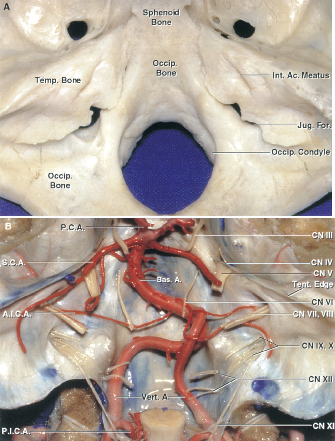
圖1.1。A,顱後窩的上視圖。後窩的骨壁由枕骨、顳骨和蝶骨形成。前顱窩以蝶背、蝶骨後部和枕骨斜坡部分為界;後麵在枕骨鱗狀部分的下部後麵;兩側分別是顳骨的岩狀和乳突部分,以及枕骨的外側部分。顳骨上方的一小部分是由頂骨的下角形成的。B,後顱窩的神經和動脈。12對顱神經中隻有2對完全走行於顱後窩外。天幕沿岩石脊附著,覆蓋後顱窩。 A., artery; Ac., acoustic; A.I.C.A., antero- inferior cerebellar artery; Bas., basilar; CN, cranial nerve; For., foramen; Int., internal; Jug., jugular; Occip., occipital; P.C.A., posterior cerebral artery; P.I.C.A., posteroinferior cer- ebellar artery; S.C.A., superior cerebellar artery; Temp., tem- poral; Tent., tentorial; Vert., vertebral.
幕的表麵與幕的下表麵相接並符合(圖1.2-1.4)。小腦表麵的前內側,由前蚓部形成的頂點是小腦的最高點。這個表麵從它的前內側到後外側邊緣向下傾斜。在小腦幕表麵,蚓部到半球的過渡是平滑的,沒有枕下表麵蚓部和半球之間的深裂。深切痕,小腦前、後切痕,在小腦幕表麵中線的前後邊緣有溝槽。腦幹與小腦前切相吻合,小腦鐮與小腦後切相吻合(圖1.2)。
前側緣,分隔小腦幕和岩麵,有平行於岩上竇的側部(前外側緣)和麵向中腦的內側(前內側緣),形成中腦和小腦裂的後緣。由前外側和前內側緣交界處形成的前角指向三叉神經後根起源的前方上方。小腦幕和枕下表麵之間的後緣也有一個側麵和一個內側部分。外側部分(後外側緣)與側竇平行,毗鄰側竇,分離枕下和幕表麵半球部分,短內側部分(後內側緣)麵對小腦後切刀,分離兩表麵蚓部部分。側角,由前外側和後外側邊緣的交界處形成,位於乙狀竇、側竇和岩上竇的交界處。脈通常會聚於前角和側角。
幕表麵半球部分包括四角小葉、單葉和上半月形小葉,蚓部包括頂葉、declive和小葉。蚓部及相關的半球部分,從上到下依次為頂葉和四角小葉,十分葉和單葉小葉,小葉和上半月形小葉。小腦幕表麵在其主要裂——小腦幕裂的位置被分為前腦幕和後腦幕。該裂隙位於半球上的四角小葉和單葉以及蚓部上的頂葉和頂葉之間,也被稱為初級裂隙。斜坡後裂將單純小葉和上半月形小葉分開。該麵葉間裂縫從中線前外側穿過,與岩麵上半部的裂縫連續。
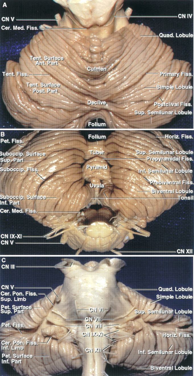
圖1.2。小腦幕、枕下和岩側表麵。A,幕表麵麵對幕的下表麵。前側蚓部是小腦幕表麵的最上部分。這個表麵向下傾斜到它的後緣和側緣。這個表麵的蚓類分支比它們相應的半球部分要好。右側列出了用於蠕蟲 和小腦幕表麵半球細分的經典命名法 ,左側列出了我們的簡化命名法。幕尖小葉和四角小葉對應小腦幕表麵的前部,幕尖小葉、單葉和部分上半月形小葉對應小腦幕表麵的後部。在我們的術語中,將小腦幕表麵分為前後兩部分的裂縫被稱為小腦幕裂,但在舊的術語中,它是主要的裂縫。這個裂縫將半球表麵在四邊形和簡單的小葉和蚓部之間分開。 The anterior part of the superior surface of the cerebellum surrounds the posterior half of the midbrain to form the cerebellomesencephalic fissure. B, suboccipital surface. The suboccipital surface is located below and between the sigmoid and lateral sinuses and is the surface that is exposed in a wide bilateral suboccipital craniectomy. The classical nomenclature applied to this surface is shown on the right, and our simplified nomenclature is on the left. The vermis sits in a large median depression, the posterior cerebellar incisura, between the cerebellar hemispheres. According to classical nomenclature, the portions of the vermis within the incisura from above to below are the folium, tuber, pyramid, and uvula. The parts of the hemispheric surface from above to below are the superior and inferior semilunar and biventral lobules and the tonsils. These lobules extend beyond the suboccipital surface to the other surfaces of the cerebellum. The prebiventral fissures between the inferior semilunar and the biventral lobules separate the hemispheres into superior and inferior parts, and the prepyramidal fissure between the pyramid and tuber separates the vermis into superior and inferior parts. We refer to the union of the prebiventral and the prepyramidal fissures that divide the suboccipital surface into superior and inferior parts as the suboccipital fissure. From below to above the corresponding vermian and hemispheric parts are the uvula and the tonsils, the pyramid and the biventral lobules, the tuber and inferior semilunar lobules, and the folium and the superior semilunar lobules. The petrosal (horizontal) fissure, the most prominent fissure on the petrosal surface, extends onto the suboccipital surface and divides the superior half of the suboccipital surface between the superior and inferior semilunar lobules. The cerebellomedullary fissure extends superiorly between the cerebellum and medulla. C, petrosal surface. The petrosal surface faces forward toward the petrous temporal bone and is the surface that is retracted to surgically expose the cerebellopontine angle. The classical nomenclature applied to this surface is shown on the right, and our simplified nomenclature is on the left. The petrosal fissure divides the petrosal surface into superior and inferior parts. The superior part is formed by the quadrangular, simple, and a small part of the superior semilunar lobules. The inferior part is formed by the inferior semilunar and biventral lobules and the tonsil. The cerebellopontine fissures are V-shaped fissures formed where the cerebellum wraps around the pons and the middle cerebellar peduncles. These fissures have a superior and an inferior limb, which meet at a lateral apex. The petrosal fissure extends laterally from the apex of the cerebellopontine fissures. Ant., anterior; Cer.Med., cerebellomedullary; Cer.Pon., cerebellopontine; CN, cranial nerve; Fiss., fissure; Horiz., horizontal; Inf., inferior; Pet., petrosal; Post., posterior; Quad., quadrangular; Suboccip., suboccipital; Sup., superior; Tent., tentorial.
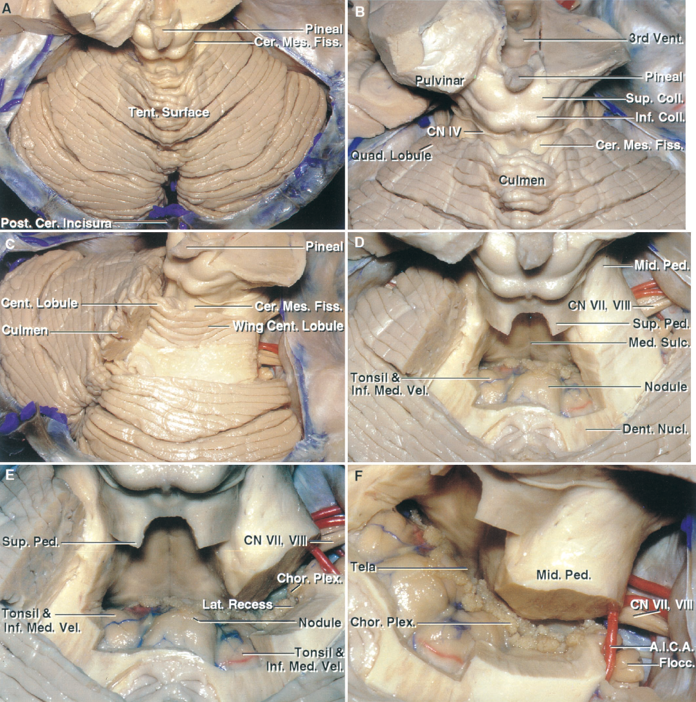
圖1.3。小腦幕表麵和小腦間裂。首先,幕部表麵正對著被移除的幕部。表麵從頂端向下傾斜到後緣和側緣。小腦幕表麵上部包圍中腦後半部分,形成小腦後裂後唇。小腦前切跡,即腦幹與小腦幕表麵前部相吻合的切跡,位於小腦前切跡,即小腦鐮與小腦相吻合的切跡,位於小腦後切跡。B,小腦脊髓裂放大圖,該裂向下延伸至中腦和小腦之間。後唇的淺部由中線的唇峰和外側的四角小葉組成。四叉腦池向尾部從鬆果體延伸至小腦裂。C,嘴部被切除,露出中央小葉和它的翅膀,它們構成小腦裂後唇的一部分。 D, the central lobule and its wings, the lingula, the superior medullary velum, and medial part of the superior cerebellar peduncles have been removed to expose the fourth ventricle. The lower half of the roof is formed in the midline by the nodule and laterally by the inferior medullary velum, which passes laterally above, but is separated from the rostral pole of the tonsils by the cerebellomedullary fissure. E, some of the middle peduncle has been removed to expose the choroid plexus extending through the lateral recess into the cerebellopontine angle below the facial and vestibulocochlear nerves. F, oblique view of the lower half of the roof formed by the inferior medullary velum and the tela choroidea in which the choroid plexus arises. The inferior medullary velum arises on the surface of the nodule and extends laterally to blend into the flocculus and, with the flocculus and nodule, forms the flocculonodular lobe of the cerebellum. A.I.C.A., anteroinferior cerebellar artery; Cent., central; Cer., cerebellar; Cer.Mes., cerebellomesencephalic; Chor., choroid; CN, cranial nerve; Coll., colliculus; Dent., dentate; Fiss., fissure; Flocc., flocculus; Inf., inferior; Lat., lateral; Mid., middle; Med., median, medullary; Nucl., nucleus; Ped., peduncle; Plex., plexus; Post., posterior; Quad., quadrangular; Sulc., sulcus; Sup., superior; Tent., tentorial; Vel., velum; Vent., ventricle.
枕下表麵位於側鼻竇和乙狀竇之間的下方,是三個表麵中最複雜的(圖1.2和圖1.5)。第四腦室和大多數小腦腫瘤的手術入路通常是繞過或穿過這個表麵。它有一個很深的垂直凹陷,即小腦後切骨,其中包含硬腦膜皺襞,即小腦鐮。蚓部折疊形成這個切骨內的皮質表麵。切骨的側壁由小腦半球的內側形成。深裂,即蚓腦裂,將蚓腦與半球分開。切骨內的蚓麵呈菱形。菱形地層的上半部分呈金字塔狀,稱為金字塔。葉和塊莖,在金字塔之上,形成蚓部的枕下部分的先端。菱形結構的下半部分,即小舌,向下投射在扁桃體之間,從而模仿口咽的情況。 The rostromedial margin of the tonsils borders the tapering edges of the uvula. The nodule, the lowermost subdivision of the vermis, is hidden deep to the uvula. The strip of vermis within the incisura is broadest at the junction of the pyramid and uvula. Inferiorly, the posterior cerebellar incisura is continuous with the vallecula cerebelli, a cleft between the tonsils that leads through the fora- men of Magendie into the fourth ventricle.
枕下表麵的半球部分由上、下半月小葉和雙腹小葉及扁桃體組成,蚓部由小葉、塊莖、錐體和小舌組成。蚓部及相關的半球部分由上至下依次為小葉和上半月形小葉、塊莖和下半月形小葉、金字塔小葉和雙腹小葉、小舌和扁桃體。
枕下表麵在它的主要裂縫,枕下裂縫,分為上部分和下部分。枕骨下裂有蚓部和半球部。蚓部裂,即錐體前裂,將塊莖和錐體分開;半球部裂,即雙腹前裂,將雙腹小葉和下半月形小葉分開。雙腹前裂和椎體前裂在半球交界處連續,共同形成枕下裂。岩裂縫是岩麵上的主要裂縫,從岩麵延伸至枕下麵,在側麵分離上半月小葉和下半月小葉,在內側分離小葉和塊莖。扁桃體雙腹裂將扁桃體和雙腹小葉分開。
扁桃體是一個半球組成部分,是阻礙進入第四腦室尾部的最突出的結構(圖1.5和圖1.6)。每個扁桃體都是枕下表麵下內側的卵球形結構,通過一束被稱為扁桃體梗的白質束與小腦的其餘部分沿其上外側邊界相連。其餘扁桃體表麵為遊離表麵。下極和後表麵麵對枕大池,在枕下表麵剩餘部分的內側下方可見。每個扁桃體的外側表麵被雙腹小葉覆蓋,但與雙腹小葉之間有一個狹窄的裂口,除了在上方的扁桃體花序梗水平處。內側、前表麵和上表麵均麵對其他神經結構,但與它們之間有狹窄的裂隙分隔。每個扁桃體的前表麵與延髓的後表麵由小腦延髓裂分開。扁桃體的內側表麵通過一個狹窄的裂縫互相麵對,這個裂縫就是小關節,它通向第四腦室。每個扁桃體上極的腹側麵向構成第四腦室頂部下半部分的三個結構(脈絡膜層、下髓膜和結節)。上極與周圍結構由小腦延髓裂的後伸分開,稱為遠側扁桃體裂或上側扁桃體裂。 The posterior aspect of the superior pole faces the uvula medially and the biventral lobule laterally.
岩骨或前表麵麵對岩骨、腦幹和第四腦室的後表麵(圖1.2和圖1.7)。岩麵側麵或半側部分靠在岩骨上,並縮回露出橋小腦角。岩麵正中或蚓部有一深縱溝,即小腦前切刀,環繞腦幹和第四腦室的後表麵。由於第四腦室位於蚓部的上、下兩部分之間,因此岩麵的左右兩部分並沒有由連續的蚓部條從一側到另一側連接起來,枕下和幕部也是如此。第四腦室口側的蚓部是舌骨、中央小葉和唇瓣,第四腦室尾側的蚓部是小結和小舌。半球表麵由中央小葉的翅膀和四邊形、單側、雙腹、上半月形小葉和下半月形小葉的前表麵、扁桃體和小葉的前表麵組成。蚓部和相關的半球部分是中央小葉和中央小葉的翅膀,唇瓣和四角小葉,小結和小葉,小舌和扁桃體。該表麵的主要裂縫岩縫,又稱水平裂縫,將岩麵分成上、下兩部分,並在上、下半月形小葉之間延伸至枕下表麵。
第四腦室是位於小腦和腦幹之間的寬闊的帳篷狀中線腔。它的喙部通過導水管與第三腦室相連,尾部通過Magendie孔與枕大池相連,側麵通過Luschka孔與橋小腦角相連。大部分顱神經在其底部附近生長。它有一個屋頂,一個地板和兩個側麵凹。它位於小腦的腹側,橋腦和延髓的背側,小腦蒂的內側。
室頂呈帳篷狀(圖1.8和圖1.9)。頂板從導水管下方的狹窄喙端向外側和後方擴展,直至頂和側隱窩水平,即其高度和寬度最大的位置,從那裏逐漸變細,直至馬琴蒂孔水平的狹窄尾端。屋頂的頂端,尖頂,把它分成高級和低級部分。上半部與下半部有明顯不同,下半部主要由薄的膜層構成,上半部由較厚的神經結構構成。
形成頂板的結構的外表麵或腦池表麵與小腦和腦幹之間的裂隙密切相關。小腦胚胎折疊在腦幹周圍形成的三個裂縫是小腦-腦裂,它向下延伸到小腦和中腦之間,並與頂部的上半部密切相關(圖1.3和圖1.4);橋腦小腦裂,由小腦在橋腦外側折疊形成,與橋腦外側隱窩密切相關(圖1.7和圖1.8);小腦延髓裂,它向上延伸於小腦和延髓之間,與頂板下半部密切相關(圖1.5和圖1.6)。
小腦的動脈和靜脈在每個裂縫中走行。小腦上動脈(SCA)和小腦脊髓裂靜脈走行於小腦脊髓裂內,小腦前下動脈(AICA)和小腦脊髓裂靜脈與小腦脊髓裂相關,小腦後下動脈(PICA)和小腦脊髓裂靜脈與小腦脊髓裂密切相關。這些動脈和靜脈將在接下來關於小腦動脈和後窩靜脈的兩章中進行回顧(10,18,19)。
每條裂縫都與相鄰的裂縫相通。橋小腦裂圍繞小腦中梗嘴麵與小腦脊髓裂尾緣連續,圍繞小腦中梗尾緣與小腦延髓裂嘴緣連續。這些裂隙將在第四腦室頂板和側隱窩的討論中進行更詳細的討論。
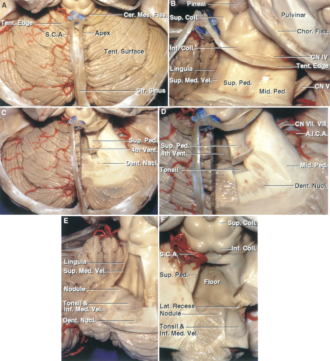
圖1.4小腦幕表麵及小腦脊裂。A,小腦幕麵麵對著小腦幕,從位於小腦幕頂下方的小腦幕頂向下傾斜。小腦間裂向前延伸,位於小腦和中腦之間。枕下表麵,蚓部是最高的部分,與枕下表麵不同,枕下表麵蚓部在小腦半球之間折疊成一個很深的裂縫,即切骨。直竇和小腦幕邊緣均被保留。SCA出於小腦裂,供應十麵。B,切除小腦裂後唇右半部分。裂隙的前壁在中線由小腦丘板和舌骨形成,外側由上小腦蒂形成。小腦中部的花梗包裹在上花梗的外側表麵。滑車神經起源於下丘下麵。 C, the right half of the lingula and superior medullary velum have been removed to expose the fourth ventricle. Additional white matter has been removed below the right superior peduncle to expose the dentate nucleus in which the superior peduncular fibers arise. D, enlarged view. The dentate nucleus appears to wrap around the rostral pole of the tonsil. E, oblique view into the fourth ventricle. Additional cerebellum has been removed to expose the nodule and rostral pole of the tonsil. The dentate nucleus wraps around the rostral pole of the tonsil. The upper half of the roof is formed by the superior medullary velum, which has the lingula layered on its outer surface. The upper part of the lower half of the roof is formed by the nodule in the midline and by the inferior medullary velum laterally. The inferior medullary velum, an almost transparent membrane, stretches laterally across the upper pole of the tonsil. F, the left half of the upper part of the roof has been removed. The velum arises on the nodule and sweeps laterally above both tonsils. The SCA courses within the cerebellomesencephalic fissure. A.I.C.A., anteroinferior cerebellar artery; Cer.Mes., cerebellomesencephalic; Chor., choroidal; CN, cranial nerve; Coll., colliculus; Dent., dentate; Fiss., fissure; Inf., inferior; Lat., lateral; Med., medullary; Mid., middle; Nucl., nucleus; Ped., peduncle; S.C.A., superior cerebellar artery; Str., straight; Sup., superior; Tent., tentorial; Vel., velum; Vent., ventricle.
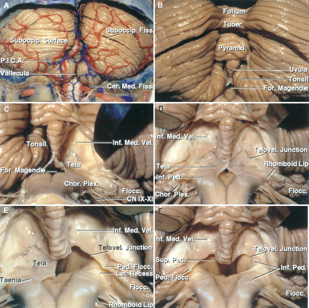
圖1.5。小腦枕下表麵和小腦延髓裂。A,枕下表麵位於乙狀竇和側鼻竇之間的下方,是寬枕骨下骨瓣切除術暴露的表麵。蚓部位於兩個半球之間的凹陷處,即小腦後切骨。小腦延髓裂沿室頂下半部分在小腦和延髓之間向上延伸。小淋巴管在扁桃體之間向上延伸,並通過馬琴蒂孔與第四腦室相通。異食癖支配著枕下表麵。B,放大圖。蚓部腦室後麵的下部是錐體和小舌。C,切除右側扁桃體,露出由下髓膜和脈絡膜形成的頂部下部。 The nodule on which the velum arises is hidden in front of the uvula. The uvula hangs downward between the tonsils, thus mimicking the situation in the oropharynx. The choroid plexus arises on the inner surface of the tela and extends through the foramen of Luschka behind the glossopharyngeal and vagus nerve. The inferior medullary velum arises on the surface of the nodule, drapes across the superior pole of the tonsil, and blends into the flocculus laterally. D, both tonsils have been removed to expose the inferior medullary velum and tela choroidea bilaterally. The telovelar junction is the junction between the velum and tela. The cerebellomedullary fissure extends upward between the rostral pole of the tonsil on one side and the tela choroidea and inferior medullary velum on the opposite side. The segment of the PICA passing through this cleft is called the telovelotonsillar segment. The rhomboid lip is a sheet-like layer of neural tissue attached to the lateral margin of the ventricular floor, which extends posterior to the glossopharyngeal and vagus nerves and joins the tela choroidea to form a pouch at the outer extremity of the lateral recess. E, the right half of the tela has been removed to expose the ventricle and the lateral recess. The inferior medullary velum extends laterally to form a peduncle, the peduncle of the flocculus, which blends into the flocculus at the outer margin of the lateral recess. F, the tela has been removed on both sides. The lateral wall of the upper half of the ventricle is formed by the superior cerebellar peduncles. The inferior cerebellar peduncles ascend along the dorsolateral medulla and form the anterior and rostral margins of the lateral recess. Cer.Med., cerebellomedullary; Chor., choroid; CN, cranial nerve; Fiss., fissure; Flocc., flocculus; For., foramen; Inf., inferior; Lat., lateral; Med., medullary; Ped., peduncle; P.I.C.A., posteroinferior cerebellar artery; Plex., plexus; Suboccip., suboccipital; Sup., superior; Telovel., telovelar; Vel., velum.

圖1.6。枕下表麵和小腦延髓裂。A、小腦延髓 裂在扁桃體和 延髓之間向上延伸。通過 切開扁桃體的蒂,切除了兩個扁桃體。切除 扁桃體暴露出下髓膜 和形成 室頂下部的脈絡膜。下小腦 梗沿後外側 髓質上升。脈絡膜叢起於脈絡膜叢的內 表麵。帶絛蟲 是脈絡膜的附著點 沿著腦室的下外側邊緣 底。延髓連接處是 下髓膜與 脈絡膜連接的部位。這個結節, 下髓膜在其上升起,隱藏在小舌深 處。 B, the tela, in which the choroid plexus arises, has been removed to expose both lateral recesses. The superior cerebellar peduncle forms the lateral wall of the upper half of the ventricle. The inferior cerebellar peduncle forms the anterior and upper margin of the lateral recess. The middle cerebellar peduncle, which forms a large prominence on the lateral surface of the pons, is separated from the ventricular surface by the superior and inferior cerebellar peduncles. C, lateral surface of the left tonsil. All of the tonsillar surfaces, except at the superolateral margin, are free surfaces. The peduncle of the tonsil, located along the superolateral margin of the tonsil, attaches the tonsil to the remainder of the cerebellum. The posterior surface of the tonsil faces the cisterna magna. The medial surface faces the other tonsil. The anterior surface faces the posterior medulla. The rostral pole faces the inferior medullary velum and tela choroidea. The lateral surface below the peduncle of the tonsil faces the biventral lobule. D, posterior view of the left tonsil. The peduncle of the tonsil is located along the superolateral margin. Dividing the narrow peduncle allows the tonsil to be separated from the remaining cerebellum. Bivent., biventral; Inf., inferior; Lat., lateral; Med., medullary; Ped., peduncle; Post., posterior; Rost., rostral; Sup., superior; Telovel., telovelar; Vel., velum.

圖1.7。腦幹、岩麵、橋小腦裂。一個,斜視圖。小腦的岩麵向前朝向岩骨,是縮回露出橋小腦角的表麵。橋小腦裂,也可稱為橋小腦角,是小腦包裹橋小腦和小腦中腳梗形成的v型裂。上、下肢在岩裂隙的前端橫向會合,岩裂隙將岩麵分為上、下兩部分。小葉和脈絡膜叢從裂下肢上方的Magendie孔向外側延伸。基底溝是位於腦橋前表麵的淺縱溝,容納基底動脈。B,放大圖。 The petrosal fissure extends laterally from the apex of the cerebellopontine fissure. The abducens nerve arises in the medial part of the pontomedullary sulcus rostral to the medullary pyramids. The facial and vestibulocochlear nerves arise just rostral to the foramen of Luschka near the flocculus at the lateral end of the pontomedullary sulcus. The hypoglossal nerves arise anterior to and the glossopharyngeal, vagus, and accessory nerves arise posterior to the olives. Cho- roid plexus protrudes from the foramen of Luschka behind the glossopharyngeal and vagus nerves. C, enlarged view of another brain- stem. The facial and vestibulocochlear nerves join the brainstem 2 or 3 mm rostral to the glossopharyngeal nerve on a line drawn dorsal to the olive along the origin of the rootlets of the glossopharyngeal, vagus, and accessory rootlets. The rhomboid lip, a thin neural membrane in the ventral margin of the lateral recess, extends laterally behind the glossopharyngeal, vagus, and accessory nerves with the choroid plexus. D, enlarged view of another cerebellopontine fissure. The cerebellopontine angle is the area situated between the superior and inferior limbs of the cerebellopontine fissure. The glossopharyngeal, vagus, and accessory nerves arise near the inferior limb, dorsal to the olive, and anterior to the choroid plexus protruding from the foramen of Luschka. The facial and vestibulocochlear nerves arise in the midportion of the fissure and the trigeminal nerve near the superior limb of the fissure. The hypoglossal rootlets arise in front of the olive and the cranial rootlets of the accessory nerve. Bas., basilar; Cer.Pon., cerebellopontine; Chor., choroid; CN, cranial nerve; Fiss., fissure; Flocc., flocculus; For., foramen; Inf., inferior; Mid., middle; Ped., peduncle; Pet., petrosal; Plex., plexus; Sup., superior.

圖1.8。f。腦幹,第四腦室,岩側小腦麵。逐步前曝光。A,岩麵向前麵對顳骨的後表麵。第四腦室位於腦橋和髓質後麵。中腦和腦橋由腦橋腦溝隔開,腦橋和延髓由腦橋腦溝隔開。三叉神經起源於腰橋。外展神經起源於橋髓溝的內側,靠近髓錐體。麵神經和前庭耳蝸神經起源於橋延髓溝的外側端,緊靠盧施卡孔。 The hypoglossal nerves arise anterior to the olives and the glossopharyngeal, vagus, and accessory nerves arise posterior to the olives. Choroid plexus protrudes from the foramen of Luschka behind to the glossopharyngeal and vagus nerves. B, right cerebellopontine angle following removal of some of the medulla. The foramen of Luschka opens into the cerebellopontine angle below the junction of the facial and vestibulocochlear nerves with the lateral end of the pontomedullary sulcus. Choroid plexus protrudes from the lateral recess and fora- men of Luschka behind the glossopharyngeal, vagus, and accessory nerves. The cerebellopontine fissure, a V-shaped fissure formed by the cerebellum wrapping around the pons and middle cerebellar peduncle, has a superior and inferior limb that define the margins of the cerebellopontine angle. The superior limb extends above the trigeminal nerve and the inferior limb passes below the flocculus and the nerves that pass to the jugular foramen. C, the part of the pons and medulla forming the left half of the floor of the ventricle has been removed to expose the fastigium, which divides the ventricular roof into superior and inferior parts. D, the right half of the pons has been removed to expose the upper half of the roof. The superior part of the roof is formed by the superior medullary velum. The rostral part of the lower half of the roof is formed by the nodule and inferior medullary velum and the caudal part is formed by the tela choroidea, a thin arachnoid-like membrane, in which the choroid plexus arises. E, the cerebellopontine fissure has upper and lower limbs, which meet at a later apex located at the medial end of the petrosal fissure, also called the horizontal fissure, which divides the petrosal surface into upper and lower halves. The junction of the pons and medulla, which forms the anterior wall of the left lateral recess, has been removed to expose the choroid plexus protruding through the lateral recess into the cerebellopontine angles. F, enlarged view. The choroid plexus protrudes laterally through the foramen of Luschka into the cerebellopontine angle below the floc- culus. Cer.Pon., cerebellopontine; Chor., choroid; CN, cranial nerve; Fiss., fissure; Flocc., flocculus; For., foramen; Inf., infe- rior; Lat., lateral; Med., medial, medullary; Mid., middle; Ped., peduncle; Pet., petrosal; Plex., plexus; Pon.Med., pontomedul- lary; Pon.Mes., pontomesencephalic; Seg., segment; Sulc., sulcus; Sup., superior; Vel., velum.

圖1.8。G-J。腦幹,第四腦室,岩側小腦麵。G,左側髓質已被切除。屋頂的上半部由上髓膜形成,外側有蚓舌層。上蓋的下半部分由起源於結節表麵的下髓膜和起源於脈絡膜叢的脈絡膜層組成。脈絡膜叢由成對的l形條紋組成,有內、外側兩段。外側段向外側延伸通過Luschka孔,內側段向縱向延伸通過Magendie孔。H,切除右側脈絡膜層和脈絡膜叢,露出右側扁桃體上極。我,右小腦扁桃體被切除了。 All of the surfaces of the tonsils are free surfaces except the superolateral margin, the site of the tonsillar peduncle, a bundle of white matter, which attaches the tonsil to the remainder of the cerebellum. The inferior medullary velum is a thin membranous layer of neural tissue that arises on the nodule and extends laterally above the rostral pole of the tonsil to blend into the flocculus and form the flocculonodular lobe of the cerebellum. The cranial loop of the PICA courses between the rostral pole of the tonsil and the inferior medullary velum. J, both tonsils have been removed. The inferior medullary velum sweeps laterally from the surface of the nodule.

圖1.9。f。後的觀點。逐步解剖檢查下髓膜、齒狀核、扁桃體、小腦髓裂和小腦脊髓裂的關係。A, pica繞過後延髓到達小腦延髓裂的下緣。左異食癖在扁桃體下極周圍活動。右側異食癖在沿扁桃體內側表麵上升之前,先下降到扁桃體下方的枕骨大孔處。B, pica在扁桃體和延髓之間上升,到達扁桃體和小舌之間的間隙,並供應枕骨下表麵。C,右異食癖的後髓段分為支配蚓部和蚓旁區的內側幹和支配半球的外側幹。D,小腦斜冠狀麵切片顯示扁桃體嘴側極與下髓膜和齒狀核的關係。 The dentate nucleus is located above the posterolateral part of the ventricular roof, near the fastigium, where it wraps around, and is separated from, the rostral pole of the tonsil by the inferior medullary velum. The left tonsil has been removed while preserving the left half of the inferior medullary velum. The SCAs course in the cerebellomesencephalic fissure. The PICA passes between the walls of the cerebellomedullary fissure formed above by the inferior medullary velum and below by the upper pole of the tonsil. E, both tonsils have been removed. The PICAs ascend through the cleft between the inferior medullary velum and rostral pole of the tonsil. F, the superior part of the ventricular roof has been removed and the nodule and the inferior medullary velum has been folded downward to expose the floor. A., artery; Cer. Med., cerebellomedullary; Cer.Mes., cerebellomesencephalic; CN, cranial nerve; Dent., dentate; Fiss., fissure; Inf., inferior; Lat., lateral; Med., medial, medullary; Nucl., nucleus; Ped., peduncle; P.I.C.A., posteroinferior cerebellar artery; S.C.A., superior cerebellar artery; Suboccip., suboccipital; Telovel. Ton., telovelotonsillar; Vel., velum; Vent., ventricle; Vert., vertebral.

圖1.9。G-J。後的觀點。G是脈絡膜總層,脈絡膜叢在其中產生,它向下折疊,露出了地板的下部。H,左側外側隱窩及Luschka孔放大圖。菱形唇是一層薄薄的神經組織,從外側隱窩的前緣向外側延伸,與脈絡膜一起在外側隱窩的外緣形成一個袋。脈絡膜叢通過側隱窩和Luschka孔延伸至橋小腦角。I,骨層已被切除,以暴露位於結節上方和下方的部分地板和下髓膜。J,結節和下髓膜已被切除,以暴露整個基底,其在中線被正中溝和顱底分為橋核部分、連接部分和髓質部分。上足梗和下足梗麵對心室表麵。 The middle cerebellar peduncle is separated from the ventricular surface by the superior and inferior peduncles. Chor., choroid; Dent., dentate; Inf., inferior; Lat., lateral; Med., median, medullary; Mid., middle; Nucl., nucleus; P.I.C.A., posteroinferior cerebellar artery; Ped., peduncle; Plex., plexus; Sup., superior; Vel., velum.

圖1.9。K、l驗視圖。K,第四腦室底部放大圖。中間溝以中線縱向分隔地板。地板的每一半都被一個不規則的溝縱向分割,溝限,加深到麵丘和舌下三角形成上凹和下凹。藍斑細胞的暗區位於局限性溝的嘴端。髓紋在外側隱窩處橫過地麵。舌下核、迷走神經核和舌後區在地板的下部一個疊在另一個上麵,形成筆尖的結構,因此,該區域被稱為scriptorius菖蒲。L,第四心室底層。小腦上梗成對的靜脈在上梗外表麵走行,並向上連接形成小腦腦裂靜脈。 The median posterior medullary vein ascends on the medulla and splits into the paired veins of the inferior cerebellar peduncle at the caudal margin of the floor. That left vein is hypoplastic. The left vein of the cerebellomedullary fissure passes along the lateral recess and ascends to join the petrosal group of veins in the cerebellopontine angle. Cer.Med., cerebellomedullary; Cer., cerebellar; CN, cranial nerve; Coll., colliculus; Emin., eminence; Fiss., fissure; Hypogl., hypoglossal; Inf., inferior; Med., median, medullary; Mid., middle; Ped., peduncle; Post., posterior; Striae Med., Stria medullaris; Sup., superior; V., vein.
第四腦室頂上部的心室表麵分為單個正中部和兩個側部(圖1.3和圖1.4)。中間部分由上髓膜形成,外側部分(也稱為外側壁)由小腦梗的內表麵形成。上髓膜是一層薄薄的白質層,橫跨上小腦梗之間,在其外表麵有舌骨,蚓部的最上麵部分。它在頂部與下髓膜相連。各側壁心室表麵的嘴端部分由小腦上梗的內側形成,尾端部分由小腦下梗形成。
小腦中梗雖然是三個小腦梗結合形成的纖維束中最大的組成部分,但其內側表麵的下、上梗纖維將其與心室表麵分開(圖1.9)。小腦下腳梗的纖維在髓質後外側上升,在三個腳梗結合形成的纖維束的後內側後轉,在外側隱窩上緣室麵和外側壁下部排列。小腦上梗的纖維起於齒狀核,在小腦中梗內側上升,形成外側壁上部的室麵。
構成頂板上部結構的腦池(外)麵也構成小腦脊膜裂的前壁。該裂向下延伸至小腦和中腦之間,從上看呈v形(圖1.3和圖1.4)。這個裂縫也被稱為小腦中央前裂。中腦背側位於v形切跡的四肢內,小腦形成外側緣,腦尖位於後部。裂縫的內壁構成了頂板上部分的外表麵,由舌骨、小腦上花序梗的背側表麵和小腦中花序梗的嘴側表麵組成。舌骨是一種細長的蚓舌,位於上髓膜的外表麵。小腦上梗在舌骨兩側形成平滑的縱向突起,然後消失在小腦丘下方的中腦。小腦中部花梗的嘴端表麵似乎包裹著小腦上部花梗的尾緣。一個淺溝,即小花梗間溝,標誌著小腦梗上和中的交界處。腦蒂間溝與腦橋腦溝(橋腦與中腦之間的橫溝)前方連續,與中腦溝外側連續,為腦蒂背側的縱裂。 The trochlear nerves arise in the cerebellomesencephalic fissure below the inferior colliculi and pass anterolateral to exit the anterior part of the fissure. The outer wall of the cerebellomesencephalic fissure is formed by the culmen and the central lobule and its wings.
頂部上部分隔腦室和腦池表麵的神經結構在上髓膜和舌骨區最薄,在小腦蒂區最厚。每個側壁的嘴側部分僅由小腦上梗形成,尾側部分由三個小腦上梗合並後形成。
從頂尖到其與底板的下邊緣連接處,屋頂的下部分向腹側傾斜,略向尾側傾斜(圖1.3-1.6)。腦室麵和腦池麵由相同的結構構成,脈絡膜層和下髓膜,除了在吻側中線處,腦室麵由結節形成,腦池麵由小舌形成。脈絡膜叢附著在脈絡膜層的心室表麵。
心室表麵分為由結節和下髓膜形成的顱部和由脈絡膜形成的尾部。下髓膜是一層膜,是小腦結節和形成原始小腦絮凝結節葉的小腦之間的唯一連接(14)(圖1.8和1.9)。它是一薄的雙側半透明蝶狀神經組織片,向內側混入結節的腦室表麵,橫向伸展,但通過小腦延髓裂狹窄的嘴側延伸與扁桃體上極分離。它融入每個側隱窩的背緣,形成每個小葉的花序梗。下髓質筋膜與上髓質筋膜在頂水平連續。尾部與脊索相連。
脈絡膜層形成頂板下段的尾部和每個側隱窩的下壁(圖1.5、1.6和1.9)。它由兩層薄的半透明膜組成,每一層的厚度相當於蛛網膜,中間夾著由脈絡膜動脈和靜脈組成的血管層。脈絡膜叢從脈絡膜層的腦室表麵投射到第四腦室。下髓膜與脈絡膜相連的線,即延綿交界處,從結節延伸至每個側隱窩。脈絡膜總層向下沿著狹窄的白色脊(帶絛蟲),從每個扁桃體的上極周圍的瞼緣交界處向下掃過,並與底部的下外側邊緣連接,它們在門柱彙合。在顱骨上,帶絛蟲向外側轉動小腦下梗,並沿外側隱窩的下緣水平通過。脈絡膜總區並沒有完全包圍第四腦室下半部分,而是有3個進入蛛網膜下腔的開口:位於側隱窩外緣的成對的Luschka孔和位於第四腦室尾尖的Magendie孔。
頂板尾半側的腦池(外)麵麵對並與小腦延髓裂密切相關(圖1.6、1.8和1.9)。這個裂縫是大腦中最複雜的裂縫之一。裂的腹壁由延髓後表麵、下延髓筋膜和脈絡膜構成。裂的背壁是由小舌在中線和扁桃體和雙腹小葉在側麵形成的。它向上延伸至側隱窩水平,並在扁桃體上極周圍與大池相連,通過與第四腦室的Magendie孔,並在與橋小腦裂周圍的Luschka孔相連。扁桃體嘴端麵對下髓膜、脈絡膜、小舌的扁桃體周部和裂上部的雙腹小葉(圖1.3-1.6)。扁桃體之間裂的那部分,脈絡膜層,和下髓膜被稱為遠側扁桃體裂,而這個裂在扁桃體上極之上的上延伸部分被稱為上扁桃體裂。
側凹是由屋頂和地板結合形成的狹窄彎曲的袋狀結構。它們在小腦梗下方橫向延伸,通過Luschka孔向橋小腦角開放(圖1.3、1.5、1.6和1.8)。每個側隱窩的腹側壁由地板與菱形唇的連接部分形成,菱形唇是一層片狀的神經組織,從地板向外側延伸,並與脈膜瓣聯合,在側隱窩的外端形成一個袋。每個側隱窩的嘴側壁由小腦梗尾緣形成。小腦下梗在底麵腹側向外側隱窩向上走行,在腦橋下部向後轉,形成吻側壁心室麵。小葉的花梗連接下髓膜和小葉在外側隱窩的背緣交叉。尾壁由從帶絛蟲側麵延伸至小葉梗的脈絡膜層形成。雙腹小葉位於側隱窩的背側。小葉在側隱窩的外端上。舌咽神經和迷走神經的細根起於側隱窩的腹側,麵神經起於側隱窩的嘴側。 The fibers of the vestibulocochlear nerve cross the floor of the recess.
每個側隱窩沿橋小腦裂進入橋小腦角(圖1.7)。這個v型裂是由小腦半球在腦橋外側和小腦中腳蒂周圍折疊形成的。它的上肢位於小腦中梗喙部和岩麵上部之間,下肢位於小腦中梗尾部和岩麵下部之間。小腦中部的花梗充滿兩肢之間的間隙。裂縫的頂端位於上、下四肢相交的外側。岩裂縫從頂端向側麵延伸。外側隱窩和Luschka孔通向下肢內側。沿著下肢的其他結構有小葉、菱形唇、從盧什卡前孔突出的脈絡膜叢、麵部神經、前庭耳蝸神經、舌咽神經和迷走神經。三叉神經起源於腦橋沿著裂縫的上肢。
橋小腦裂上肢在三叉神經上方與小腦裂外側部相通,下肢在外側隱窩水平與小腦延髓裂外側部相通。小葉在橋小腦和小腦延髓裂隙彙合處向橋小腦角突入。前庭耳蝸神經和麵神經在小葉前上進入腦幹,舌咽神經絲和迷走神經在小葉前下交叉。
後窩的脈絡膜叢由兩個倒l形的條紋組成,它們出現在脈絡膜層的心室表麵,位於中線兩側(圖1.3和圖1.8)。與正中平麵相鄰的成對縱肢為內側節段。起源於內側節嘴端的橫肢稱為外側節。整個結構呈現出字母T的形式,然而,它的垂直分支是雙的。
內側節段位於椎頂中線附近,外側節段通過外側隱窩和Luschka孔延伸至橋小腦角。內側段從扁桃體前結節水平延伸到Magendie孔水平。每個內側節又細分為嘴側或結節部分和尾側或扁桃體部分。結節部與側節交界處最寬。扁桃體的部分在扁桃體的前麵通過Magendie孔向下延伸。內側節的嘴端和尾端常融合。
外側段形成一個橫向的邊緣,附著在內側段的嘴側部分,並通過外側隱窩平行於瞼緣交界處延伸到橋小腦角。每個外側段又細分為內側或花序梗部分和外側或絮狀部分。花序梗部分形成一個狹窄的邊緣,與內側段的嘴端連續,並附著在覆蓋小腦梗下側隱窩的脈絡膜上。在小腦梗外側緣,絮狀部分與梗狀部分連續,通過Luschka孔突出至小腦梗下方的橋小腦角。
腦幹和腦室底被認為是一起的,因為腦幹形成第四腦室底。後顱窩的腦幹由中腦、腦橋和髓質組成(圖1.7-1.9)。中腦由腦梗、被蓋和頂蓋組成。它以視神經束和腦梗之間的溝為界,以橋腦橋的溝為界。腦梗間窩是腦梗之間的楔形凹陷,其底部有後側穿孔物質。動眼神經的細根起源於眶間窩的深處,並形成後穿孔物外側的眶壁。在椎間窩尾部有一個小凹陷,即盲腸上孔。腦橋腦溝從上盲腸孔繞著腦梗延伸至中腦外側溝,中腦外側溝是被蓋和腦梗之間的垂直溝。
腦橋的腹部從一側到另一側以及從上到下都是凸出的,並且每一側都是連續的,中間有小腦梗。它有一條淺的中線溝,即基底溝,從上邊界延伸到下邊界。三叉神經後根在小腦前角下方的小腦中梗上部出現。橋腦由橋腦溝從延髓向下分隔,橋腦溝從盲腸下孔(中線窩)向外側延伸至橄欖上窩(橄欖嘴側的凹陷)。麵神經和前庭耳蝸神經的細根起源於這個窩的上方舌咽神經的細根和迷走神經起源於它的背側。
髓質前部由麵對斜坡、枕骨大孔前緣和齒狀突喙部的髓錐體形成(圖1.7和圖1.8)。中間溝在金字塔之間的前中線將上髓質分開,並在金字塔交疊處的下髓質處消失,但在交疊處的下方再次出現,並與脊髓的前中間裂尾側連續。髓質外側表麵主要由下橄欖形成,下橄欖位於金字塔外側,並被前外側(橄欖前)溝與金字塔隔開。舌下神經的小根起源於前外側溝。外側表麵後方由舌咽根、迷走神經和後外側(橄欖後)溝背側的副神經的出口劃定,該後外側溝沿橄欖後緣走行,並與下方的脊髓後外側溝相連。外展神經從橋延髓溝口部向金字塔部延伸。延髓的後表麵分為上、下兩部分。上部分位於第四腦室底下半部分的中線,外側由下小腦梗組成。後表麵下段在中線被後正中溝分為兩半,每半部分由纖薄束和結節向內側組成,楔形束和結節向外側組成。髓質後正中溝在中線分隔成對的薄筋膜束,上止於第四腦室的門部,並在下方與脊髓後正中溝相連。 The posterior intermediate sulcus, which separates the gracile and cuneate fasciculi, is continuous inferiorly with the posterior intermediate sulcus of the spinal cord. The lower medulla blends indistinguishably into the upper spinal cord at the level of the C1 nerve roots (Figs. 1.5–1.7).
地板呈菱形(圖1.9)。腹底嘴部三分之二在腦橋後,尾部三分之一在延髓後。顱尖位於腦導水管水平;它的尾端,即閂,位於椎管殘端的喙端,在馬琴蒂孔前;側角經側隱窩和Luschka孔進入橋小腦角。連接側隱窩孔的一條線位於尾椎骨與底椎骨長度中三分之一的連接處,也位於橋腦與髓質連接處。
基底分為三個部分:上部分或橋腦部分,中間部分或連接部分,下部分或髓質部分。上部呈三角形:其頂點位於腦導水管處,其基底由一條假想的線連接小腦梗下緣,其側肢由腦梗內側表麵形成。中間部分是位於小腦梗下緣與帶絛蟲側隱窩下方的脈絡膜附在帶絛蟲上的條帶。中間部分延伸至側凹。下部呈三角形,並在側麵受帶絛蟲的限製,帶絛蟲標記著底板的下外側邊緣。它的尾端,obex,在Magendie孔的前麵。
底在縱向上由嘴端至尾端由中溝對稱分成兩半。溝限,另一個縱向溝,將地板的每一半分成一個凸起的中間地帶,稱為中間隆起,與中線和外側區域接壤,稱為前庭區。
每個正中隆起,即溝限和正中溝之間的條帶,從上到下包含麵神經丘,一個與麵神經相關的圓形突起,以及覆蓋舌下核和迷走神經核及後區的三個三角形區域。所述三個三角形區域是成對的,並沿所述正中溝堆疊,以使所述地板尾部部分具有羽毛或筆尖結構;因此,這個區域被稱為菖蒲。在橋腦橋水平,中位隆起的寬度等於整個橋底的一半,因此溝限與這部分橋底的側限相對應。
局限性溝不連續,在基底的橋腦和髓質部分最突出,在那裏兩點加深形成窩,稱為窩,在基底的交界部分最不明顯。兩個窩中的一個,即上窩,位於基底的橋腦部分,另一個,即下窩,位於基底的髓質部分。在上凹處,正中隆起形成拉長的腫脹,即麵神經丘,它覆蓋著外展神經核和麵神經根的上行段。在底側緣每個溝限的嘴端有一個淺藍色的灰色區域,藍斑,它的顏色是由於一組色素沉著的神經細胞。舌下三角位於下凹的內側,覆蓋在舌下神經的nu上。在下凹的尾部舌下三角和前庭區域的下部之間是一個三角形的暗區,迷走神經三角,它覆蓋在迷走神經的背核上。一條半透明的脊,即分離的索,穿過迷走神經三角的下部。骨後區在正中隆起的下端緊鄰門喙處,在分離索和薄薄結節之間形成一個小的舌狀區域。
前庭區域是地板正中隆起和界限溝外側的部分,在地板的中間部分最寬,形成一個圓形的隆起,延伸到側隱窩。白線,即髓質紋,從外側隱窩區域橫過舌下三角上方的小腦下梗,向中線方向移動,消失在正中溝中。前庭神經核位於前庭區域的下方。聽覺結節由下耳蝸背核和前庭耳蝸神經耳蝸部分形成,在前庭區外側部分突出。
第四腦室的每一壁都有外科重要的動脈關係:心室壁與上半部心室頂密切相關;異食癖與屋頂的下半部分密切相關;AICA與側隱窩和Luschka孔密切相關;基底動脈和椎動脈產生許多到達第四腦室底部的穿通分支(5,7,9,10,18,19)(圖1.9和1.10)。AICA的脈絡膜分支支配著橋小腦角的脈絡膜叢和側隱窩鄰近部分,PICA支配著橋小腦角的脈絡膜叢和側隱窩內側部分(7)。
第四腦室腔內沒有大靜脈。與第四腦室最密切相關的靜脈是小腦與腦幹之間的裂隙和小腦梗上的靜脈(21)。小腦脊髓裂和小腦上梗的靜脈走行於頂板上部,小腦脊髓裂和小腦下梗的靜脈走行於頂板下半部分,橋小腦裂和中梗的靜脈走行於外側隱窩周圍的側壁和橋小腦角。這些血管關係將在後麵關於小腦動脈和後窩靜脈的兩章中進行更詳細的探討。

圖1.10。模擬。腹側入路進入第四腦室。A,小腦延髓裂在扁桃體後方和延髓前部之間向上延伸。小淋巴管在扁桃體之間打開進入第四腦室。B,兩個扁桃體向外側收縮,露出構成室頂下部的下髓膜和脈膜層。蚓部的結節,下髓起源於此,隱藏於小舌深處。C,小腦延髓裂左側放大圖。脈絡膜動脈沿著脈絡膜總層走行,脈絡膜叢從這裏伸入第四腦室的頂部。小腦延髓裂靜脈穿過下髓膜,是小腦延髓裂內最大的靜脈。 The interrupted line shows the site of the incision in the tela to provide the exposure seen in the next step. The telovelar junction is the line of attachment of the tela to the velum. D, the tela choroidea has been opened extending from the foramen of Magendie to the junction with the inferior medullary velum. The uvula has been displaced to the right side to provide this view extending from the aqueduct to the obex. A., artery; Cer.Med., cerebellomedullary; Chor., choroidal; Fiss., fissure; For., foramen; Inf., inferior; Med., medul- lary; P.I.C.A., posteroinferior cerebellar artery; Telovel., telovelar; V., vein; Ve., vermian; Vel., velum.
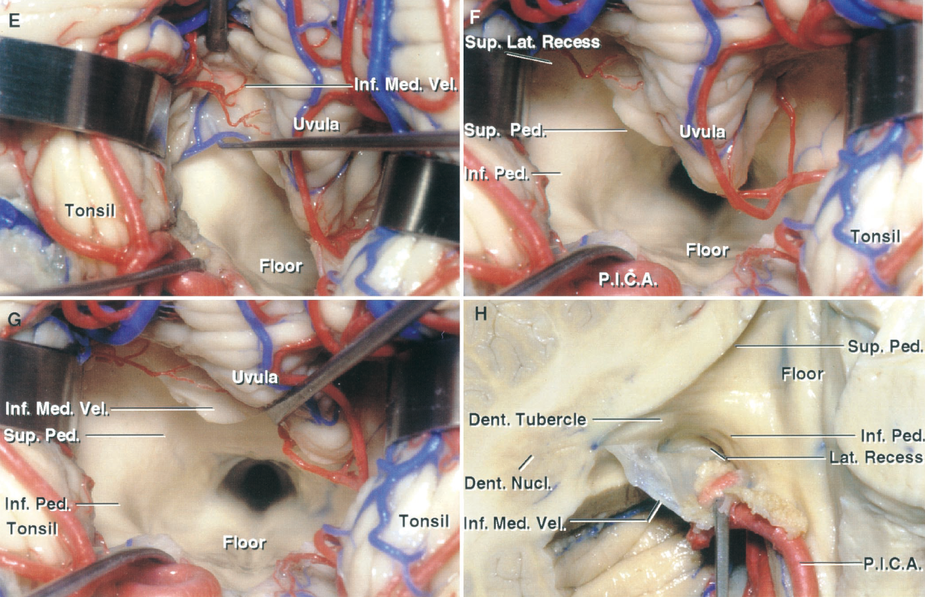
圖1.10。情況。腹側入路進入第四腦室。E,位於第四腦室內的神經鉤的尖端通過薄如紙的下髓膜可見。F,下髓膜的左半部分被切開,露出上外側隱窩和由上、下梗形成的心室麵。G,小舌向右縮回,露出整個心室和大部分心室頂。H,右側小腦矢狀分割小腦蚓部,橫切小腦蒂。扁桃體被切除,下髓膜和PICA顱環被向下移位,以暴露出通往外側隱窩的開口。齒狀核在第四腦室頂上外側隱窩靠近下髓膜附著處形成一個突出,即齒狀結節。凹痕。, dentate; Inf., inferior; Lat., lateral; Med., medullary; Nucl., nucleus; P.I.C.A., posteroinferior cerebellar artery; Ped., peduncle; Sup., superior; Vel., velum.
進入小腦和第四腦室的手術入路可能需要切開蚓部、切除部分腦半球、切除扁桃體、打開下髓膜、將腫瘤與基底和頂板分離、解剖小腦蒂和深部核,以及收縮或切除小腦小葉。Horsley指出,可以犧牲大量的小腦組織,而很少或沒有明顯的功能喪失(13)。進入第四腦室的常見方法是在枕下表麵切開蚓部,如Dandy(3)和Kempe(15)所推薦的。Dandy指出,隻要操作者小心地避開齒狀核(3),蚓部中心可以打開,以進入第四腦室腫瘤,而不會造成功能障礙。蚓部的小病變不會引起症狀或缺陷,但小舌、小結和小葉的較大病變,累及前庭係統相關的小腦纖維,會引起平衡障礙,伴有軀幹性共濟失調、蹣跚步態、四肢隨意運動時,保持直立姿勢而不出現共濟失調時頭部和軀幹的擺動(8,11,12,16)。從腦幹到絮凝結節葉的前庭投射損傷也可引起眼球震顫,出現在凝視的各個方向。小腦緘默症是小腦腫瘤切除後可能出現的一種短暫並發症,通常發生在兒童,特征是清醒的患者言語輸出不足,但言語理解完好,有時伴有口咽失用(2,4,24)。雖然沉默的確切解剖底物仍不清楚,但大多數發生在切除中線腫瘤後,包括蚓部(2,4,24,26)。蚓部的下半部分,包括錐體、小舌和小結節。
可能需要半球切除的病變的頂部側麵部分或側隱窩的第四腦室。Frazier切除了半球外側部分,沒有永久性後遺症(6)。單側切除齒狀核外側部分的半球可導致同側肢體自主運動共濟失調、張力低下和adiadochokiksnesia,其運動的頻率、範圍、方向和力量都存在誤差,這些誤差往往是一過性的(8,11,12,16)。 如果消融涉及齒狀核,這些紊亂更嚴重和持久,此外,有四肢隨意運動的意圖震顫。在對頂尾部進行手術時,應記住齒狀核位於扁桃體上極的嘴側,並包裹在腦室上外側隱窩附近,靠近下髓膜。當切除延伸到小腦半球副蚓部時,就會導致構音障礙,而左半球損傷比蚓部或右半球損傷更容易發生構音障礙(17)。半球病變眼震與眼休息點朝未受影響一側10 - 30度相關,當看病變一側時,眼震振蕩更大。與單側半球病變相比,蚓類病變或延伸至對側半球的病變可產生更明顯的症狀,並伴有站立、行走和說話障礙。小腦幕表麵前部的病變導致維持直立姿勢的肌肉張力增加。如果這一區域的外側受損,高張力主要出現在同側肢體。
所有小腦梗集中於側壁和頂板,此處可能受損。在腦室內手術時,由於小腦上、下兩腳直接位於腦室表麵,因此更易損傷;在靠近外壁的手術中,如橋小腦角的手術,小腦中腳梗更容易受到損傷,因為它是室壁池麵的主要部分。小腦中梗的病變可引起同側肢體自主運動時的共濟失調和共濟失調,其張力減退類似於半球外側的水壩期所產生的張力減退。小腦上梗病變可引起嚴重的同側意圖震顫、共濟失調和運動分解。這種症狀是輕微的,如果隻出現部分腳梗,症狀會迅速消退。小腦下梗部分引起的平衡紊亂類似於絮凝結節葉消融引起的平衡紊亂,伴有軀幹性共濟失調和蹣跚步態。
切除或輕操作附著於第四腦室底的腫瘤的後果包括術中血壓降低、呼吸暫停、和/或呼吸頻率增加和術後複視、言語和吞咽障礙,以及與胃腸道出血、吸入性肺炎和電解質紊亂相關的咳嗽反射不良(1)。
第四腦室的病變對神經外科醫生來說是一個特殊的挑戰,因為嚴重的缺陷可能會隨著心室壁和底部結構的損傷而出現。在過去,手術進入第四腦室是通過分割小腦蚓部或切除部分小腦半球獲得的(1,3,15)。在檢查小腦延髓裂和裂壁時,我們發現,在大多數情況下,單獨打開小腦延髓裂可以充分暴露腦室,而不會裂開蚓部(20,22,23)(圖1.10)。下髓膜,另一薄如紙的層,也可以打開,如果打開腱膜不能提供足夠的暴露。單獨打開腱膜可以進入整個地板和所有的室腔,除了可能的頂、上外側隱窩和屋頂的上半部。打開下髓膜可進入後半部分和頂部的上半部。向外側向Luschka孔延伸的端孔打開外側隱窩,暴露與隱窩相鄰的花梗麵。第四腦室的腫瘤可能將這兩層半透明的膜拉伸和變薄到人們可能意識不到的程度,從而暴露出第四腦室腫瘤。單獨切開腱膜和腓腸肌後,未見損傷報告。然而,暴露在腦室壁上的其他結構可能產生上述缺陷,包括齒狀核、小腦梗、第四腦室底和異食癖。 During an operation on the caudal part of the roof, one should remember that the dentate nuclei are located just rostral to the superior pole of the tonsils underlying the dentate tubercles in the posterolateral part of the roof where they are wrapped around the superolateral recesses near the lateral edges of the inferior medullary velum (Figs. 1.9 and 1.10). All of the cerebellar peduncles converge on the lateral wall and roof where they may be damaged. The superior cerebellar peduncle is more likely to be injured during operations on lesions involving the superior part of the roof above the level of the dentate tubercles; the inferior peduncles are most susceptible to damage in exposing lesions within the lateral recess; and the middle cerebellar peduncle is susceptible to injury in procedures near the external wall of the superior half of the roof, such as those in the cerebellopontine angle, because the middle peduncle forms a major part of the cisternal surface of the ventricular wall. The consequences of removal or gentle manipulation of tumors attached to the floor of the fourth ventricle have been reviewed.
在經脈絡膜層或下髓膜入路時,異位竇經常暴露,但在第四腦室的手術入路中很少出現異位竇閉塞。在第四腦室頂板水平閉塞PICA遠端分支至髓支可避免髓梗綜合征,但可產生類似迷路炎的綜合征,包括旋轉性頭暈、惡心、嘔吐、無法獨立站立或行走,以及無闌尾測音障礙的眼震(11)。小腦髓裂不常顯露AICA的主幹,但也可向側隱窩的脈絡膜分支和脈絡膜叢發送脈絡膜分支。
作者:Albert L. Rhoton, Jr, MD
內容來自Rhoton AL, Jr.後顱窩:顯微外科解剖和手術入路。神經外科2000; 47:1196。doi.org/10.1097/00006123 - 200105000 - 00065.經牛津大學出版社授權代表神經外科醫生大會。©神經外科醫生大會。
神經外科188bet手机app圖譜很榮幸能夠繼承Albert L. Rhoton, Jr, MD的遺產。
請登錄發表評論。
一定要在社交媒體上關注我們的精彩內容並保持更新生活與科恩醫生的會議,關於手術技術的問題,以及更多!


您必須登錄才能查看這些材料。
的188bet手机app這幾乎完全取決於你的捐款。
沒有你們的大量捐贈,我們無法繼續進行地圖集。
請承諾每年至少捐款250美元給地圖集。如果沒有這個承諾,Atlas將很快需要付費訂閱,世界上許多病人的治療依賴於它的外科醫生將無法獲得它。
現在請捐!
沒有你們的大量捐贈,我們無法繼續進行地圖集。請承諾每年至少捐款250美元給地圖集。
如果沒有這個承諾,Atlas將很快需要付費訂閱世界各地的許多外科醫生將無法獲得它,他們的病人的治療依賴於它。現在請捐!