


你可以做出改變。
的188bet手机app幾乎完全取決於你的捐款。
我們不能繼續阿特拉斯沒有重大捐贈。
請提交至少每年250美元捐贈到阿特拉斯。沒有這一承諾,阿特拉斯將很快需要付費訂閱,將成為訪問世界各地許多外科醫生的病人的護理依賴於它。
現在請捐!

最後更新:2021年4月9日
頸靜脈孔很難理解和訪問手術(3、11、15、19、24、28)。很難概念化,因為不同大小和形狀不同的頭蓋骨,從一邊到另一邊同樣的頭蓋骨,並從其顱內與顱外端在同一孔,和由於其複雜的不規則形狀,其彎曲,形成由兩根骨頭,無數的神經和靜脈通道,通過它(圖9.1)。困難暴露這個孔是由它的深度位置和周圍結構,如頸動脈在前麵,麵部神經外側,舌下神經內側,椎動脈劣等,所有這些屏蔽孔,需要仔細的管理。
頸靜脈孔分為三個部分:兩個靜脈和神經或intrajugular隔間。靜脈隔間組成一個更大的後外側的靜脈通道,乙狀結腸,收到乙狀竇的流動,和一個小入靜脈通道,堅硬的部分,接收下堅硬的竇的排水。堅硬的部分形成一個靜脈特征也收到舌下運河支流彙合,岩斜裂縫和椎靜脈叢。堅硬的部分流入乙狀結腸部分通過開放內側牆之間的頸靜脈球舌咽神經前方迷走和輔助神經後方。intrajugular或神經的部分,通過舌咽神經、迷走神經,和輔助神經,位於乙狀結腸和堅硬的部分之間intrajugular過程的現場,顳部及枕部的骨頭,由一種纖維加入或骨的橋。舌咽神經、迷走神經和輔助神經穿透硬腦膜的內側邊緣intrajugular顳骨的過程達到的內側牆頸內靜脈。手術方法訪問的各個方麵孔和鄰近地區是postauricular transtemporal, retrosigmoid,極端的橫向transcondylar, preauricular subtemporal-infratemporal方法。

頸靜脈孔位於顳骨和枕骨(無花果。9.1和9.2)。正確的孔通常大於左側。在先前的研究中,我們觀察到右孔大於左在68%的情況下,等於在12%,小於20% (24)。周圍的孔配置乙狀結腸偽劣堅硬的鼻竇。它可以被視為一個時間和枕骨骨之間的空隙。遍曆頸靜脈孔的結構,乙狀竇和頸靜脈球下堅硬的鼻竇,腦膜咽升和枕動脈的分支,舌咽神經、迷走神經,和副神經節神經,舌咽神經的鼓膜的分支(雅各布森的神經)、迷走神經的耳分支(阿諾德的神經)和耳蝸導水管。
孔坐落,其長軸定向後外側的入,給它一個前外側的邊緣形成的顳骨和後中的保證金由枕骨。從顱內端,直接向前,內側,向下。不能透過孔高於或低於直接觀看的頭骨因為它的屋頂,形成的下表麵堅硬的顳骨的一部分。孔,當從顱內一邊在前方向,後麵有一個大橢圓橫向組件,稱為s形的部分,因為它收到乙狀竇的排水係統,和一個小內側部分,稱為堅硬的部分,因為它收到劣質堅硬的竇的排水。下麵的視圖通過孔直接揭示了顳骨的一部分形成頸靜脈球的穹頂,而不是一個明確的開放。
乙狀結腸的結和堅硬的部分是對方表麵骨日珥,顳部及枕部的骨頭,稱為intrajugular過程,加入了一種纖維,一般或更少,和骨的橋,intrajugular隔,分離乙狀結腸和堅硬的孔的一部分。
雖然利潤率的頸靜脈孔形成堅硬的顳骨的一部分,枕骨髁的部分,這些骨骼的其他部分也有重要的關係到頸靜脈孔。岩斜裂縫,裂縫之間的橫向邊緣clival枕骨的一部分和堅硬的顳骨的一部分,相交雙側孔的邊緣,和occipitomastoid縫合,縫合的顳骨乳突的部分和髁的枕骨的一部分,相交後外側的邊緣。
,顳部及枕部骨骼的intrajugular過程劃分的前部和後部邊緣之間的孔乙狀結腸和堅硬的部分。顳骨的intrajugular過程突出到了頸靜脈孔從枕骨相反的過程,並且可能很少達到較小的intrajugular枕骨的過程,把頸靜脈孔分為兩個骨小孔。intrajugular嶺嶺,向前延伸的intrajugular過程沿著內側顳骨頸靜脈球邊緣(圖9.1)。舌咽神經課程在其內側邊緣。偶爾,脊延伸的邊緣內側向鄰顳骨的一部分創建一個深溝的神經課程或者它可能到達顳骨形成一條運河,環繞的舌咽神經通過頸靜脈孔。
乙狀竇的排水是向前進的乙狀結腸部分孔,高圓頂休會,頸靜脈窩,形成一個屋頂在頸靜脈球的頂部(無花果。9.1和9.3)。休息,有其峰會略側乙狀竇的入口,通常是更大的在右邊的頭骨,反映了大乙狀竇。休會的圓頂通常是光滑的,因為它符合頸靜脈球,但峰會也可能成脊狀和不規則的。一個小三角休會,錐體窩,向前延伸的內側intrajugular過程沿著前壁顳骨的堅硬的孔的一部分。耳蝸的外部孔徑小溝,這房子perilymphatic管和管狀硬腦膜的延長,打開到錐體的前尖窩。舌咽神經進入這窩下麵的耳蝸導水管連接頂峰。
頸枕骨髁的部分的過程中,延伸在頸靜脈孔和連接clival枕骨的鱗狀骨部分,形成後中的孔的牆。這個過程橫向延伸從枕髁的後一半以上的麵積和舌下滲透的運河。頸的上表麵過程的枕骨區域superomedial孔提供了一個橢圓形的突出,頸靜脈結節,這是舌下上方的運河。頸靜脈結節通常有一個淺皺紋的網站通過舌咽神經、迷走神經,並在其表麵輔助神經。乙狀竇課程發展的終端上頸表麵過程深鉤形槽,乙狀溝,導演內側頸靜脈孔的乙狀結腸部分。
頸靜脈孔的側壁上,在外部邊緣幾毫米,後麵的點occipitomastoid縫線穿過孔的外側邊緣,是一個很小的孔,乳突小溝,淺槽從內側到外側的前壁部分乙狀結腸乳突小管(無花果。9.2和9.3)。耳迷走神經分支(阿諾德的神經)課程沿著槽和進入小管。神經穿過乳突和下側的出口骨頭tympanomastoid縫合的一部分。現場的intrajugular嶺顳骨頸脊,一個小運河,鼓小管,直接向上,導致鼓膜的分支產生的劣質舌咽神經神經節(雅各布森的神經)鼓室(無花果。9.2)。從下麵看顱外頸靜脈孔的孔,可以認識到的舌咽神經課程沿著內側intrajugular過程和山脊到達下麵的麵積鼓膜的小管。

點擊這裏查看這張圖片的交互模塊和相關內容。
圖9.1。模擬。骨的關係。,之間的頸靜脈孔位於顳枕的骨頭。不能看到從上麵直接通過孔,如圖所示,因為它是直接顳骨下向前發展。乙狀溝是沿著乳突和穿過occipitomastoid縫合的地方是向前的上表麵枕骨頸過程和進入後下的孔通過堅硬的顳骨的一部分。B,視圖後和上級直接顯示形狀的孔,未見在直接上級的觀點。孔有一個更大的橫向乙狀結腸乙狀竇倒空和一個更小的一部分入堅硬的部分通過下堅硬的鼻竇清空。這兩部分是分開的骨頭枕葉和顳intrajugular流程。舌咽神經、迷走神經和輔助神經穿過intrajugular部分之間的孔位於乙狀結腸和堅硬的部分。 The foramen is asymmetric from side to side with the right side often being larger as shown. The cochlear aqueduct opens just above the anterior edge of the petrosal part. The vestibular aqueduct opens into the endolymphatic sac, which sits on the back of the temporal bone superolateral to the sigmoid part of the jugular foramen. C, jugular foramen viewed from directly below. One cannot see directly through the foramen from below because the foramen is covered above by the part of the petrous temporal bone forming the jugular fossa, which houses the jugular bulb. The entrance into the carotid canal is located directly in front of the medial half of the jugular foramen. The stylomastoid foramen is located lateral and the anterior half of the occipital condyle medial to the jugular foramen. The posterior condylar foramen is transversed by an emissary vein, which joins the sigmoid sinus. The hypoglossal canal passes above the middle third of the occipital condyle and opens laterally into the interval between the jugular foramen and carotid canal. D, the view directed from anterior and backward reveals the shape of the jugular foramen. The roof over the foramen formed by the jugular fossa of the temporal bone is shaped to accommodate the jugular bulb. The posterior margin of the foramen is formed by the jugular process of the occipital bone, which connects the basal (clival) part of the occipital bone to the squamosal part. The petroclival fissure intersects the anteromedial margin of the petrosal part of the foramen. Ac., acoustic; Car., carotid; Coch., cochlear; Cond., condyle; Fiss., fissure; For., foramen; Hypogl., hypoglossal; Int., internal; Intrajug., intrajugular; Jug., jugular; Mast., mastoid; Occip., occipital; Pet., petrous; Petrocliv., petroclival; Post., posterior; Proc., process; Sig., sigmoid; Squam., squamosal; Stylomast., stylomastoid; Temp., temporal; Vest., vestibular.
點擊這裏查看這張圖片的交互模塊和相關內容。
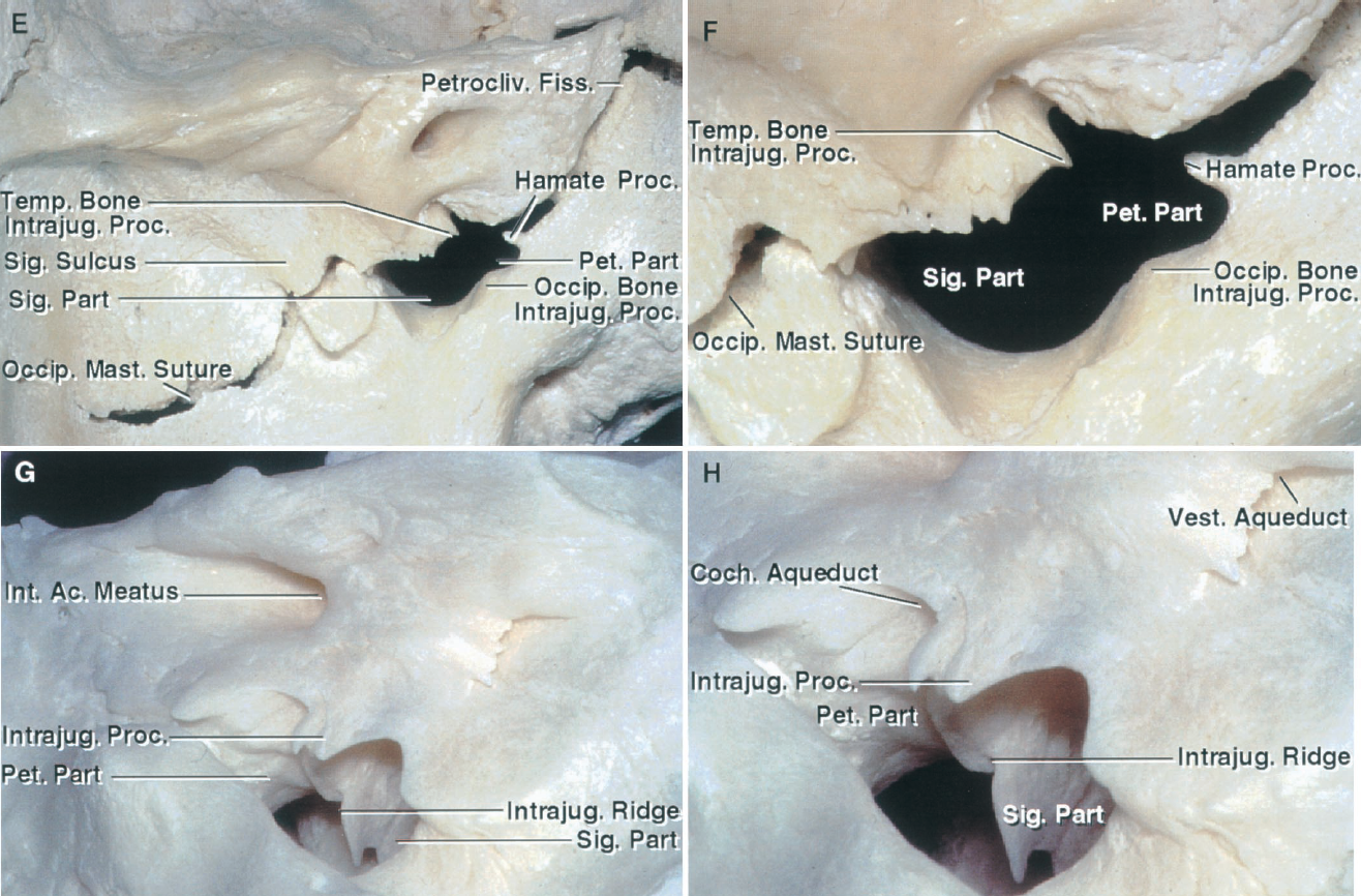
圖9.1。情況。E和F,另一個頸靜脈孔。左:E,縫合線被迫開放給孔的關係岩斜和occipitomastoid縫合線。頸靜脈孔有一個較大的橫向部分,乙狀結腸,收到乙狀竇的排水係統,和一個更小的內側部分,堅硬的部分,接收下堅硬的竇的排水。枕骨的intrajugular過程比C所示更加突出和項目向前向intrajugular顳骨的過程。鉤狀的過程通常沿著內側邊緣延伸的堅硬的部分孔到鄰近的顳骨的一部分,但在這種情況下,縫合線被迫開放,留下一個鉤狀的過程和顳骨之間的時間間隔。F,放大視圖。G和H,另一個頸靜脈孔。G的intrajugular過程顳骨項目為乙狀結腸之間的間隔和堅硬的部分孔。 A ridge, the intrajugular ridge, extends forward from the intrajugular process along the medial side of the jugular bulb. The glossopharyngeal nerve passes forward along the medial side of the intrajugular process and ridge. The vagus and accessory nerves enter the dura on the medial side of the process, but quickly descend and do not pass forward along the medial edge of the ridge as does the glossopharyngeal nerve. The jugular process of the occipital bone often has a small prominence on its surface that projects toward the intrajugular process of the temporal bone, and in some foramina, the intrajugular processes of the two bones are joined by an osseous bridge that converts the foramen into two osseous foramina. In this case, the intrajugular process of the occipital bone is absent. H, enlarged view. The cochlear aqueduct opens above the petrosal part of the foramen and the site where the glossopharyngeal nerve enters the intrajugular part of the foramen on the medial side of the intrajugular process. The vestibular aqueduct opens onto the posterior surface of the temporal bone superolateral to the jugular foramen.
圖9.2。骨的關係。側麵圖。莖突向下項目和麵神經退出莖突乳突的孔在側麵,和兩塊側頸靜脈孔。下頜髁塊訪問在前麵的孔和椎動脈提升從後麵通過C1橫突限製訪問。C1坐在橫突和經常縮進頸內靜脈的後壁。B,劣質的頸靜脈孔。頸靜脈孔位於外側前枕髁的一半。的顳骨頸靜脈球形成圓頂。的頸過程枕骨頸靜脈孔的後緣。 The jugular foramen and carotid canal are separated by a narrow bony ridge, which is penetrated medially by the tympanic canaliculus through which passes the tympanic branch of the glossopharyngeal nerve (Jacobson’s nerve). This branch of the nerve passes forward across the promontory in the medial part of the tympanic cavity, then crosses the floor of the middle fossa as the lesser petrosal nerve, and eventually reaches the otic ganglion, providing parasympathetic innervation to the parotid gland. The anterior wall of the sigmoid part of the foramen is the site of a shallow groove across which the auricular branch of the vagus nerve (Arnold’s nerve) passes to enter the mastoid canaliculus. It exits the mastoid through the tympanomastoid suture. C, lateral view of the left temporal bone. A small fiber (arrow) placed in the tympanic canaliculus, shown in B, exits the canaliculus in the middle ear where the fibers of the tympanic branch of the glossopharyngeal nerve cross the promontory, and then regroup to cross the floor of the middle fossa as the lesser petrosal nerve. The styloid process projects downward lateral to the jugular foramen. Aur., auricular; Br., branch; Canalic., canaliculus; Car., carotid; CN, cranial nerve; Cond., condyle; Ext., external; Fiss., fissure; For., foramen; Jug., jugular; Mandib., mandibular; Occip., occipital; Petrotymp., petrotympanic; Proc., process; Trans., transverse; Tymp., tympanic.
顱內,堅硬的孔的一部分位於約5毫米以下porus內部運河和5毫米以上的舌下神經管的顱內孔(無花果。9.2和9.4)。下麵的側孔位於邊緣,大約在矢狀麵通過側麵的內部聲音道。頸靜脈結節,圓形突出位於基底的結和枕骨髁的地區,位於約8毫米內側頸靜脈孔的內側邊緣。耳軟骨囊,它位於岩石的顳骨的一部分,其中包含的半規管和耳蝸,位於優於頸靜脈球的穹頂。
枕髁位於沿側緣的前一半的枕骨大孔區下方內側頸靜脈孔。
舌下運河,它通過枕骨髁的部分在枕骨髁部以上的地區,位於中間的小孔(無花果。9.1和9.3)。顱內的舌下運河位於頸靜脈結節約5毫米以下inferomedial堅硬的部分下麵的頸靜脈孔和幾個毫米的下部岩斜裂縫。更詳細的審查包括在這一章far-lateral方法。
前頸靜脈孔的邊緣,顱外,是由狹窄的山脊顳骨,頸脊,分離孔和頸動脈管(無花果。9.1和9.2)。打開鼓小溝附近或內側頸脊的一部分。莖突和莖突乳突的孔位於外側的外口頸靜脈孔、莖突是位於莖突乳突的稍微入孔。麵神經退出莖突乳突的約5毫米孔外側側頸靜脈孔的邊緣。頸靜脈孔的前邊緣位於後方的一部分鼓骨形成顳下頜關節的後壁和前偽劣的外耳道。的陰道過程鼓骨,將頸動脈管和乙狀結腸孔從關節窩的一部分,是附件莖突的頭骨基地。莖突項目從陰道向下鼓骨的過程,橫向孔。二腹肌溝是後方的莖突沿著乳突的內側緣。訪問頸靜脈孔外側阻塞乳突和莖突的過程,阿特拉斯的橫突、下頜支(無花果。9.3和9.4)。
鼓室,位於內側的鼓膜,坐落在外側頸靜脈球和尖銳的直角的曲線,稱為橫向彎曲,垂直和水平段交界處的堅硬的頸動脈(圖9.4)。幾個結構可能暴露在手術過程中病變的頸靜脈孔的垂直和水平段頸內動脈的堅硬的部分,咽鼓管,張量定音鼓肌肉。耳蝸和半規管位於堅硬的部分上麵的顳骨頸靜脈球的穹頂(圖9.4)。麵神經顳骨,經常屏蔽病變在頸靜脈孔,通過乳突外側頸靜脈球下降。內淋巴的囊位於的後表麵堅硬的骨頭之間的兩層硬腦膜在角落裏的乙狀竇改變了曆史的進程從垂直方向到水平(無花果。9.3和9.5)。

點擊這裏查看這張圖片的交互模塊和相關內容。
圖9.3。後頸靜脈孔的高級視圖。乙狀溝使急轉彎前流入的乙狀結腸部分頸靜脈孔。下堅硬的竇延伸沿岩斜裂縫和進入堅硬的孔的一部分。的神經進入intrajugular部分孔位於乙狀結腸和堅硬的部分。概述了區域顯示了近似的站點B, F。乙,乙狀竇降臨在乙狀溝,使一把鋒利的前轉向進入頸靜脈孔。頸靜脈球向上延伸下岩石的顳骨內部聲學道。上麵的內淋巴的囊位於較低的部分乙狀竇的顳骨並打開上麵通過前庭導水管到門廳。舌咽神經、迷走神經和輔助神經穿透硬腦膜的內側intrajugular過程。 C, the dura covering the jugular foramen and the jugular bulb have been removed. The nerves penetrate the dura on the medial side of the intrajugular process of the temporal bone. The intrajugular ridge extends forward along the medial side of the jugular bulb. D, enlarged view. The glossopharyngeal nerve passes forward along the medial side of the intrajugular ridge, but the vagus and accessory nerves, although entering the dura on the medial side of the intrajugular process, almost immediately turn downward and do not course along the medial edge of the intrajugular ridge in the medial wall of the jugular bulb, as does the glossopharyngeal nerve. The auricular branch of the vagus nerve (Arnold’s Nerve) arises from the vagus nerve, passes along the groove in the anterior wall of the jugular fossa, and penetrates the mastoid canaliculus in the lateral wall of the fossa. E, the nerves entering the jugular foramen have been displaced downward. The intrajugular process of the temporal bone projects backward to join the intrajugular process of the occipital bone, thus forming an osseous bridge that divides the foramen into two parts. The vagus and accessory nerves pass lateral to the osseous bridge and the inferior petrosal sinus descends below the bridge to open into the internal jugular vein. F, the hypoglossal nerve has been exposed on the lateral side of the occipital condyle. It exits the hypoglossal canal and joins the glossopharyngeal, vagus, and accessory nerves below the jugular foramen in the interval between the internal carotid artery and internal jugular vein. A., artery; Ac., acoustic; Aur., auricular; Br., branch; Car., carotid; CN, cranial nerve; Cond., condyle; Endolymph., endolymphatic; Gang., ganglion; Inf., inferior; Intrajug., intrajugular; Jug., jugular; Occip., occipital; Pet., petrosal, petrous; Petrocliv., petroclival; Proc., process; Sig., sigmoid; Sup., superior; Temp., temporal; Vert., vertebral; Vestib., vestibular.
顱內孔,頸靜脈孔硬腦膜分為三個部分:堅硬的隔間坐落入,乙狀結腸室坐落後外側的,和intrajugular或神經室坐落在堅硬的和乙狀結腸部位intrajugular過程的現場,顳部及枕部的骨頭,intrajugular隔,舌咽神經、迷走神經,輔助神經(無花果。9.3和9.5)。的硬腦膜intrajugular孔的一部分,入位於乙狀結腸的部分,有兩個特點穿孔、舌咽神經道,通過舌咽神經傳遞,並通過迷走神經和迷走神經導管,輔助神經傳遞(無花果。9.5和9.6)(24)。道都是位於內側的intrajugular流程和隔膜。舌咽神經和迷走神經導管始終由一個硬鋁隔分開寬度從0.5到4.9毫米不等(13)。唯一的硬膜內的網站舌咽神經始終不能區分迷走神經是近端硬鋁隔。密切舌咽神經和迷走神經的起源在腦幹,和兩者之間的arachnoidal粘連蛛網膜下腔可能使分離困難的過程中除了在該地區近端硬鋁隔。上級舌咽神經神經節很容易可見顱內大約三分之一的神經。上可以看到的迷走神經節顱內神經的隻有六分之一。雖然腦和脊髓的部分副神經最頻繁進入迷走神經的道在一起,硬鋁隔分開。
上和橫向利潤率intrajugular孔的一部分的網站特點厚硬腦膜的褶皺形成的屋頂或唇項目下級和內側部分覆蓋舌咽神經和迷走神經的道(無花果。9.5和9.6)。這個結構,稱為頸硬腦膜的褶皺,在雙方在一個僵化的標本(31)13日,16日,17日,24日。唇項目最顯著舌咽神經道,可比,但小於,後唇的內部聲音道。主要是骨或纖維,可能項目最多2.5毫米/舌咽神經道的邊緣。迷走神經的唇不太突出,突出最多1毫米的側緣迷走神經的道。
舌咽神經、迷走神經和輔助神經產生於髓質中延伸出來的一條線位於後邊緣的劣質橄欖油postolivary溝(無花果。9.3和9.5)。舌下神經出現了中延伸出來的一條線,沿著前退出腦幹利潤率較低的三分之二的preolivary溝橄欖,橄欖油和髓之間的槽金字塔。
舌咽神經,點它穿透硬鋁舌咽神經道,突然轉身向前,然後向下和課程通過頸靜脈孔槽從下麵的錐體窩的耳蝸導水管,沿著內側intrajugular嶺。神經退出後頸靜脈孔,向前,穿過側麵的頸內動脈深莖突。隨著神經橫頸靜脈孔,它擴展的網站其優越的偽劣神經節(圖9.5)。在外部頸靜脈孔的孔,它引起鼓膜的分支(雅各布森的神經),遍曆的鼓膜的小管進入鼓室引起鼓膜的神經叢,纖維的課程在海角淺溝槽和重組形成較小的堅硬的神經,提供副交感神經支配的腮腺的耳神經節。
迷走神經延伸進入硬腦膜的小班,稱為迷走神經的道,不如舌咽神經道,它是由一個硬鋁隔分開(無花果。9.5和9.6)。它由副神經加入進入硬腦膜。延伸出來後收集顱內孔的孔,迷走神經擴張優越的神經節,約長2.5毫米,結束在顱外孔的孔。它坐落在硬腦膜,頸靜脈孔,在那裏,沿著內側intrajugular過程的顳骨,向下。優越的神經節,迷走神經與副神經、通信部分的混合到神經節。耳的分支(阿諾德的神經)出現在上級迷走神經的神經節,加入了一個分支的劣質舌咽神經神經節(圖9.3)。耳分支通過橫向的淺槽上頸靜脈球的前壁及頸靜脈窩的側壁,在進入乳突小管,提升向垂直(乳突)的麵神經管,發出一個提升部門的麵部神經穿過外側之前將向下退出顳骨tympanomastoid裂縫。
迷走神經的主幹(或者,更準確地說,上級神經節)課程前偽劣如下它穿過的midportion intrajugular顳骨的過程(無花果。9.3和9.5)。顱內孔的孔,顳骨的intrajugular過程分離乙狀竇的神經節。在大多數情況下,在該地區立即硬腦膜下intrajugular水平的過程,沒有纖維樂隊舌咽神經和迷走神經的神經節。
迷走神經退出頸靜脈孔垂直,保留副神經的親密關係(無花果。9.3 - 9.5)。在這兩個神經退出頸靜脈孔,它們坐落在舌咽神經後中的牆上頸內靜脈。隨著迷走神經外側的外口舌下運河,它加入了舌下神經內側。迷走神經開始擴大網站的劣質迷走神經的神經節在孔,長度大約為2.5厘米。

點擊這裏查看這張圖片的交互模塊和相關內容。
圖9.4。模擬。逐步剝離周圍的結構表麵, 頸靜脈孔。, 耳朵周圍的皮膚和頭皮一直反映暴露麵積 側頸靜脈孔。胸鎖乳突肌 背後暴露和腮腺 前的耳朵。枕大神經和到達皮下 枕動脈組織通過 依戀之間的斜方肌和胸鎖乳突肌 項線。外部 聲道位於一個小 向前深頸靜脈球。B,去除表麵的 麵部神經肌肉和腮腺暴露,顳肌和嚼肌 肌肉,二腹肌後腹的 ,頸內靜脈。胸鎖乳突肌 一直反映落後暴露 副神經進入深 表麵。C,下頜支 髁,內側和外側翼狀的 肌肉,和二腹肌後腹的 揭露莖突,位於 側頸靜脈孔。進入 內部頸動脈提升麵前的頸動脈管頸 孔。 Both the jugular foramen and carotid canal are situated behind the tympanic part of the temporal bone, which forms the posterior wall of the condylar fossa. The tensor and levator vela palatini muscles are attached to the eustachian tube in the area below the horizontal segment of the petrous carotid. The infratemporal fossa is located below the greater wing of the sphenoid. The mandibular nerve passes through the foramen ovale to enter the upper part of the infratemporal fossa. Branches of the ascending pharyngeal artery pass through the jugular foramen to supply the surrounding dura. The hypoglossal nerve passes forward across the external and internal carotid artery. D, the styloid process has been removed to expose the glossopharyngeal, vagus, accessory, and hypoglossal nerves descending between the internal carotid artery and the internal jugular vein in the area immediately below the jugular foramen. The glossopharyngeal nerve descends along the lateral side of the internal carotid artery. The accessory nerve passes backward across the lateral surface of the internal jugular vein. The hypoglossal nerve passes through the hypoglossal canal, which is located below and medial to the jugular foramen, and descends with the nerves exiting the jugular foramen. The occipital artery gives rise to a meningeal branch, which passes through the jugular foramen to supply the surrounding dura, and to the stylomastoid artery, which passes through the stylomastoid foramen with the facial nerve. A., artery; Asc., ascending; Aur., auricular; Br., branch; Cap., capitis; Car., carotid; Chor. Tymp., chorda tympani; CN, cranial nerve; Cond., condylar; Dors., dorsal; Eust., eustachian; Ext., external; Fiss., fissure; Gl., gland; Gr., greater; Inf., inferior; Int., internal; Jug., jugular; Laryn., laryngeal; Lat., lateral, lateralis; Lev., levator; Long., longus; M., muscle; Mast., mastoid; Men., meningeal; N., nerve; Obl., oblique; Occip., occipital; Pal., palatini; Pet., petrosal, petrous; Pharyn., pharyngeal; Post., posterior; Proc., process; Pteryg., pterygoid; Rec., rectus; Retromandib., retromandibular; Scap., scapulae; Seg., segment; Semicirc., semicircular; Sig., sigmoid; Squamotymp., squamotympanic; Sternocleidomast., sternocleidomastoid; Stylogloss., styloglossus; Stylomast., stylomastoid; Stylophar., stylopharyngeus; Submandib., submandibular; Sup., superior; Temp., temporal; Tens., tensor; TM., temporomandibular; Trans., transverse; Tymp., tympanic, tympany; V., vein; Vel., veli; Vent., ventral; Vert., vertebral.

點擊這裏查看這張圖片的交互模塊和相關內容。
圖9.4。情況。E,上級偽劣斜被反映出更多的暴露表麵的肌肉。C1橫突和腹直肌外側capitis休息與頸內靜脈的後表麵。腹直肌外側capitis高度的頸過程枕骨頸靜脈孔的後緣。縮回肩胛提肌暴露的部分提升通過C2椎動脈橫孔前腹側支的C2神經根。椎動脈,因為它通過內側沿阿特拉斯的後拱的上表麵,位於枕骨下的三角形之間的地板上偽劣斜肌和腹直肌肌後主要。F,頸內動脈已流離失所的後方暴露咽升的分支,通過破裂孔、頸靜脈孔、供應的舌下神經管周圍的硬腦膜。頭骨的鼓索出口內側髁的窩的一部分,首先通過岩鼓,然後沿著squamotympanic縫合線。G,鼓骨形成前低,保證金的外耳道被移除,但鼓膜的溝的鼓膜高度一直保存了下來。 The surface of the temporal and occipital bones surrounding the jugular foramen and carotid canal have an irregular surface that serves as the attachment of the upper end of the carotid sheath. The mastoid segment of the facial nerve and the stylomastoid foramen are situated lateral to the jugular bulb. The chorda tympani arises from the mastoid segment of the facial nerve and courses along the deep side of the tympanic membrane crossing the neck of the malleus. It exits the skull by passing through the petrotympanic and squamotympanic sutures and joins the lingual branch of the mandibular nerve distally. The carotid ridge separates the carotid canal and jugular foramen. Meningeal branches of the ascending pharyngeal and occipital arteries enter the jugular foramen. The glossopharyngeal, vagus, and accessory nerves pass through the jugular foramen on the medial side of the jugular bulb. H, the tympanic ring and bone lateral to the tympanic cavity have been removed. The internal carotid artery has been displaced forward out of the carotid canal to expose the carotid sympathetic nerves that ascend with the artery. The glossopharyngeal, vagus, accessory, and hypoglossal nerves exit the skull on the medial side of the internal carotid artery and jugular vein. The glossopharyngeal and hypoglossal nerves pass forward along the lateral surface of the internal carotid artery, and the accessory nerve descends posteriorly across the lateral surface of the internal jugular vein. The vagus nerve descends in the carotid sheath.

點擊這裏查看這張圖片的交互模塊和相關內容。
圖9.4。i n。我,側乳突和鼓室前移除鼓膜的戒指。麵神經鼓室的的部分通過以下側半規管,向下的乳突段退出莖突乳突的孔。莖突乳突的孔和乳突段位於外側頸靜脈球。上麵的半圓形凹槽位於頸靜脈球。J,探針放在咽鼓管,通過向下、向前,和內側鼓室和前麵的岩石的頸動脈。第三個三叉部門通過卵圓孔未閉的咽鼓管的外側。K,放大圖的鼓膜的環鼓膜移除。張鼓脹的肌肉通過向後上方的咽鼓管和引起肌腱急轉彎側向附著在錘骨trochleiform過程。 The chorda tympani crosses the inner surface of the tympanic membrane and neck of the malleus. The round window opens into the vestibule. The stapes sit in the oval window. The promontory is located lateral to the basal turn of the cochlea. L, the floor of the middle fossa and the tympanic sulcus have been removed to expose the jugular bulb and petrous carotid. The greater petrosal nerve courses along the floor of the middle fossa on the upper surface of the petrous carotid. The deep petrosal nerve arises from the sympathetic bundles on the internal carotid artery. The deep and greater petrosal nerves join to form the vidian nerve, which passes forward through the vidian canal to join the maxillary nerve and pterygopalatine ganglion in the pterygopalatine fossa. The pharyngobasilar fascia and upper part of the longus capitis have been reflected downward to expose the lower margin of the clivus. M, the jugular bulb has been removed from the jugular fossa located below the vestibule and semicircular canals. The vertical segment of the petrous carotid has been removed. The cochlea, which has been opened, is located above the lateral genu of the petrous carotid. The tympanic segment of the facial nerve passes posteriorly below the lateral semicircular canal. N, the retrosigmoid and presigmoid dura have been opened. The lateral wall of the vestibule and cochlea have been removed. The vestibule, semicircular canals, and cochlea are exposed above the jugular bulb and lateral genu of the petrous carotid.
雖然腦和脊髓的部分副神經最頻繁進入迷走神經的道,他們可能很少被硬鋁隔分開。脊髓部分提升向枕骨大孔表麵爬行的硬腦膜,甚至可能被埋在下麵的硬腦膜枕骨大孔(無花果。9.3、9.5和9.6)。在硬腦膜的頸靜脈孔的孔,神經通常的迷走神經。副神經通常進入相同的硬腦膜的小班迷走神經和通常遵循和混合的迷走神經在優越的迷走神經的神經節。迷走神經的神經節的副神經離開後,出口之間的頸靜脈孔和橫向下降間接頸內動脈和頸內靜脈,然後向後外側靜脈表麵達到肌肉。大約有30%的神經沿著內側,而不是橫向表麵頸內靜脈(8)。
舌下神經不遍曆頸靜脈孔(無花果。9.3 - -9.5)。然而,它連接神經退出頸靜脈孔下方頭骨在頸動脈鞘和運行。神經退出下側的舌下運河的一部分,通過相鄰迷走神經,是頸內動脈和頸內靜脈之間的水平橫突的地圖集,它會突然向前沿著側麵的頸內動脈的舌頭,隻留下下的頸袢的主要血管。
可能參與動脈病理異常在頸靜脈孔包括頸上部和頸內動脈的堅硬的部分,後方指揮頸外動脈的分支,和上部椎動脈(圖9.4)。
頸內動脈,幾乎直向上,後入頸外動脈和頸內靜脈,進入頸動脈管(圖9.4)。在顱底,頸內靜脈課程後頸內動脈,頸脊分開了。舌咽神經位於外側和迷走神經,配件和舌下神經內側。
頸內動脈進入後頸動脈管頸交感神經和周圍的靜脈叢,它提升短距離(垂直段),達到下麵的區域,身後的耳蝸,結果入在一個直角(橫向彎曲的網站)和橫向(水平段)課程向堅硬的頂端(圖9.4)。破裂孔的內側邊緣,它大幅上行的內側彎曲進入海綿竇的後部分。
前頸外動脈提升頸內動脈。近端終端分岔到上頜骨和顳淺動脈,它產生六個枝子,這可以分為前部和後部組根據他們的方向。後者與頸靜脈孔。
咽升動脈,第一個分支後,通常供應提供了最突出的頸靜脈孔周圍的腦膜(圖9.4)(18)。它出現在分岔或從最低的外部或內部頸動脈的一部分。很少來自枕動脈的起源。它向上課程之間的內部和外部的頸動脈,引起眾多分支鄰近肌肉,神經和淋巴結。腦膜分支通過破裂孔分布到硬腦膜襯砌中間窩和通過頸靜脈孔或供應的舌下神經管周圍的顱後窩硬腦膜。咽升動脈也產生下鼓室的動脈,在鼓室通過鼓膜的小管舌咽神經的鼓膜的分支。
枕動脈,第二個最大的分支後,來自頸外動脈的後表麵和課程斜向上二腹肌後腹之間的肌肉和頸內靜脈(圖9.4)。腦膜的分支,通過頸靜脈孔或進入後窩髁的運河,腫瘤可能做出重大貢獻的頸靜脈孔。
耳後動脈,最後一個分支後,出現以上二腹肌後腹的肌肉和腮腺和莖突之間傳播。乳突的前邊緣,它分為耳和枕分支,分別分配給postauricular和枕骨區域。莖突乳突的分支,它出現在莖突乳突的孔,進入莖突乳突的孔提供麵部神經。其損失會導致麵部麻痹雖然腦膜中動脈的吻合與堅硬的分支。耳後分支可能共享一個公共與枕動脈主幹,或有時是缺席,在這種情況下,枕動脈引起莖突乳突的動脈。前組的成員,其起源可能可視化暴露頸靜脈孔的病變,包括甲狀腺上、舌、動脈和麵部。

點擊這裏查看這張圖片的交互模塊和相關內容。
圖9.5。,後的左頸靜脈孔的顱內方麵。舌咽神經、迷走神經和輔助神經皮爾斯頸靜脈孔的硬腦膜的屋頂。舌咽神經和迷走神經分離的一個狹窄的硬腦膜的隔膜。頸硬腦膜的向下折疊項目和內側外側和頸靜脈孔的上緣的網站進入硬腦膜神經孔的屋頂。麵部和蝸神經和複雜的動脈進入內部聲學道。的subarcuate分支下小腦動脈進入subarcuate窩。內淋巴的囊位於硬鋁層之間的側頸靜脈孔。髓質連接的橋靜脈下堅硬的竇內側頸靜脈球。B,硬腦膜已經從顳骨的後表麵中刪除。 The intrajugular processes of the temporal and occipital bones, which are connected by a fibrous bridge, the intrajugular septum, separates the sigmoid and petrosal parts of the foramen. The glossopharyngeal, vagus, and accessory nerves enter the intrajugular part of the foramen by penetrating the dura on the medial side of the intrajugular process of the temporal bone. C, the glossopharyngeal nerve enters the jugular foramen below the cochlear aqueduct. The vagus nerve enters the jugular foramen behind the glossopharyngeal nerve. The auricular branch of the vagus nerve (Arnold’s nerve) arises at the level of the superior ganglion and passes around the anterior wall of the jugular bulb. The accessory nerve is formed by multiple rootlets, which arise from the medulla and spinal cord. The accessory rootlets collect together to form a bundle that blends into the lower margin of the vagus nerve at the level of the jugular foramen. The lower vagal and accessory roots pass across the surface of the jugular tubercle. D, enlarged view. The glossopharyngeal nerve expands at the site of the superior and inferior ganglia. The superior ganglion of the vagus nerve is located at the level of or just below the dural roof of the foramen, and the inferior ganglion is located below the foramen at the level of the atlanto-occipital joint. A., artery; Atl., atlanto-; Aur., auricular; Br., branch; Bridg., bridging; Car., carotid; CN, cranial nerve; Coch., cochlear; Cond., condyle; Endolymph., endolymphatic; Gang., ganglion; Glossophar., glossopharyngeal; Hypogl., hypoglossal; Inf., inferior; Int., internal; Intrajug., intrajugular; Jug., jugular; Labyr., labyrinthine; Lat., lateral; Occip., occipital; Pet., petrosal; Proc., process; Sig., sigmoid; Subarc., subarcuate; Sup., superior; Temp., temporal; Vert., vertebral.

點擊這裏查看這張圖片的交互模塊和相關內容。
圖9.6。Retrosigmoid頸靜脈孔的方法。,細節顯示垂直的頭皮切口和右retrosigmoid顱骨切開術。小腦已經發展到了暴露神經在正確的小腦橋腦角。舌咽神經和迷走神經的神經分離的硬鋁隔在硬腦膜的頸靜脈孔的屋頂。舌咽神經進入舌咽神經道和迷走神經進入迷走神經導管輔助神經的分支。兩道都很淺而內部聲學道。上級和橫向兩道項目利潤率下降和內側神經進入道。舌下延伸出來的椎動脈取代腦神經十二後方,這樣他們混合副神經的延伸。B,另一個標本顯示關係的菱形孔的嘴唇和脈絡叢凸Luschka舌咽神經和迷走神經。 The choroid plexus protrudes laterally behind the glossopharyngeal nerves. The rhomboid lip is a thin layer of neural tissue that forms the ventral margin of the foramen of Luschka at the outer end of the lateral recess. C and D, enlarged view of two jugular foramina. The glossopharyngeal and vagus nerves are consistently separated by a dural septum at the level of the roof over the jugular foramen. The jugular dural fold projects downward and medially over the lateral edge of the glossopharyngeal and vagal meatus and over the site at which the nerves penetrate the dura. A., artery; A.I.C.A., anteroinferior cerebellar artery; Chor., choroid; CN, cranial nerve; Glossophar., glossopharyngeal; Jug., jugular; Plex., plexus; Vert., vertebral.
椎動脈,因為它提升到通過阿特拉斯的橫孔,位於頸靜脈孔下麵和後麵(圖9.4)。分支方法中遇到頸靜脈孔包括腦膜病變,脊髓後,posteroinferior小腦動脈。
頸靜脈球和相鄰頸內靜脈的一部分接收排水顱內和顱外的來源,其中包括乙狀結腸偽劣堅硬的鼻竇,椎靜脈叢,舌下靜脈叢的運河,後髁的使者靜脈,靜脈追逐低等方麵的岩斜裂縫(無花果。9.4和9.5)。
乙狀竇是最大的渠道流入頸靜脈孔(無花果。9.1和9.3 - -9.5)。流下來的乙狀溝後,竇先前地向頸靜脈孔,穿過occipitomastoid縫合立即近端孔。從那裏,岩石的下麵的竇直接向前顳骨頸靜脈球的現場。向上的上緣的頸靜脈球膨脹創建一個圓形窩的下表麵下麵的顳骨內部聽覺運河。頸靜脈球的穹頂可能向上擴展的後壁內部聽覺運河運河的上邊緣的水平。燈泡通常是更大的在右邊,反映了大直徑乙狀竇。從頸靜脈球的水平,流動是指導下行鼓膜的背後的骨頭和頸動脈管進入頸內靜脈。
孔還接收來自下堅硬的流入竇靜脈彙合在堅硬的孔的一部分。下堅硬的鼻竇,課程顱內岩斜裂縫表麵,將海綿竇和基部的靜脈叢在其上端和頸靜脈球在其下端(無花果。9.3和9.5)。下堅硬的鼻竇,進入頸靜脈孔的堅硬的部分,形成叢狀的彙合與舌下靜脈叢的運河,劣質岩斜靜脈,支流從椎靜脈叢和後髁的導靜脈。這彙合,填補了堅硬的孔的一部分,通常由一個主要渠道,2 - 3毫米直徑,和幾個較小的頻道,直徑小於1毫米。它流入內側頸靜脈球通過一個或兩個方麵之間的靜脈壁開口舌咽神經和迷走神經或頸內靜脈在顱外的孔。
下岩斜靜脈課程沿著岩斜裂縫的顱外表麵和下堅硬的竇的鏡像,這課程在顱內的表麵裂縫(圖9.5)。它流入靜脈彙合在低端劣質堅硬的竇或略低於顱外頸靜脈孔的孔甚至上麵,通過骨石穴之間,顳部及枕部的骨頭。
橋靜脈,這課程後舌咽神經、迷走神經,從背外側和輔助神經髓質乙狀竇的低端,在大約三分之一的小腦橋腦角(圖9.5,見圖3.12)。很少,橋靜脈從腹側下緣的髓質下堅硬的竇前的神經。
幾個肌肉中遇到的手術方法提供重要的地標的頸靜脈孔和該方法的章節中詳細綜述了枕骨大孔和顳骨(圖9.4)。這些包括胸鎖乳突肌,位於表麵上在外側的脖子,頭夾肌,longissimus肌,肩胛提肌,斜角肌的肌肉更深層的肌肉層。
更多的在前麵二腹肌後腹的肌肉,出現在二腹肌溝位於內側乳突和longissimus。莖突及其附著的肌肉出現在三角區域有界二腹肌後腹的,外部聽覺運河,和下頜支。反映出腹肌暴露了橫向atlas的過程,這是由眾多肌肉的附件,包括優勢和劣勢斜,形成的上、下邊緣枕骨下的三角形。腹直肌外側capitis肌肉肌肉頸靜脈孔最密切相關。它垂直延伸頸內靜脈橫突的阿特拉斯枕骨頸過程。
後脖子上斜方肌,夾肌,半棘肌。半棘肌肌肉,下麵三個肌肉出現劣質項線和枕骨大孔的邊緣:腹直肌肌後主要和次要以及優越的斜肌。枕骨下的三角形,麵積定義的反對腹直肌肌後主要以及優越的利潤和下斜肌,是椎動脈的課程上阿特拉斯的後表麵。

點擊這裏查看這張圖片的交互模塊和相關內容。
圖9.7。模擬。Postauricular頸靜脈孔的接觸。,細節顯示了頭皮切口的網站。位於腦部retroauricular切口提供訪問乳突切開術,頸部解剖和腮腺位移。頭皮皮瓣向前一直反映暴露胸鎖乳突肌和後腮腺的一部分。B,膚淺的肌肉和二腹肌後腹一直反映暴露頸內靜脈和優越的依戀和下斜橫C1的過程。暴露麵神經乳突切開術已經完成,乙狀竇,膠囊的半規管。C,乳突切開術的放大圖。頸靜脈球暴露下的半規管。 The chorda tympani arises from the mastoid segment of the facial nerve and passes upward and forward. The tympanic segment of the facial nerve courses below the lateral canal. D, enlarged view of the caudal part of the exposure shown in C. The facial nerve and styloid process cover the extracranial orifice of the jugular foramen. The facial nerve crosses the lateral surface of the styloid process. The stylomastoid artery arises from the postauricular artery. The rectus capitis lateralis attaches to the jugular process of the occipital bone behind the jugular foramen. A., artery; Aur., auricular; Cap., capitis; Car., carotid; Chor. Tymp., chorda tympani; CN, cranial nerve; Coch., cochlear; Gl., gland; Gr., greater; Inf., inferior; Int., internal; Intrajug., intrajugular; Jug., jugular; Laryn., laryngeal; Lat., lateral, lateralis; M., muscle; Med., medial; Mid., middle; N., nerve; Obl., oblique; Occip., occipital; Pet., petrosal, petrous; Post., posterior; Proc., process; Rec., rectus; Semicirc., semicircular; Sig., sigmoid; Sternocleidomast., sternocleidomastoid; Stylomast., stylomastoid; Sup., superior; Symp., sympathetic; Tr., trunk; Trans., transverse; V., vein.
從橫向的postauricular transtemporal方法訪問該地區,通過乳突,從下麵,通過頸部(圖9.7)(2、4、5)。位於腦部postauricular皮膚切口提供了接觸乳突切開術和頸部解剖。外耳道保存或切斷,取決於前病理異常的程度。頸部解剖完成最初的控製權的主要血管,分支機構提供腫瘤。頸內動脈、頸外動脈的分支,頸內靜脈和頸動脈鞘的低顱神經暴露。大量鑽井的乳突切開術infralabyrinthine地區訪問頸靜脈球。乳突切開術局限於有限區域背後的莖突乳突的孔和麵神經乳突段,結合切除毗鄰顳骨頸過程的一部分,將提供訪問後和頸靜脈孔的後外側的方麵。的三個障礙暴露側頸靜脈孔的一半,麵部神經,莖突,和腹直肌外側capitis肌肉被置換處理麵部神經,消除莖突,將腹直肌肌外側肌肉。前達成的擴展病理異常的犧牲外部和中耳結構。感音神經性聽力可以保存維護的鐙骨足板卵圓窗避免打開迷宮。retrosigmoid達成的顱內病變的擴展添加一個枕下顱骨切除術後或presigmoid方法。 The lesion can be removed by a transtemporal infralabyrinthine approach directed through the temporal bone below the labyrinth without the neck dissection, if the extracranial extension of the lesion is not prominent. The exposure can be extended by opening the otic capsule (translabyrinthine approach).

點擊這裏查看這張圖片的交互模塊和相關內容。
圖9.7。情況。E、外耳道已經切斷了和中耳結構已被移除,除了鐙骨,離開在橢圓形窗口。頸靜脈孔的外側邊緣暴露了完成乳突切開術,置換麵神經在前麵,壓裂莖突在其基礎和反映它尾。腹直肌外側capitis已經脫離枕骨頸過程。岩石的頸圍在頸動脈管靜脈叢。乙狀竇的F,一段,頸靜脈球,頸內靜脈已被移除。頸靜脈球的側壁被移除,同時保留內側牆和暴露的劣質堅硬的竇到頸靜脈球。消除靜脈壁暴露了舌咽神經、迷走配件,和舌下神經,深藏的靜脈。主要流入從堅硬的融合是舌咽神經和迷走神經。 G, the medial venous wall of the jugular bulb has been removed. The intrajugular ridge extends forward from the intrajugular process, which divides the jugular foramen between the sigmoid and petrosal parts. The glossopharyngeal, vagus, and accessory nerves enter the dura on the medial side of the intrajugular process, but only the glossopharyngeal nerve courses through the foramen entirely on the medial side of the intrajugular ridge. The vagus nerve also enters the dura on the medial side of the intrajugular process, but does not course along the medial side of the intrajugular ridge. H, the intrajugular process and ridge have been removed to expose the passage of the glossopharyngeal, vagus, and accessory nerves through the jugular foramen. The tip of a right-angle probe identifies the junction of the cochlear aqueduct with the pyramidal fossa, just above where the glossopharyngeal nerve penetrates the dura.
可以切除病理異常主要位於硬膜內的retrosigmoid方法(圖9.6)。橫向枕骨下的顱骨切除術使乙狀竇背後的硬腦膜。輕輕打開硬腦膜,小腦是高架遠離顳骨的後表麵暴露的水箱小腦橋腦角和顱神經的顱內方麵進入頸靜脈孔,舌下運河,和內部聲學道。
延長修改retrosigmoid方法,far-lateral方法,這個問題的另一個章節的主題,可能選擇如果腫瘤延伸到前麵的枕骨大孔或側下腦幹(10、30、32、33)。在這種方法中,從後麵打開頸靜脈孔。打開硬腦膜和小腦高暴露的顱內擴展病理異常低的斜坡和枕骨大孔。一些變體,根據病理異常的位置和範圍,包括鑽井頸靜脈結節硬膜外的骨頭和刪除上麵沒有擾亂髁(21歲,33)。頸靜脈結節的硬膜外的減少艾滋病在最小化達到所需的腦幹區域的收縮前髓質和pontomedullary結。
的preauricular subtemporal-infratemporal方法,詳細介紹了在這一章顳骨(參見無花果。8.10和8.18),使頸靜脈孔前方。也許選擇腫瘤擴展頸內動脈的堅硬的部分,通過咽鼓管,或通過的多孔的部分岩石的頂端(29)。出現耳前hemicoronal頭皮切口延長到至少耳屏的水平,可能到頸部區域,根據病理發現的程度和是否需要一個頸部解剖。顴弓被刪除或反射與顳肌向下,照顧保護麵神經額支的。額顳葉骨皮瓣,其中可能包括上級或橫向軌道邊緣,高架,關節窩的下頜髁關節囊是脫臼下級或刪除。硬腦膜升高,和中間的骨窩內側關節窩中刪除,直到頸動脈管打開。咽鼓管和張量定音鼓肌肉,哪門課程前頸動脈管,犧牲在這過程中,照顧保護低顱神經,因為他們退出頸靜脈孔。基部莖突劃分和頸內動脈反射在前麵進入斜坡和頸靜脈孔前的方麵。鑽井可以擴展到後窩通過之森的三角形或斜坡到側頸內動脈(14)。
腫瘤是最常見的病變影響頸靜脈孔;大多數是chemodectomas(頸靜脈腫瘤),纖維瘤,腦膜瘤,與一小部分其他腫瘤,如軟骨肉瘤和摘要脊索瘤(12日25)。頸靜脈腫瘤發生在頸動脈外膜的圓頂或腫大的沿著舌咽神經的鼓膜的分支或耳迷走神經分支的頸靜脈孔(9)。腫瘤性質相同的出現在乳突鼓室或這些神經的分支被稱為血管球tympanicum腫瘤。小頸腫瘤仍局限在頸靜脈孔內。然而,腫瘤可以擴展如下:1)沿著咽鼓管在鼻咽和通過小孔底部的頭骨,2)在頸動脈中央窩,3)通過頸靜脈孔的顱內孔或在舌下運河兩側顱後窩,4)通過外殼定音鼓的地板中間窩,5)通過圓窗和內部聲小腦橋腦角道,和6)顱外頸靜脈孔的孔頸上部區域。
神經瘤出現要麼從舌咽神經、迷走神經或附屬的神經,從蛛網膜顆粒和腦膜瘤頸靜脈球或靜脈竇。盡管每個腫瘤特征的入侵和破壞模式,的基本解剖環境類似於頸腫瘤。
頸靜脈孔的方法可以分為三組:1)橫向組織直接通過乳突骨,2)通過顱後窩後組織指導,和3)一群前直接通過鼓膜的骨頭。這個分類是基於顳骨解剖的塊,排除鱗狀部分,被認為是一個不規則的金字塔,在其基礎上乳突表麵。此外,中央窩的方法可以被歸類為“卓越集團”和頸部解剖向上“劣質的頸靜脈孔組。“然而,後者的方法通常是不適合單獨使用時病理異常的頸靜脈孔。
外側的方法直接通過一個乳突切開術,單獨使用或與其他方法相結合,是病變的路線最常選擇的擴展通過頸靜脈孔(7、12、22)。因為耳下的頸靜脈孔位於膠囊,這個集團的基本方法叫做infralabyrinthine方法。麵部神經經常調換在前麵鑽骨頭不如迷宮。避免損傷麵神經的關鍵點在外側的方法(1)。即使有特別的照顧,一定程度的瞬態麵部麻痹症是常見的,可能是因為幹擾神經的血管。外科領域可以擴大在前麵犧牲外耳道和中耳結構或內側鑽探的耳軟骨囊(translabyrinthine方法)或者耳蝸(transcochlear方法)。
的postauricular transtemporal方法,結合頸部解剖時,提供令人滿意的頸靜脈孔的接觸,乳突氣房,鼓室,顱外的結構在頸動脈鞘。去除莖突和換位的麵部神經促進廣泛的顱外頸靜脈孔的孔,並提供訪問的下部岩石的頸內動脈的一部分。更廣泛接觸的顱外的腫瘤可以通過去除阿特拉斯的橫突下頜髁或脫臼或成就。顱內腫瘤的延伸接近retrosigmoidally或presigmoidally後添加側枕下顱骨切除術或顱骨切開術(4、6、10、26日27)。
這組包括retrosigmoid方法及更廣泛的far-lateral transcondylar變體。這些方法是適合的顱內腫瘤。傳統retrosigmoid方法提供了訪問小腦橋腦角和顱內頸靜脈孔的孔。然而,擴展腫瘤通過枕骨大孔或內側到斜坡的這種方法。far-lateral和transcondylar修改訪問這些領域,提供了一個從下麵仰視圖打開後外側的四分之一的枕骨大孔和刪除後枕髁的一部分。後和頸靜脈孔的後外側的邊緣可以通過刪除訪問的頸過程的一部分枕骨位於頸靜脈孔和乳突的一部分位於麵神經乳突段和莖突乳突的孔。平視圖向中線斜坡得到額外的硬膜外的鑽井頸靜脈結節,雖然鑽前的這些神經可能會破壞神經穿過頸靜脈結節(21日,23)。
的preauricular subtemporal-infratemporal方法是這群的主要變異方法。它使用的路徑前外耳道和鼓膜的骨頭,暴露的移除或位移的關節窩和顳下頜關節。單獨的方法可以訪問的前一部分頸靜脈孔後反映頸內動脈的堅硬的部分在前麵。進一步廣泛的開采將使中間上斜坡前部。然而,這種方法通常是結合橫向方法訪問一個前擴展病理學(22)。費斯等人稱之為方法顳顬骨下的窩的方法相結合,根據前B或C型曝光(4)的延伸。
最優方法的選擇需要了解的性質和病變的延伸。的組合兩個或三個方法可能需要分階段或組合在一個操作程序(4、25)。術前栓塞通常會減少失血和血管腫瘤。術中電生理監測具有重要的幫助避免神經損傷,定位神經軌跡在腫瘤,或在預測術後神經功能(3,20)。精心策劃的重建需要減少術後並發症,尤其是腦脊液漏,達到滿意的美容效果。
貢獻:阿爾伯特·l·Rhoton Jr
內容從Rhoton, Jr .顱後窩:顯微外科解剖和手術方法。神經外科2000;47:1196。doi.org/10.1097/00006123 - 200105000 - 00065。牛津大學出版社的許可的國會代表的神經外科醫生。©國會的神經外科醫生。
神經外科188bet手机app圖譜榮幸維護的遺產阿爾伯特·l·Rhoton Jr。
請登錄發布評論。
一定要按照我們在社交媒體上激動人心的內容,保持更新生活科恩博士與手術技術問題,和更多!


你必須登錄查看這些材料。
的188bet手机app幾乎完全取決於你的捐款。
我們不能繼續阿特拉斯沒有重大捐贈。
請提交至少每年250美元捐贈到阿特拉斯。沒有這一承諾,阿特拉斯將很快需要付費訂閱,將成為訪問世界各地許多外科醫生的病人的護理依賴於它。
現在請捐!
我們不能繼續阿特拉斯沒有重大捐贈。請提交至少每年250美元捐贈到阿特拉斯。
沒有這一承諾,阿特拉斯將很快需要付費訂閱並將成為訪問世界各地許多外科醫生的病人的護理依賴於它。現在請捐!