

你可以有所作為。
的188bet手机app這幾乎完全取決於你的捐款。
如果沒有你們的大量捐贈,我們就無法繼續開展地圖集。
請承諾每年至少捐贈250美元給Atlas。如果沒有這種承諾,Atlas將很快需要付費訂閱,世界各地的許多外科醫生將無法獲得它,他們的病人的護理依賴於它。
請立即捐款!

最後更新:2021年4月9日
摘要目的:本研究旨在闡明海綿竇(CS)內側壁的邊界、關係和組成。
方法: 40個CSs,在x3至x40倍鏡下檢查,從外側到內側逐步解剖,暴露內側壁。4個CSs從中線到外側進行解剖。
結果:內側壁由鞍部和蝶部兩部分組成。鞍部是一塊薄板,將腦垂體窩與腦脊液腔分隔開來。這部分雖然很薄,但在所有研究的屍體標本中都沒有穿孔或缺陷。蝶骨部分是由蝶骨本體上頸動脈溝內襯的硬腦膜形成的。在所有的屍體標本中,內側壁似乎是由一層硬腦膜形成的,不像外側壁那樣容易分成兩層。在52.5%的病例中,海綿內頸動脈被確定與垂體直接接觸,僅被內側壁的薄鞍部分與垂體分離。在39 / 40個CSs中,CS中的靜脈叢和間隙延伸到海綿內頸動脈和襯頸動脈溝的硬腦膜之間的狹窄空間,形成了內側壁的蝶竇部分。垂體側麵軸向分為上三分、中三分、下三分。海綿內頸動脈在27.5%的CSs中沿著所有上、中、下三分之一的部分外側,在32.5%的CSs中沿著下三分之一,在35%的CSs中隻沿著下三分之一,在5%的CSs中在腺和鞍底以下。在40例CSs中,18例垂體腺將內側壁的鞍部向外側移位,並靠在海綿內頸動脈上,6例垂體腺有舌狀的外側突出,延伸到海綿內頸動脈壁的一部分。 No defects were observed in the sellar part of the medial wall, even in the presence of these protrusions.
結論下丘腦有一個可辨認的內側壁,它將下丘腦與垂體鞍和垂體囊分開。內側壁有兩段,鞍骨和蝶骨,由一層硬腦膜組成,不能像CS外側壁那樣分成兩層。在本研究中,內側壁與鄰近結構之間的關係表現出明顯的變異性。
海綿竇(CS)對位於頭部中心附近,蝶鞍和蝶骨體的兩側。每個竇都有硬腦膜壁包圍著靜脈空間頸動脈的一段和它的分支,外展神經和交感神經叢,當然。竇從前麵的眶上裂延伸到後麵鞍背外側的區域(20)。
每個CS有四堵牆:側壁、內側和後壁,以及屋頂或上壁。外側壁和內側壁沿著眶上裂向前連接,沿著上頜神經的上緣向下連接,形成一個狹窄的邊緣,類似於船的龍骨。CS的解剖和入路已被廣泛研究(2,7,9 - 14,17 - 25,27 - 29)。然而,很少有報道討論內側壁(7,8,32),它不僅構成腦下垂體窩的內側邊界,也構成垂體窩的外側壁。最近的一些報道認為垂體內側壁是不存在的,是垂體囊將垂體與CS隔開(8,32)。由於垂體腺和腺瘤經常越過鞍區邊界進入CS(7,15,32),因此,內側壁的性質在決定垂體腺瘤的生長方向和籌劃垂體手術方麵起著重要作用。本研究的目的是為了闡明內側壁的性質和邊界及其與周圍結構的關係。
22個成人屍體標本的44個CSs在動脈和靜脈灌注彩色矽後,在x3到x40的放大倍率下檢查內側壁。在20頭(40 CSs)中,從外側到內側逐步進行剝離,暴露內側壁。逐步切除的結構包括側壁和上壁、前臥突、顱神經和頸動脈海綿狀內段。在中線切開兩個頭(4個CSs),從中線到外側逐步進行剝離。獲得了選定的測量值(表1;圖1)。

圖1.CS內側壁測量圖(定義見表1)。V2,三叉神經的第二分支;V3,三叉神經的第三分支。(圖片由AL Rhoton, Jr.提供)
腦脊膜壁是由硬腦膜襯在頭顱骨內表麵形成的。覆蓋內表的硬腦膜由兩層組成:內襯骨的內膜層,也被稱為顱硬腦膜外膜或外膜;還有一個腦膜層,也叫內膜或內層,它麵對著大腦。在中顱窩外側部分,腦膜層和內膜層緊密粘附,但在三叉神經外側,它們分離為兩層(圖2)。在上頜神經上緣,也就是腦脊膜的最下端,腦膜層向上延伸形成腦脊膜外側壁的外側部分,它包裹著向內側延伸的前岩狀褶皺,形成了CS的頂部和膈鞍的上層。在上頜神經上緣和頸動脈溝下緣的內切層分為兩層。一層附著在蝶骨上,覆蓋頸動脈溝和鞍底,另一層向上延伸,構成CS和膈鞍的外側壁和頂的內層。在我們的標本中,薄鞍部很容易從垂體囊分離。垂體窩與腦脊液腔之間的內側壁鞍部未見缺損。形成內側壁的薄硬腦膜層不能像外側壁一樣分成內外兩層。 Thus, the single layer in the sellar part of the medial wall was thought to represent a continuation of the meningeal dural layer that faces the brain (Fig. 2, A–C). At the level of the sellar floor, the thin sellar part of the medial wall comes to rest and joins with the endosteal layer on the middle fossa floor that extends medially to line the sellar floor. Thus, two layers line the sellar floor and the lower surface of the pituitary gland, one that is adhered to the sphenoid bone and the other that comes from the diaphragm and wraps around the pituitary gland. It is easy to dissect one layer from the other, similar to the lateral wall of the CS (peeling), but in the majority of cases, this “virtual” space became “real” in that the intercavernous sinuses course between the two layers (Fig. 2A). Therefore, with the exception of the paired lateral aspects of the sella and pituitary gland that are covered by one layer, two layers cover the other sellar surfaces.

圖2。腦下垂體和腦下皮質的冠狀麵圖。硬腦膜被分為腦膜層(橙色)和髓膜層(綠色)。這兩層在顱中窩底緊密粘附,但在第二三叉神經區(V2)的上緣,即CS的最下端,它們分離為兩層。腦膜層向上延伸形成腦脊膜外側壁和頂板的外層和膈鞍的上層。在上頜神經上緣水平的內切層分為兩層。一層向上延伸構成頸動脈外側壁和鞍底的內層,另一層緊貼蝶骨,覆蓋頸動脈溝和鞍底。從膈膜的遊離邊緣,一層薄薄的硬腦膜向下延伸包裹,但很容易與腦垂體分離。我們的解剖顯示腦膜層形成了內側壁的鞍部,內膜層(綠色層)形成了內側壁的蝶竇部。A,下海綿間竇連接成對的海綿竇。 These intercavernous sinuses extend across the midline between the meningeal dural layer covering the inferior aspect of the pituitary gland and the endosteal layer covering the osseous sellar floor. B, no inferior intercavernous sinus, and the meningeal and endosteal layers of dura fuse into a single layer on the sellar floor. C, the ease of separating the meningeal layer covering the inferior aspect of the pituitary gland from the endosteal layer covering the bony sellar floor. D, the intracavernous carotid indenting and deforming the lateral surface of the pituitary gland to create protrusion of the gland above the artery. E, lateral tongue-like protrusions of the pituitary gland extending above the intracavernous carotid. F, an empty sella. The superior portion of the sellar part of the medial wall, formed by the meningeal layer (orange), separates the contents of the CS from the extension of the chiasmatic cistern into the sella. A., artery; Car., carotid; CN, cranial nerve; Inf., inferior; Intercav., intercavernous; Pit., pituitary; Sphen., sphenoid; V1, first division of the trigeminal nerve; V2, second division of the trigeminal nerve; V3, third division of the trigeminal nerve; III, oculomotor nerve; IV, trochlear nerve. (Images courtesy of AL Rhoton, Jr.)
脊膜和鞍膈的外側壁和頂的腦膜層和內膜層向前延伸至前顱窩,向後覆蓋鞍背和斜坡。腦膜層也在前方繼續形成上(遠端)硬腦膜環,圍繞頸動脈和視神經鞘,而內膜層則在前方和內側繼續形成頸動脈周圍的下(近端)硬腦膜環(圖3和圖4)。腦膜外側壁很容易分裂為兩層,內層和外層。把硬腦膜的外層抬高,它麵對著大腦,因此是腦膜層,露出內層或內膜層。腦內膜覆蓋在腦脊骨外側壁的腦神經。這些層也延伸到竇頂,在那裏麵向大腦的外層可以從內層剝離。

圖3。照片顯示左腦脊骨逐步剝離。左顱底上外側視圖。切除左側前凸突以暴露視神經支柱。骶髂脊位於眶上裂後方,三叉神經第二段上緣和蝶骨舌部上方,鞍膈下方,鞍背前方。蝶骨上與CS相關的骨結構為(虛線):前臥突、視神經支柱、頸動脈溝、舌、鞍背和後臥突。B, CS外側壁從穹幕邊緣向下延伸並與覆蓋Meckel 's洞穴和中窩的硬腦膜融合。動眼神經和滑車神經進入CS的頂部。頸動脈在前床突的內側出CS。C,硬腦膜的外層(腦膜層)已經從腦脊膜的側壁剝離,薄薄的內層(神經內膜層)保存了下來。 The lateral wall of Meckel’s cave and the anterior clinoid process have been removed to expose the clinoidal segment of the carotid artery. This exposes the oculomotor, trochlear, and ophthalmic nerves in the roof and lateral wall of the CS and passing forward through the superior orbital fissure. The thin layer covering Meckel’s cave consists in part of the arachnoid membrane extending forward from the posterior fossa and surrounding the trigeminal nerve to the level of the trigeminal ganglion. D, the thin inner layer of dura covering the lateral wall (endosteal layer) has been removed. The oculomotor, trochlear, and ophthalmic nerves converge on the superior orbital fissure. The optic strut separates the optic canal and superior orbital fissure. The dura extending medially off the upper surface of the anterior clinoid forms the upper dural ring around the internal carotid artery, and the dura lining the lower margin of the clinoid extends medially to form the lower dural ring. The clinoidal segment of the carotid artery, located between the upper and lower dural ring, is enclosed in a dural sheath, referred to as the carotid collar. E, the ophthalmic nerve has been depressed to expose the abducens nerve, which courses medial to the ophthalmic nerve. The oculomotor nerve splits into superior and inferior divisions just behind the level of the superior orbital fissure. F, the ophthalmic and abducens nerves have been depressed to expose the venous space of the cavernous sinus and the cavernous segment of the internal carotid artery. G, the cranial nerves have been displaced, and the intracavernous carotid has been removed to expose the sellar and sphenoidal parts of the medial wall. The superior and inferior margins of the removed segment of the carotid artery that coursed along all the pituitary gland are shown (yellow dotted line). H, another specimen in which the sellar part of the medial wall of the left CS has been opened and reflected away from the capsule of the pituitary gland. The sellar part of the medial wall separates easily from the pituitary capsule that is attached to the gland. A., artery; Ant., anterior; Bas., basilar; Car., carotid; Cav., cavernous; Clin., clinoid, clinoidal; CN, cranial nerve; Div., division; Fiss., fissure; For., foramen; Gang., ganglion; Gr., greater; Inf., inferior; Lat., lateral; M., muscle; Ophth., ophthalmic; Orb., orbital; Pet., petrosal; Pit., pituitary; Post., posterior; Rec., rectus; Seg., segment; Sphen., sphenoidal; Sup., superior; V, trigeminal nerve. (Images courtesy of AL Rhoton, Jr.)

圖4。照片顯示右側脊髓脊髓的階梯式剝離。A,前臥位和動眼神經的後部,眼神經和上頜神經都被切除了。CS向下延伸到海綿內頸動脈和蝶骨體上的頸動脈溝的下方並延伸到上頜神經的上緣。硬腦膜從前凸肌上表麵向內延伸形成頸內動脈周圍的硬腦膜上環,位於前凸肌下緣的硬腦膜向內延伸形成下硬腦膜環。頸動脈的斜位段位於硬腦膜上環和下環之間。B,海綿內頸動脈水平段上方的靜脈空間已經被清空。通過內側竇壁可觀察到垂體。下外側幹起於海綿內頸動脈水平段中部的外側並從外展神經的上方和外側穿過。C,海綿狀內頸動脈,下外側幹起點遠端,已被切除以暴露內側壁和內靜脈空間。 The posterior clinoid and dorsum sellae also have been removed to expose the posterior lobe of the pituitary gland. The inferior hypophyseal and dorsal meningeal arteries arise from the meningohypophyseal trunk. D, the part of the intracavernous carotid above the level of the superior edge of the abducens nerve has been removed to expose the sphenoidal part of the medial wall. The inferior hypophyseal artery runs in the dura that covers the posterior inferior part of the anterior lobe and pierces the pituitary capsule at the level of the posterior lobe. The superior and inferior margins of the intracavernous carotid are marked (yellow dotted line). In this case, the horizontal segment of the intracavernous carotid courses below the level of the sellar floor and the pituitary gland. E, enlarged view of the medial wall. In this specimen, the intercavernous sinuses are located along the anterior and inferior margins of the pituitary gland. The dorsum sellae has been removed. The sellar part of the medial wall (dotted line) covers the lateral surface of the anterior lobe but not the posterior lobe, which sits in the concavity of the dorsum sellae behind the sellar part of the medial wall. F, the pituitary gland and the sellar part of the medial wall have been removed from the sella. The sellar part of the medial wall (outer margin, yellow dotted line) does not extend lateral to the posterior lobe but blends into the lateral margin of the dorsum in front of the posterior lobe, which sits in the concavity of the dorsum. A., artery; Ant., anterior; Car., carotid; Cav., cavernous; Clin., clinoid, clinoidal; CN, cranial nerve; Diaph., diaphragm; Dors., dorsal; Hyp., hypophyseal; Inf., inferior; Lat., lateral; Lig., ligament; Men., meningeal; Ophth., ophthalmic; Petroling., petrolingual; Pit., pituitary; Post., posterior; Seg., segment; Tr., trunk; V, trigeminal nerve. (Images courtesy of AL Rhoton, Jr.)
內側壁由鞍部和蝶部兩部分組成。鞍部將蝶鞍和腦垂體與竇內的靜脈分隔開來。蝶骨部分是由蝶體外側頸動脈溝內襯的硬腦膜形成的。內側壁位於蝶鞍和頸動脈溝的外側(圖3-5)。它的前界限沿著一條線延伸,從視神經支柱與蝶骨體的連接處開始,沿著眶上裂的內側向下延伸到圓孔的上邊緣。上界限位於鞍膈水平,由一條向後延伸的線形成,從視神經支柱與蝶骨體交界處的上邊緣延伸到後床突。在下方,內側壁的下邊緣從圓孔的上邊緣向後延伸,穿過蝶骨舌的前部,到達岩石斜坡裂隙的上端處的後極限。它的後邊緣位於連接後床突和岩斜坡裂隙上界限的一條線上(圖2和3A)。鞍和蝶兩個區域很容易識別(圖4-7)。

圖5。顯示顳下頜關節內側壁的照片。A,內側壁鞍部與垂體外表麵接觸;內側壁的蝶骨部分覆蓋在上頜神經上緣上方的蝶骨體。海綿內頸動脈的後垂直和水平段被切除,但外展神經被保留。B,一個微型解剖器通過鞍膈放置,在內側壁的鞍部和垂體之間。C,放置了三個微型解剖器。一個放置在內側壁鞍部與垂體之間,另一個放置在垂體上方,傾斜的微解剖器與覆蓋鞍背凹麵的內膜層接觸,並與鞍底的內膜層混合。垂體後葉位於鞍背前凹麵,位於內側壁與鞍背外側緣連接點的後麵。D,內側壁的鞍部被打開了葉子從腺體中縮回。 The pituitary capsule is tightly attached to the gland and is a separate structure from the medial wall of the CS. E, superolateral view. The pituitary gland and the diaphragm sellae have been retracted to show the sellar part of the right medial wall. A., artery; Car., carotid; Clin., clinoid; CN, cranial nerve; Diaph., diaphragm; Ophth., ophthalmic; Pit., pituitary; Post., posterior; Sphen., sphenoidal; VI, abducens nerve; V1, first division of the trigeminal nerve; V2, second division of the trigeminal nerve. (Images courtesy of AL Rhoton, Jr.)

圖6。照片顯示從內側到外側的階梯夾層。A,中線水平的矢狀切麵。部分鼻中隔被切除了。蝶竇位於蝶骨體內。貝殼被保存了下來。B,垂體和蝶竇的放大視圖。蝶竇的粘膜被切除了。C,垂體被切除,露出內側壁的鞍部。通過內側壁的鞍部可以觀察到海綿狀內頸動脈內側靜脈空間和神經叢的藍色。 The anterior intercavernous sinus crosses anterosuperior to the pituitary gland and the basilar sinus crosses posterior to the gland and dorsum. D, the bone forming the lateral wall of the sphenoid sinus has been removed to expose the sphenoidal part of the medial wall (yellow dotted line). A rectangular window of dura has been removed to show the venous space inside the CS (yellow arrow) that separates the intracavernous carotid from the dura forming the sphenoidal part of the medial wall. The posterior vertical segment of the intracavernous carotid can be observed through the dura of the medial wall. The horizontal segment of the intracavernous carotid courses lateral to the venous space. A., artery; Ant., anterior; Car., carotid; CN, cranial nerve; Diaph., diaphragm; Eust., Eustachian; Inf., inferior; Intercav., intercavernous; Mid., middle; Pit., pituitary; Post., posterior; Seg., segment; Sphen., sphenoid; Sup., superior; Vert., vertical. (Images courtesy of AL Rhoton, Jr.)

圖7。顱腦內側壁照片。A,側壁和頂部、前、後側凸、一段顱神經和部分海綿狀頸內動脈被切除,暴露出與腦脊膜內壁直接接觸的靜脈空間。靜脈腔最厚的部分位於頸動脈溝的上緣和下緣附近。B,海綿內頸動脈內側的靜脈空間已經部分被清空以暴露內側壁的鞍部和蝶部。海綿內頸動脈的上緣和下緣(黃色虛線)沿著垂體腺的下三分之一。C-F,另一個標本的左內側壁。C,內側壁鞍部側麵放大圖。頸動脈外側壁、顱神經和海綿內頸動脈已被切除。靜脈腔內的物質向下收縮以顯示內側壁的鞍部(黃色虛線)。 Intercavernous sinuses cross the anterior and posterior margins of the pituitary gland and connect both CS (yellow arrows). D, the material in the venous space has been removed to expose the sellar and sphenoidal parts of the medial wall. E, the sellar part of the medial wall is located at the area lateral to the anterior lobe but not the posterior lobe because the posterior lobe sits in the concavity of the anterior surface of the dorsum sellae behind the medial wall. The inferior hypophyseal artery runs in the posterior inferior part of the sellar part of the medial wall to pierce the pituitary capsule at the level of the posterior lobe. F, the endosteal layer of the dura lining the sellar floor (red arrows) separates easily from the meningeal layer that forms the sellar part of the medial wall (yellow arrows) and extends below the pituitary gland. The intercavernous sinuses cross the midline between the meningeal and endosteal layers of dura to communicate the paired CS. A., artery; Ant., anterior; Car., carotid; Cav., cavernous; Clin., clinoid; CN, cranial nerve; Hyp., hypophyseal; Inf., inferior; Intercav., intercavernous; Ophth., ophthalmic; Pit., pituitary; Post., posterior; Seg., segment; Sphen., sphenoidal. (Images courtesy of AL Rhoton, Jr.)
內側壁的鞍部與鞍部外側壁形成(圖3-7)。所有標本均與垂體囊直接接觸,但容易與垂體囊分離(圖3H和圖5,B-D)。形成CS內側壁的硬腦膜非常薄,不能被分成兩層,就像鞍上、下、前、後壁較厚的硬腦膜一樣。除了腦垂體的兩個側麵都被一層非常薄的硬腦膜覆蓋外,腦垂體的其他四個表麵(上、下、前、後)都被可分為兩層的硬腦膜覆蓋,海綿間竇在兩者之間流過。垂體包膜,與腦脊膜的內側壁分離,是一種非常薄的半透明膜,與腺體緊密相連。
CS中心內側壁鞍部上下長度平均為7.24±1.23 mm,中心前後長度平均為8.52±1.25 mm(表1;圖1)。一個標本(兩個CSs)的蝶鞍是空的,在這種情況下,內側壁的鞍部分將CS的內容物從交叉池向下延伸進入蝶鞍(圖2F和8)。
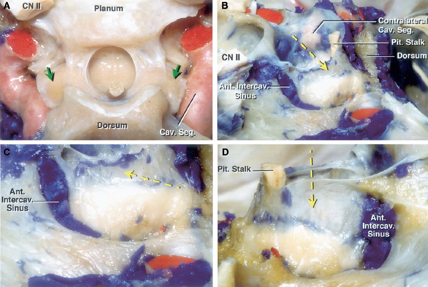
圖8。三個標本的照片。海綿內頸動脈(綠色箭頭)上方的垂體雙側突起標本的後上位視圖。頸動脈外側壁,前斜突,顱神經,頸動脈斜突段和頸動脈脊上段都被切除。B,另一標本左骶管上外側視圖,蝶鞍部分空(黃色箭頭)。左側海綿內頸動脈,靜脈腔內的物質,以及顱神經都被切除了。通過內側壁觀察到對側(右)頸動脈海綿狀內動脈。交叉池延伸到鞍區(黃色箭頭),靠在鞍區內側壁的上半部分。C,巨大的前海綿間竇穿過鞍壁的硬腦膜層。內側壁的鞍部與交叉池的下延伸部直接接觸,進入部分空鞍區(黃色箭頭)。 D, lateral view of the right medial wall of the specimen shown in C. This specimen presents a large anterior intercavernous sinus with extension of the chiasmatic cistern into the partial empty sella (yellow arrow). Ant., anterior; Cav., cavernous; CN, cranial nerve; Intercav., intercavernous; Pit., pituitary; Seg., segment. (Images courtesy of AL Rhoton, Jr.)
蝶骨部分的結構比形成鞍部的簡單四邊形硬腦膜更複雜(圖2-7)。可以確定三個部分或子區域。前部分是由硬腦膜在頸動脈床側內側的頸動脈溝襯裏形成的。這部分分別受到硬腦膜上環(遠端)和下環(近端)的限製,它們由硬腦膜從前床突的上表麵和下表麵向內側延伸並環繞頸動脈形成(圖4)。中間部分由硬腦膜內襯位於蝶鞍底外側邊緣以下的頸動脈溝形成(圖3-5)。後部分沿後床突和鞍背外側邊界延伸,止於岩斜坡裂隙上緣(圖4D)。內側壁的蝶部和鞍部僅由一層硬腦膜構成,但蝶部由骨內膜層構成,鞍部由腦膜層構成(圖2)。
鞍底中心到上頜神經上緣的平均距離為11.27 - 2.13 mm。頸動脈溝最前邊界到鞍背(岩斜裂隙)的平均距離為19.21 1.37 mm(表1;圖1)。

在21例(52.5%)的CSs中,海綿內頸動脈與腦垂體直接接觸,僅被CS薄內側壁的鞍部分開。在剩下的19例CSs(47.5%)中,在海綿頸動脈內和腦下垂體之間有靜脈空間和眶脂肪的後延伸。後者的動脈與腺體之間的最短距離平均為2.55±1.16 mm。
在40個CSs中有39個CS的靜脈間隙延伸到頸動脈海綿內和頸動脈溝硬腦膜之間。靜脈組成部分的厚度和長度在標本中有所不同(圖4C和圖7,A和B)。靜脈空間或叢最厚的部分在頸動脈溝邊緣附近(平均,1.92±0.51 mm),最薄的部分在頸動脈溝中部附近(平均,0.78±0.2 mm)。
intracavernous頸動脈的位置,相對於sellar內側牆的一部分,明顯不同(無花果。3 g、4 d、7 b,和9)。腦下垂體的橫向方麵是縱向分為優越,中間,偽劣三分之二(圖9)。intracavernous頸一起追逐隻有劣質第三14 CSs(35%)(圖7 b),以及部分劣質和13個CSs中間三分之二(32.5%),以及部分的三分之二11 CSs(27.5%)(圖3 g)。在2例CSs(5%)中,海綿內頸動脈沿蝶骨延伸至鞍底和垂體腺下方(圖4D)。垂體下動脈沿內側壁鞍部後下方延伸至後葉(圖4E和圖7E)。

圖9。右側頸動脈側位圖顯示了海綿內頸動脈和內側壁鞍部之間的不同關係。A,海綿內頸動脈在頸動脈溝上,與內側壁鞍部沒有任何接觸。B,海綿內頸動脈在腺下三分之一和內側壁的外側。C,海綿狀內頸動脈延伸到腺體中部和下三分之一部分和內側壁的外側。D,海綿內頸動脈在腺體的三分之一和內側壁的外側。一個,動脈;螞蟻。前;的車。, carotid; Cav., cavernous; Clin., clinoid; Fiss., fissure; For., foramen; Orb., orbital; Pit., pituitary; Post., posterior; Seg., segment; Sphen., sphenoidal; Sup., superior. (Images courtesy of AL Rhoton, Jr.)
垂體由前葉和後葉組成(圖4,C-F)。內側壁的鞍部覆蓋前葉的外側表麵(圖5A),但後葉位於鞍背凹陷處的前葉和內側壁的後麵(圖4,C-F)。後葉位於鞍背前凹麵,在鞍背內側壁的鞍部沿鞍背內表麵外側緣與硬腦膜融合。
在我們的標本中,腦垂體的形狀變化明顯。在40例CSs中,18例(45%)垂體腺有側突。13例(32.5%)突出位於腺體外側表麵的上三分之一處,3例(7.5%)突出位於中間三分之一處,1例(2.5%)突出位於下三分之一處,1例(2.5%)突出位於所有三分之一的前部。在6例CSs中,海綿內頸動脈使腺體凹陷,舌狀延伸在動脈上方側向突出(圖2、D和E和8A)。內側壁的鞍區部分,甚至在覆蓋突出的區域,是完整的,沒有一個缺陷,腺體通過疝出。
CS內側壁鞍部周邊與蝶竇部交界處,是不同大小和分布的靜脈竇的多發部位,這些靜脈竇穿過連接成對CSs的中線(圖2A, 4E, 6C,和8,B-D)。海綿間竇最常穿過蝶鞍和腺的前緣和下緣(27css)。其他類型包括7個CSs的前、下、後緣竇;在三個CSs的前緣和後緣,隻在三個CSs的前緣。海綿間竇位於硬腦膜和硬腦膜內膜層之間,硬腦膜內襯鞍前、後壁和底(圖2A)。
在40例檢查的CSs中,我們觀察到腦下垂體外側表麵和CS之間有硬腦膜壁。這與之前的一些研究結果形成對比,後者認為腺體和CS之間沒有硬腦膜壁(8,32)。Yokoama等(32)在10具成人屍體的30個切片中,觀察到了3個內側壁鞍部的小組織缺損,作者認為這些缺損是腺瘤擴展的重要部位。在手術顯微鏡提供的x3到x40放大倍率下檢查的40 CSs中,我們沒有發現這樣的缺陷。
在Umansky和Nathan(28,29)的研究之後,人們普遍認為CS的側壁和頂部是由硬腦膜形成的,硬腦膜可以分裂成兩層。然而,對內側壁的特征了解甚少。內側壁在兩個不同區域(鞍區和蝶區)的劃分和鞍區厚度有助於了解垂體腫瘤的擴散途徑。內側壁的鞍部是由一層非常薄的單層形成的,與另一層較厚的內側壁分開為兩層形成對比,並且垂體窩外側沒有與前、下、後窩表麵相似的骨壁,這一發現解釋了垂體瘤向內側壁延伸的趨勢。內側壁單層薄的特性也解釋了在磁共振成像掃描中顯示內側壁的困難(6,8,15)。
此外,本研究結果支持覆蓋鞍骨表麵的硬腦膜層是兩層類型:一層是麵向骨的內膜型,一層是麵向腺的腦膜型。內膜層起源於上頜神經的上緣,包裹著腺麵向鞍骨麵部分的硬腦膜是膈肌中腦膜層的延續(圖2)。因此,在腺和鞍骨壁之間有兩層硬腦膜。在我們的解剖中,CS的內側壁似乎是鞍膈的腦膜硬腦膜的延續。除了內側壁的鞍部和蝶部外,其他四個表麵覆蓋著兩層硬腦膜。這層雙層襯砌著骨鞍壁,使沿著鞍前、下、後邊緣的靜脈竇得以穿插。在我們的標本中,內側壁單層鞍部沒有靜脈竇交叉。
海綿內頸動脈和腦脊液內側壁之間的關係在垂體腫瘤的治療中很重要。在超過一半(52.5%)的標本中,海綿內頸動脈和正常垂體之間直接接觸,隻有薄薄的內側壁將兩者隔開。在6例CSs中,一個巨大的腦垂體突起向外側延伸,穿過海綿內頸動脈上表麵。海綿內頸動脈也可能壓痕和壓迫垂體腺的外側表麵,導致垂體腺的突出擴散到動脈周圍(圖2D和8A)。垂體腺瘤侵襲CS的術前診斷是多項研究的主題(6,15,16)。Knosp等人(15)提出了一種基於冠狀磁共振成像掃描顯示垂體腺和海綿狀內頸動脈的分類。我們的假設是,部分動脈壁周圍腺體的突出表明海綿樣擴張,但在我們研究的6個CSs中,在動脈周圍有正常腺體的舌狀突出。Cottier等人(6)提出使用靜脈間隙的CS作為評估CS侵犯的一種方法。如果在冠狀麵觀察到腺瘤與海綿頸動脈內之間的內側靜脈間隙,則認為頸動脈無侵犯。然而,我們確定52.5%的海綿內頸動脈與內側壁的鞍部薄部分直接接觸。 Cottier et al. (6) also proposed that invasion of the CS was highly probable if the venous plexus along the carotid sulcus was not observed. This cavernous venous plexus was observed in 97.5% (39 of 40 CSs) of the CSs we studied. Therefore, the thinness of the medial wall and the variability in the shape, size, and distribution of the venous plexus render it an unreliable method of identifying CS invasion on computed tomographic or magnetic resonance imaging scans.
海綿狀頸動脈橫向放置在垂體下三分之一的水平比位於中三分之一和上三分之一外側的頻率更高,這表明暴露或損傷海綿狀頸動脈的風險最大的是暴露沿著內側壁鞍部的下三分之一的區域。在經蝶竇手術中打開鞍前硬腦膜時,需要注意避免對海綿內頸動脈的損傷,特別是當動脈突出進入腺體時。應避免使用鋒利的刀在硬腦膜切口的角落切開硬腦膜(圖10A)。資深作者(ALR)用刀進行一個短的垂直硬腦膜中線切口作為初始步驟。一個小的,鈍的,直角環形刮匙通過小的垂直硬腦膜開口插入,硬腦膜與腺體或腫瘤的前表麵分離。硬腦膜取出後,選擇一把45度角短吻鱷剪刀,而不是小刀,從一個角到另一個角切開硬腦膜,剪成x形,因為尖刀可能會損傷暴露處遠外側角的頸動脈(圖10)。用45度角剪刀的遠端刀片將鞍硬腦膜從腺體或腫瘤中取出,這樣就可以觀察到硬腦膜內的剪刀刀片不會延伸到硬腦膜深處的任何結構。在研究標本中,我們還注意到垂體下動脈與內側壁鞍部的後下區接觸,在那裏有損傷的風險。
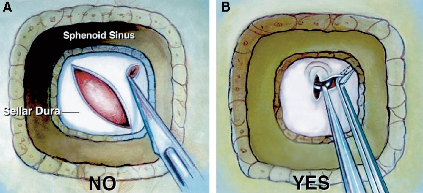
圖10。圖示經蝶竇入路至蝶鞍後硬腦膜開口。第一,在鞍前角使用刀切開硬腦膜應避免暴露因為海綿內頸動脈會壓進腺體或腫瘤的外側可能在硬腦膜外側切開時被刀損壞。B,高級作者(ALR)從中線處的一個短垂直切口開始打開硬腦膜。一個小的,鈍的,直角環形刮匙通過小的垂直硬腦膜開口插入,將硬腦膜與腺體和腫瘤分開。取出硬腦膜後,用一把45度角的剪刀從一個角到另一個角以x形切口將硬腦膜切開。用插入硬腦膜內的剪刀將硬腦膜從腺體抬高,這樣就可以通過硬腦膜觀察到硬腦膜刀片,以確保沒有其他結構被切割。(圖片由AL Rhoton, Jr.提供)
腦下垂體在腦下垂體窩內被一薄包膜覆蓋,與腦下垂體內側壁分離。垂體囊的性質和起源尚未完全確定。不同的作者支持不同的理論(3-5,26,30,31)。Wisloki(30,31)和Ciric(4)分別認為垂體囊來源於硬腦膜或腦膜蛛網膜,而Chi和Lee(3)則認為垂體囊來源於拉特克囊(1,8,18,27,32)。
脊膜內側壁有兩個節段(鞍部和蝶部),由一層薄薄的硬腦膜組成,手術無法將其分離成兩層。這堵牆與周圍結構之間的關係表現出明顯的變異性。
貢獻者:Alexandre Yasuda,醫學博士,Alvaro Campero,醫學博士,Carolina Martins,醫學博士,Albert L. Rhoton, Jr,醫學博士,Guilherme C. Ribas,醫學博士
內容來自Yasuda A, Campero A, Martins C, Rhoton AL, Jr, Ribas GC。海綿竇內側壁:顯微外科解剖。神經外科2004; 55:179 - 190。doi.org/10.1227/01.NEU.0000126953.59406.77.經牛津大學出版社代表神經外科醫師協會批準。©神經外科醫生協會。
神經外科188bet手机app圖譜很榮幸能夠繼承Albert L. Rhoton, Jr . MD的遺產。
請登錄發表評論。
一定要在社交媒體上關注我們,獲取精彩內容並保持更新生活科恩醫生的會議,關於手術技術的問題,以及更多!


您必須登錄才能查看此材料。
的188bet手机app這幾乎完全取決於你的捐款。
如果沒有你們的大量捐贈,我們就無法繼續開展地圖集。
請承諾每年至少捐贈250美元給Atlas。如果沒有這種承諾,Atlas將很快需要付費訂閱,世界各地的許多外科醫生將無法獲得它,他們的病人的護理依賴於它。
請立即捐款!
如果沒有你們的大量捐贈,我們就無法繼續開展地圖集。請承諾每年至少捐贈250美元給Atlas。
如果沒有這個承諾,Atlas將很快需要付費訂閱世界上許多病人的護理都依賴於它的外科醫生將無法使用它。請立即捐款!