

你可以有所作為。
的188bet手机app這幾乎完全取決於你的捐款。
如果沒有你們的大量捐贈,我們就無法繼續開展地圖集。
請承諾每年至少捐贈250美元給Atlas。如果沒有這種承諾,Atlas將很快需要付費訂閱,世界各地的許多外科醫生將無法獲得它,他們的病人的護理依賴於它。
請立即捐款!

最後更新:2021年4月7日
摘要目的:進一步確定頸動脈腔的顯微解剖及其與鄰近結構的關係。
方法:在屍體標本上用3 ~ 40倍放大鏡觀察洞穴及其關係。
結果:該洞穴是硬腦膜內袋,位於20個斜突旁區域中的19個,延伸至遠端硬腦膜環水平以下,介於ICA壁和圍繞ICA的硬腦膜頸圈之間。遠端硬腦膜環與毗鄰前臥突和視神經支柱的ICA的前側壁和外側壁緊密粘附,但與頸動脈洞穴所在的頸動脈溝上部的動脈的內側壁和後側壁沒有緊密粘附。垂體上動脈常在洞穴中出現。洞穴的深度和周向長度平均分別為2.4 mm (1.5 - 5mm)和9.9 mm (4.5 - 12mm)。發源於洞穴水平的動脈瘤,雖然在放射學研究中表現為延伸至前臨上邊緣以下,但可延伸至蛛網膜下腔並可能是蛛網膜下腔的來源。
結論:在手術治療洞穴動脈瘤時,需要準確了解洞穴與ICA、硬腦膜環和頸動脈頸圈的關係。
頸動脈洞,由Kobayashi等人在1989年命名,是一個小的隱窩或袋,延伸到內頸動脈壁(ICA)內側遠端(上)硬腦膜環水平以下ICA通過硬腦膜近端和遠端環進入蛛網膜下腔和基底池,硬腦膜環由硬腦膜從前床突的上下表麵向內側延伸包圍動脈形成。ICA的床突段位於硬腦膜近端和遠端環之間,位於內側,通過移除前床突暴露出來遠端環似乎在動脈周圍形成了一個緊密的頸圈,但在手術顯微鏡下仔細檢查發現,通常有一個隱窩,頸動脈洞沿著頸動脈後內側延伸到動脈壁和遠端硬膜環之間。關於頸動脈腔的顯微外科解剖的報道很少。本研究的目的是進一步確定頸動脈腔的顯微解剖及其與鄰近結構的關係。
在10具屍體標本中注射彩色矽膠後,用3 ~ 40倍的手術顯微鏡檢查20個旁凸區。頸動脈洞穴是位於ICA後內側外側的一個小隱窩,從近端延伸到遠端硬腦膜環(圖1)。測量ICA周圍洞穴的長度和每個洞穴的垂直深度。從上方觀察時,按順時針方向記錄頸動脈洞的位置,12點鍾為頸動脈前方,3點鍾為頸動脈右側外側,6點鍾為頸動脈後方,9點鍾為頸動脈內側。左邊的洞穴被轉換為右邊,通過時鍾係統來描述洞穴的位置其他測量包括眼動脈起點到ICA上升穿過遠端硬腦膜環的前緣之間的距離;眼動脈起點與視神經下外側穿過視神經鞘進入眶尖的位置之間的距離;頸動脈近端和遠端硬腦膜環之間或ICA外側之間的頸動脈斜突段的長度;以及眼動脈起源的直徑。

圖1所示。鞍區優越的視野。A,視神經和交叉向前反射,露出眼動脈。垂體上動脈起源於左髂內動脈的脊狀肌上段。左側ICA已向外側縮回,露出頸動脈洞,其延伸至遠端硬腦膜環以下,進入頸動脈頸和動脈壁之間的空間。一個垂體上動脈出現在洞穴的上部。B,頸動脈腔放大圖。C,切除前床突形成的右側床突間隙的上視圖。床突空間近端由硬腦膜形成的頸動脈動眼膜定義,硬腦膜將床突下表麵與動眼神經隔開,並向內側延伸環繞頸動脈形成近端硬腦膜環,遠端由硬腦膜從環繞頸動脈的床突上表麵向內側延伸形成遠端硬腦膜環。從前到後,床突空間內的結構包括視神經支柱、圍繞ICA的頸動脈頸圈(近端環和遠端環之間)和海綿竇頂的前部。 D, the right carotid artery has been retracted laterally to expose the carotid cave. A., artery; Car., carotid; Cav., cavernous; Clin., clinoid; CN, cranial nerve; Dist., distal; Hyp., hypophyseal; Memb., membrane; Oculo., oculomotor; Ophth., ophthalmic; Pit., pituitary; Prox., proximal; Seg., segment; Sup., superior; ICA, internal carotid artery. (Images courtesy of AL Rhoton, Jr.)
頸動脈洞穴位於頸動脈麵向頸動脈溝的一側,這是蝶骨體外側的一個淺槽,ICA沿此方向前進(圖2)。海綿內頸動脈靠在頸動脈溝上,並被內鼻竇壁的硬腦膜與頸動脈溝隔開。頸動脈溝起於頸動脈管顱內端鞍背下方和外側。經過最初的短垂直切麵後,頸動脈溝向前轉向,在蝶鞍外側邊緣下方的蝶鞍體上形成凹槽,然後向上轉向,在蝶鞍前壁外側邊緣的前方,沿著視神經支柱的後邊緣和前床突的內側邊緣延伸。頸動脈溝,在通氣良好的蝶骨中,形成一個蛇形凸起,在蝶竇的側壁垂體窩下可見。蝶竇外側壁的骨在某些部位可能很薄,甚至缺失,從而使襯在竇溝內的動脈和硬腦膜靠在竇粘膜上。

圖2。A,頸動脈洞穴的骨關係,上位視圖。頸內動脈的床突段,即穿過前床突內側的段,幾乎被外側的前床突、前方的視神經支柱和內側的蝶骨頸動脈溝所包圍。頸動脈腔位於ICA壁,麵向頸動脈溝。前突基部外側附著於由蝶小翼形成的蝶脊內側端,內側附著於蝶小翼的前、後根。小翼前根從前突基部向內側延伸至蝶骨體,形成視神經管的頂部。後根,稱為視神經支柱,向視神經下方內側延伸至蝶骨體,形成視神經管的底部。B、放大上位視圖。交叉溝是一對視神經管之間的淺凹陷,後麵是鞍結節,前麵是蝶平麵。鞍結節位於中線,沿脊形成交叉溝的後緣。 The middle clinoid process projects upward on the medial side of the terminal part of the carotid sulcus toward the tip of the anterior clinoid process. An osseous bridge may extend from the middle clinoid to the anterior clinoid, thus creating a bony ring, referred to as a caroticoclinoidal foramen, through which the ICA passes. The dura lining the upper margin of the anterior clinoid extends medially above the optic nerve to form the falciform ligament (blue arrow) and slightly downward to line the upper margin at the optic strut and form the anterior part of the distal dural ring (green line). The margins of the left carotid sulcus are shown (interrupted lines). C, oblique posterior view. The carotid sulcus (interrupted lines) begins below and lateral to the dorsum sella, turns forward in a shallow groove below the lateral edge of the sellar floor, and turns upward to end medial to the anterior clinoid process. D, oblique posterior view of the right optic strut, the bridge of the bone that extends from the inferomedial aspect of the base of the anterior clinoid to the body of the sphenoid bone and separates the optic canal from the superior orbital fissure. The clinoid segment of the ICA rests against the posterior margin of the strut. The dura lining the upper margin of the anterior clinoid extends medial above the optic nerve to form the falciform ligament (blue arrow) and slightly downward to line the upper margin at the optic strut and form the anterior part of the distal dural ring (green arrow). The posterior surface of the strut widens as it slopes medially. E, superior view. The lesser sphenoid wing and the base of the left anterior clinoid, have been removed to unroof the optic canal and upper and posterior margin of the optic strut. The posterior margin of the optic strut is shaped to accommodate the anterior surface of the anterior bend of the intracavernous carotid. The pneumatization of the sphenoid sinus may extend through the strut into the anterior clinoid. The lateral wall of the sphenoid sinus forms the medial wall of the optic canal. F, superior view of specimen with bilateral caroticoclinoidal foramen and interclinoidal osseus bridges. An osseous bridge connects the tips of the anterior and middle clinoid processes bilaterally, thus creating a bony ring around the artery called a caroticoclinoidal foramen, on each side. There are also interclinoidal osseus bridges connecting the anterior and posterior clinoid processes on both sides. Ant., anterior; Car., carotid; Car. Clin., caroticoclinoidal; Chiasm., chiasmatic; Clin., clinoid; Fiss., fissure; For., foramen; Inf., inferior; Interclin., interclinoidal; Lac., lacerum; Less., lesser; Mid., middle; Orb., orbital; Plan., planum; Post., posterior; Rotund., rotundum; Sphen., sphenoid, sphenoidale; Sup., superior; Tuberc., tuberculum; ICA, internal carotid artery. (Images courtesy of AL Rhoton, Jr.)
前突從蝶骨的小翼向後突出。前突基部有3個位點與相鄰的蝶骨相連。在前方和外側,前床突的基部與蝶小翼的內側邊緣相連。在內側有2個附著點,一個在蝶小翼的前根,另一個在後根。蝶小翼前根從視神經上方的前突基部向內側延伸,形成視神經管頂。後根,稱為視神經支柱,向視神經下方內側延伸至蝶骨體,形成視神經管的底部。前床突基部的內側邊緣形成視神經管的外側邊緣。視神經管的內側緣是由蝶骨體的鄰近部分形成的。前瘤突是幕前內側部和前岩瘤和前岩瘤間硬腦膜褶皺的附著部位。4
視神經支柱是一個小的骨橋,從前臥突基部的下內側向內側延伸到頸動脈溝前麵的蝶骨體。它將眶上裂頂部的內側部分與視神經管分開。支柱的上表麵形成視神經管的底部,下表麵形成眶上裂的超內側邊緣。該支柱位於眶尖前方與眶上裂和視神經管後方的交界處。它的橫截麵呈三角形,有上、下、後三麵。視神經支柱後表麵的形狀是為了適應頸動脈床突段前彎的前表麵,當視神經支柱在前床突內側上升時,頸動脈床突段前彎靠在視神經支柱的後表麵上。
髂內關節瘤的床突段,定義為前床突的內側段,其外側、內側和前方被骨結構緊緊包圍,隻在骨和動脈之間留下狹窄的空間。床突段的外側壁與前床突的內側表麵有緊密的硬腦膜附著,前壁與視神經支柱的後側有硬腦膜附著。頸動脈床突段的後部與海綿竇和非硬骨結構接觸。在內側,床突段麵對蝶體上頸動脈溝的遠端,在那裏動脈的硬腦膜錨定小於外側和前方動脈麵對床突和視神經支柱的地方。
硬腦膜之間的關係對於規劃入路到斜突旁區和洞穴的手術很重要(圖3和圖4)。硬腦膜從前凸突基部的上表麵向內側延伸,直接向內側延伸,並在前凸突上表麵的軸向水平與ICA相連。硬腦膜也從床突上表麵向內側延伸,在前床突上表麵軸向水平的視神經上方,延伸到小翼的前根和蝶平麵的後邊緣。然而,硬腦膜從前凸突的上表麵向內側延伸到視神經支柱的上表麵並形成遠端硬腦膜環的前部,在向內側推進時向下傾斜,因此遠端硬腦膜環的內側實際上位於比前凸突和視神經管的上表麵低的軸位。就在小翼前根的後麵是硬腦膜褶皺,鐮狀韌帶延伸到視神經上方就在神經進入視神經管的近端。鐮狀韌帶向內側與覆蓋蝶平麵的硬腦膜混合。位於視神經支柱上表麵的硬腦膜向ICA的後方和內側延伸,靠近頸動脈溝遠端,形成上環的後方和內側部分。遠端硬腦膜環與近端硬腦膜環在前床突的後尖端連接,形成一個單一的硬腦膜層,在後方與膈鞍混合(圖4)。

圖3。鞍上區上方視圖。A,頸動脈通過前床突的內側進入顱腔並在視神經下方。位於前突上表麵的硬腦膜向內側延伸,向兩個方向延伸:上伸展部穿過視神經上方,在蝶骨小翼前根處形成鐮狀韌帶;下延伸略微向下延伸到視神經支柱的上邊緣形成硬腦膜遠端環的前部。B,視神經管頂部的硬腦膜和前凸肌被切除。鐮狀韌帶沿小翼前根後緣延伸至視神經上方,向內側與覆蓋蝶平麵的硬腦膜融合。C,視神經和交叉被抬高,暴露垂體柄、眼動脈和垂體上動脈。頸動脈動眼膜已被切除以顯露頸動脈動眼膜由位於頸動脈下緣的硬腦膜形成,它將頸動脈下突與動眼神經分開並向內側延伸形成近端硬腦膜環。硬腦膜從前凸突的上表麵向內側延伸到視神經支柱的上表麵,也形成了遠端硬腦膜環的前部,遠端硬腦膜環定義了ICA的上緣。 This dura forming the proximal ring slopes downward as it proceeds medially, so that the medial part of the distal dural ring actually lies at the level of the lower rather than the upper surface of the anterior clinoid. The lateral part of the distal ring near the origin of the ophthalmic artery tightly adheres to the lateral wall of the ICA. D, the clinoid segment of the ICA, located between the proximal and distal dural rings, has been exposed by removing the anterior clinoid. The ICA between the proximal and distal ring is enclosed in a thin layer of dura referred to as the carotid collar. The proximal ring is loosely applied to the clinoid segment and allows the clinoid venous plexus, a thin venous plexus that courses inside the carotid collar and outside the carotid wall, to communicate inside the ring with the anterior part of the cavernous sinus. E, the right ICA has been retracted laterally to expose the carotid cave. The dura along the posterior edge of the carotid cave contains the anterior intercavernous sinus. F, the clinoid segment has been retracted anteriorly to expose the part of the cave adjacent the diaphragm sellae. G, oblique anterior superior view. The roof of the sphenoid sinus has been removed to expose the medial side of the right ICA. A green piece has been inserted into the carotid cave. The cave, the short downward directed pouch inside the carotid collar, extends below the level of the distal dural ring between the arterial wall and the carotid collar. H, the distal dural ring and the carotid collar have been divided and the dural flaps retracted with white silk to expose the carotid cave, the space between the carotid collar and the outer carotid wall that opens upward into the intradural space. A., artery; Ant., anterior; Br., branch; Car., carotid; Ch., choroidal; Clin., clinoid; CN, cranial nerve; Comm., communicating; Diaph., diaphragma; Dist., distal; Falc., falciform; Hyp., hypophyseal; Intercav., intercavernous; Lent. Str., lenticulostriate; Lig., ligament; Memb., membrane; Oculo., oculomotor; Ophth., ophthalmic; P., posterior; Perf., perforating; Pit., pituitary; Plex., plexus; Prox., proximal; Rec., recurrent; Seg., segment; Sphen., sphenoid; Subst., substance; Sup., superior; ICA, internal carotid artery. (Images courtesy of AL Rhoton, Jr.)
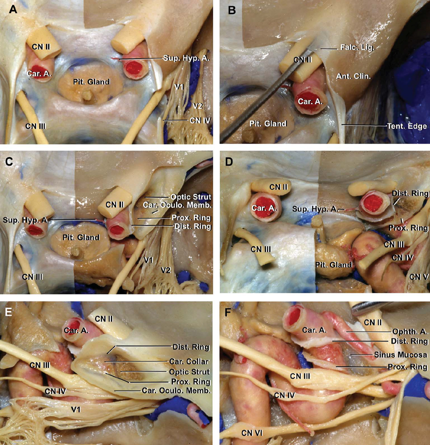
圖4。A,鞍區優越的視角。硬腦膜頂、前臥突、視神經管和右側海綿竇外側壁被切除。右上垂體動脈殘端被保存下來。膈肌缺失暴露垂體的上緣。B,鐮狀韌帶下方的解剖器進入視神經管。C,在保留頸動脈動眼膜和硬腦膜近端和遠端環的情況下,切除前床突。在切除前凸突所形成的空間中暴露出視神經支柱和側突段。硬腦膜形成近端環和遠端環在動脈外壁連接形成硬腦膜頸圈,頸動脈頸圈,環繞ICA。頸動脈腔位於ICA的內側和後部,向下延伸至遠端硬腦膜環和頸動脈頸和動脈外壁之間。 D, posterior superior view. The proximal and distal dural rings converge as they extend along the posterior margin of the carotid where they blend into a single layer forming the diaphragm sellae. E, lateral view of the proximal and dural rings. The proximal and distal dural rings converge posteriorly where both rings blend into the dural layer forming the diaphragm sellae. The clinoid segment of the carotid artery is surrounded by the carotid collar. F, lateral view of the cavernous sinus and the proximal and dural rings. The distal dural ring is formed by the dura extending medially from the upper surface of the anterior clinoid process. The proximal ring is formed by the dura that separates the lower margin by the anterior clinoid from the oculomotor nerve and extends medially around the ICA. In this specimen, the mucosa lining the sphenoid sinus extends into the optic strut, and in some cases, the sinus may pneumatize through the strut into the anterior clinoid process. A., artery; Ant., anterior; Car., carotid; Clin., clinoid; CN, cranial nerve; Dist., distal; Falc., falciform; Hyp., hypophyseal; Lig., ligament; Memb., membrane; Oculo., oculomotor; Ophth., ophthalmic; Pit., pituitary; Prox., proximal; Sup., superior; Tent., tentorial; ICA, internal carotid artery. (Images courtesy of AL Rhoton, Jr.)
位於前突下緣並將前突與動眼神經分開的硬腦膜向內側延伸包圍ICA並形成近端硬腦膜環。這層膜被稱為頸動脈動眼膜因為它將頸突下表麵與顱神經III的上緣分開。這層膜向內側向前延伸到視神經支柱的下表麵,形成近端環的前部。頸動脈動眼膜在動脈內側與頸動脈溝的硬腦膜混合,但在動脈內側麵向頸動脈溝形成的近端環不像在動脈前緣和外側緣形成的近端環那樣明顯。
外側的部分遠端環緊緊堅持ICA的側壁鞍突的上邊緣的水平,但是在ICA的患者,硬腦膜轉下行之前牢牢地固定在ICA牆,從而創建一個休會,頸動脈洞穴,在遠端環和外頸動脈壁之間的蛛網膜可以推動創建一個蛛網膜下腔和硬膜內的水平以下的遠端環(圖3和5)。洞穴,向下突出的隱窩,在頸動脈外側和頸動脈溝內側硬腦膜之間的上環水平以下延伸可變距離。洞穴可能向下延伸到下環附近,並可能是蛛網膜下腔產生動脈瘤的出血的位置,即使在血管造影上,它的頸部位於前床突上緣的水平以下。3.

圖5。A,前視圖。蝶鞍和視交叉前的冠狀麵。遠端硬腦膜環已被保留。蝶竇壁被切除,露出蝶鞍前壁和ICAs。視頸隱窩延伸到視神經支柱。右側硬腦膜近端環向內側延伸至頸動脈隱窩下方,在動脈內側不像遠端環那樣形成明顯的近端環。基底靜脈叢是海綿竇之間最大的交流通道。B,右側鞍旁區和延伸至視神經支柱的視頸隱窩的放大視圖。硬腦膜沿視神經支柱的下緣向後延伸形成近端硬腦膜環。 The dura on the upper surface of the optic strut extends around the ICA to form the distal dural ring. The abducens nerve passes around the lateral surface of the carotid and ascends medial to the ophthalmic nerve in the cavernous sinus. C, enlarged view after removing the dura in the sellar region and along the medial wall of the right cavernous sinus. The opticocarotid recess, a pneumatized diverticulum of the sphenoid sinus extends laterally into the optic strut, which separates the optic nerve in the optic canal from the nerves passing through the superior orbital fissure. The dura forming the proximal dural ring does not form as distinct a ring on the medial side of the artery adjacent the carotid sulcus as it does along the anterior and lateral surface of the artery. D, enlarged view. The distal dural ring encases the carotid artery just below the level of the origin of the ophthalmic artery. The proximal ring blends into the dura surrounding the artery on the side of the carotid sulcus. E, a triangular piece of green material has been inserted into the carotid cave located between the dura forming the collar around the artery and the outer wall of the ICA. The proximal ring is not as distinct on the medial side of the artery as is the distal ring. F, the distal dural ring and upper medial part of the carotid collar has been incised and retracted with black sutures to expose the arterial wall in the carotid cave. G, transnasal exposure of the left parasellar region in another specimen. One superior hypophyseal artery arises in the carotid cave proximal to the distal ring and ascends to exit the cave and pass to the pituitary stalk. Another superior hypophyseal artery arises above the level of the distal dural ring. A triangular piece of green material has been placed inside the cave. H, the carotid collar, below the level of the distal ring, has been opened to expose the origin of the superior hypophyseal artery in the carotid cave. The proximal ring is not as distinct on the medial side of the ICA as is the distal ring. A., artery; Bas., basilar; Car., carotid; Cav., cavernous; CN, cranial nerve; Dist., distal; For., foramen; Gyr., gyrus; Lac., lacerum; Med., medial; Ophth., ophthalmic; Opticocar., opticocarotid; Pit., pituitary; Prox., proximal; Rec., recess; Seg., segment; ICA, internal carotid artery. (Images courtesy of AL Rhoton, Jr.)
在本研究中檢查的20個頸突旁區中有19個存在頸動脈洞穴。這些洞穴位於ICA的後內側,並在遠端環水平向上打開進入硬膜內空間。洞穴位於3點至11點之間,沿動脈周長,平均最大深度為7點,在頸動脈溝對麵的區域,在ICA周圍的周位中點附近。在19個有頸動脈洞穴的鞍旁區,沿動脈周長的平均深度和長度分別為2.4 mm(範圍1.5 - 5mm)和9.9 mm(範圍4.5 - 12mm)。
頸環是由低環將向上的硬腦膜包圍ICA的段近端和遠端之間的環(圖3 - 5)。頸領不嚴格遵守ICA的牆,直到達到上部硬腦膜的戒指,它融入,是連續上硬腦膜的戒指,這是緊密相連的外牆ICA除了在該地區麵臨的洞穴。側突靜脈叢是一條細長的靜脈叢,位於頸動脈頸和頸內動脈側突段的外壁之間,在近環和外壁之間進入海綿竇的前部。形成硬腦膜頸圈的硬腦膜很薄,通過薄硬腦膜頸圈可以看到動脈壁和床狀靜脈叢。頸動脈頸圈在床突尖端後方消失,在那裏,位於前床突上下表麵的硬腦膜融合成單一硬腦膜層,形成海綿竇頂的後部,並融入膈鞍區(圖4)。
頸動脈頸和上、下環向下傾斜,因為它們從前床突向內側延伸。遠端硬腦膜環在向後內側方向前進時向下傾斜,因此前外側部分是遠端硬腦膜環的最高部分。3.ICA外側的上下環之間的分離大於ICA後側環的連接和融合。沿ICA側麵上下環之間的距離平均為3.6 mm(範圍為2.5-5.2 mm)。
頸動脈腔內的兩根起源於ICA的動脈分別是眼動脈和垂體上動脈(圖3和圖5)。後者可能起源於頸動脈腔。垂體上動脈是一組1 - 5個(平均為2個)小分支,發源於ICA的眼段,終止於垂體柄和垂體腺,但也發送分支到視神經、交叉和第三腦室底。5 - 7在本研究中,20個斜旁區中有15個有位於頸動脈腔附近腹內側的小動脈。這些小動脈,即垂體上動脈,位於遠端硬腦膜環的下方,在20個被檢查的頸動脈旁的7個區域。
眼動脈通常從ICA前彎上側的內側半部分起於遠端環之上,並在視神經下向前穿過。眼動脈起點平均直徑1.9 mm(範圍1.2-2.1 mm)。眼動脈起點與毗鄰硬腦膜遠端環的ICA之間的距離平均為3毫米(範圍2-4毫米)。眼動脈進入視神經管顱內端,穿過視神經管底的硬腦膜,從視神經下外側進入眼眶。動脈起點到硬腦膜穿透點之間的距離平均為8.6毫米(範圍6-13毫米)。視神經管內的眼動脈有時會向視神經顱內段發出一個返支。在2% - 8%的病例中,眼動脈可能出現在眶外,從ICA的床突或海綿狀段內,在這種情況下,動脈通常通過眶上裂或視支的異常孔進入眼眶。5、8、9眼動脈很少起源於中腦膜動脈或基底動脈。10 - 12眼動脈起源於硬腦膜遠端環下的斜突段,位於本研究中20個斜突旁區中的1個。
動眼神經穿過海綿竇頂,沿著前突下內側緣走行(圖3 - 5)。硬腦膜近端環將動眼神經與前突下緣分開。動眼神經被海綿竇頂的一個短池包圍,直到到達前床突的後尖池的末端才與外側竇壁牢固地結合在一起。
滑車神經在動眼神經進入點的後外側進入海綿竇頂,並在動眼神經的下方外側壁的後部分進入。滑車神經在床前突基部水平沿動眼神經外側表麵向上移動,並在動眼神經上表麵和床前突下緣的硬腦膜之間向內側旋轉,直至眶內側和上斜肌。
頸動脈洞穴是指在頸動脈頸圈麵向頸動脈溝的部分和ICA壁後內側部分之間從近端延伸到遠端環的小隱窩,Kobayashi等人首次描述了它,他們注意到它與該區域出現動脈瘤的關係。1, 13、14頸動脈洞穴已在68%至77%的屍體標本中被確認。15日16遠端硬腦膜環由硬腦膜向內延伸至前凸肌上表麵,向後延伸至視神經支柱上表麵,向外側延伸至頸動脈溝遠端,向前延伸至蝶鞍膈和後凸肌上表麵。3.Hitotsumatsu等15值得注意的是,遠端硬腦膜環的後內側,即頸動脈洞的位置,不與任何骨結構接觸。頸動脈骨溝的遠端通常止於遠端硬腦膜環的近端,這為頸動脈洞的形成提供了條件,這一發現與我們的研究一致。硬腦膜與頸動脈腔相鄰,包含海綿竇和前海綿間竇。頸動脈洞穴不應與側突間隙混淆,側突間隙是通過移除前側突而形成的,位於ICA外側,海綿竇前部上方。17
在我們研究的15個頸旁區中,在遠端環水平附近有向內的分支,7個在遠端硬腦膜環水平以下的頸動脈洞中有小分支。這些分支都是垂體上動脈,起源於硬膜內棘突旁肌內側壁,數量從1到5,而文獻中為1.8到2.2。5、6田中等18據報道,起源於垂體上動脈的動脈瘤在床側或斜下平麵向內側或內側投射。所有位於斜下位的動脈瘤都起源於垂體上動脈的起點。因此,頸動脈洞穴動脈瘤構成了起源於頸動脈硬膜內最近端的上垂體病變亞群。18一些研究者將頸動脈洞穴動脈瘤定義為最近端的硬膜內ICA病變,因為它們嵌在洞穴中。1、3
眼動脈通常起於視神經下方和遠端硬腦膜環上方,起於頸動脈脊上肌上表麵的內側三分之一處,並在視神經的前外側下方進入視神經管和視神經眶。然而,眼動脈的硬膜外或硬膜間起源也有報道。8日,19在之前的一項研究中,85.7%的眼動脈來自硬膜內ICA, 7.6%來自硬膜外ICA, 6.7%來自近端和遠端環之間的硬膜間水平。20.夾閉眼動脈瘤頸部和上垂體動脈瘤通常需要切除前臥突,動眼動脈和視神經,並至少分離部分遠端硬腦膜環。18眼動脈的起源可能很難從上環上方的區域與起源於近端環和遠端環之間的床突段或海綿竇中近端環以下的區域區分開來。因此,在切割遠端硬腦膜環時,如果遠端硬腦膜環上方看不到眼動脈,應特別注意避免損傷眼動脈。
入路可通過翼點入路或眶顴入路。打開睫狀體裂和頸動脈池暴露前臥突和頸動脈脊上。從這裏開始,通過幾個步驟來幫助沿著ICA和視神經周圍的近端擴展暴露。這些步驟包括去除床突,打開部分遠端硬腦膜環,打開視神經管和打開鐮狀韌帶。
前臥突是暴露病變延伸至頸動脈腔的障礙。對於是在硬膜外還是硬膜內進行側位切除術,目前尚無共識。硬腦膜外斜位切除術的支持者注意到硬腦膜在鑽孔過程中起到了保護動脈瘤的屏障作用,同時減少了骨粉塵進入蛛網膜下腔的可能性。21日,22日對於長前瘤或巨大的斜突旁動脈瘤的患者,建議硬膜內斜突切除術。
如果要在腸外切除前臥突,關鍵的一步是在眶上裂的外側邊緣切開眶膜上硬腦膜褶皺。位於眶上裂外側邊緣的腦膜眶周硬腦膜將硬腦膜固定在鄰近的顱底上,防止床突的後部分暴露。在腦膜眶周硬腦膜皺襞分離後,可以將額側和顳側硬腦膜向後剝開,露出前臥突的後尖,進行臥突切除術。鑽穿床突的中央鬆質骨,隻留下外層皮質骨的薄殼。用精致的刮匙將前凸突的剩餘外殼與周圍硬腦膜分離,注意避免損傷沿內邊緣的頸動脈和視神經,以及沿下邊緣的動眼神經。頸動脈斜突的後端可能在頸動脈後麵向中斜突的內側突出,它可能通過骨橋與中斜突相連,從而形成一個完整的骨環,稱為頸動脈斜突孔,圍繞在海綿竇頂的動脈周圍。前臥位也可向後臥位延伸,並與前臥位和後臥位之間的臥位骨橋相連(圖2)。將視神經管剝去骨架,打開鐮狀韌帶和視神經鞘,可暴露視神經下方的眼動脈起源和ICA內側洞內的病變。打開遠端硬腦膜環的外側部分有助於暴露頸動脈斜突旁。最好在遠端環與頸動脈頸圈交界處外切開硬腦膜,留下一小塊硬腦膜套附在動脈上,而不是將遠端環的貼壁外側部分與動脈壁分開,這樣可能會損傷動脈,打開到頸動脈頸圈內與海綿竇相通的靜脈空間。遠端環的切口限於ICA的前、外側和前內側區域,這可能產生足夠的暴露,可以在眼動脈瘤頸部放置夾子,或在眼動脈起源以下的頸動脈上放置臨時夾子。 Opening the ring along the posterior part of the artery bordering the carotid cave will commonly open into the junction of the anterior intercavernous and the cavernous sinuses with brisk bleeding that can usually be controlled with gentle packing of a hemostatic agent. The most commonly used clip on a cave aneurysm is an angle ring (fenestrated) clip with straight or slightly curved blades advanced from distal to proximal along the ICA and around the neck of the aneurysm (Figure 6). Care is required to avoid occluding the origins of the ophthalmic, superior hypophyseal, posterior communicating, anterior choroidal, and perforating arteries that may not be seen on the lateral side of the exposed ICA.

圖6。右頸動脈洞穴動脈瘤的上外側切麵。動脈瘤頸位於遠端硬腦膜環下方的洞穴中就在垂體上動脈起源的上方。前臥突被切除,形成遠端環的外側和前緣的硬腦膜被打開。B,這種類型動脈瘤最常見的夾應用是從動脈瘤頸的遠端到近端的角度開孔夾。洞穴的邊緣已被收回,露出動脈瘤頸。夾正在向動脈瘤頸部推進。需要注意避免阻塞起源於ICA的眼動脈、上垂體、後交通、前脈絡膜和穿通動脈的起源。C,夾已經閉合動脈瘤已經放氣。在夾層前環包圍動脈的水平上,ICA上有一個陰影。 A., artery; A.Ch.A., anterior choroidal artery; Car., carotid; CN, cranial nerve; P.Comm.A., posterior communicating artery; Dist., distal; Hyp., hypophyseal; Prox., proximal; Sup., superior; ICA, internal carotid artery.
鞍上腫瘤(如鞍結核腦膜瘤)累及視神經管已被報道發生在多達20%的患者中。23日,24日似乎鞍上腫瘤,特別是那些累及鞍結節、遠端硬腦膜環內側和視神經管的腫瘤,可能會延伸到頸動脈洞穴,但尚未有報道,可能是因為對洞穴缺乏認識。
頸動脈洞穴位於內腔內膜的後內側,鞍膈的前外側邊緣,在遠端硬腦膜環的下方。它在ICA外壁和遠端硬腦膜環和頸動脈頸圈之間向下延伸。垂體上動脈和垂體上動脈瘤的頸部起源於頸動脈洞。似乎涉及鞍結節和視神經管的腫瘤也可能擴展到洞穴中。
貢獻者:Wonil Joo, MD, Takeshi Funaki, MD, Fumitaka Yoshioka, MD,和Albert L. Rhoton, Jr, MD
內容來自Joo W, Funaki T, Yoshioka F, Rhoton AL, Jr.頸動脈洞穴的顯微外科解剖。神經外科(黑格斯敦)2012; 70: ons300-ons312。doi.org/10.1227/NEU.0b013e3182431767.經牛津大學出版社代表神經外科醫師協會批準。©神經外科醫生協會。
神經外科188bet手机app圖譜很榮幸能夠繼承Albert L. Rhoton, Jr . MD的遺產。
請登錄發表評論。
一定要在社交媒體上關注我們,獲取精彩內容並保持更新生活科恩醫生的會議,關於手術技術的問題,以及更多!


您必須登錄才能查看此材料。
的188bet手机app這幾乎完全取決於你的捐款。
如果沒有你們的大量捐贈,我們就無法繼續開展地圖集。
請承諾每年至少捐贈250美元給Atlas。如果沒有這種承諾,Atlas將很快需要付費訂閱,世界各地的許多外科醫生將無法獲得它,他們的病人的護理依賴於它。
請立即捐款!
如果沒有你們的大量捐贈,我們就無法繼續開展地圖集。請承諾每年至少捐贈250美元給Atlas。
如果沒有這個承諾,Atlas將很快需要付費訂閱世界上許多病人的護理都依賴於它的外科醫生將無法使用它。請立即捐款!