


你可以有所作為。
的188bet手机app幾乎完全取決於你的捐款。
如果沒有你們的大量捐贈我們無法繼續製作《地圖集》。
請承諾每年至少向Atlas捐贈250美元。如果沒有這種承諾,Atlas將很快需要付費訂閱,世界各地的許多外科醫生將無法使用它,他們的病人的護理依賴於它。
請立即捐款!

最後更新:2021年5月5日
摘要目的:本文的目的是描述海綿竇的解剖結構,並提供在這個複雜區域進行手術時的使用指南。臨床病例被用來說明通往海綿竇及其內容物的路徑,並演示海綿竇如何被用作暴露更深結構的通路。
方法:在動脈和靜脈注射彩色矽膠後,用×3至×40放大鏡檢查30例屍體海綿竇。記錄動眼神經和滑車神經入口與後斜突之間的距離。逐步解剖海綿竇,以展示硬膜內和硬膜外通路,並附有這些入路的術中照片。
結果:海綿竇的解剖結構是複雜的,因為非常重要的神經和血管結構的高密度。選擇性的病例表明,詳細的海綿竇解剖知識可以提供更安全的手術和低發病率。
結論:精確了解海綿竇的骨質關係和神經血管內容,以及顱底和顯微外科技術的使用,使神經外科醫生能夠以降低發病率和死亡率的方式接近海綿竇,改變該區域選定病變的自然史。完全切除海綿竇腦膜瘤已被證明是困難的,在許多情況下,不可能不引起顯著的發病率。然而,手術複位這種病變可增加後續治療成功的機會。
海綿竇的顯微外科解剖已被廣泛描述(15,17,18,20,23,26,27,29,30,34,40,43,46,48)。Browder(4)和Parkinson(27)實施了第一個海綿竇入路治療頸動脈海綿竇瘺,Taptas(44)、Dolenc(7)和Umansky(46)是研究該區域的先驅。目前,海綿竇入路用於基底尖動脈瘤(11,37),頸動脈-眼動脈瘤(8),垂體腺瘤(9,13),一些三叉神經瘤(5),以及該區域的其他腫瘤(13,14,16,31,33,35,38)。盡管海綿竇的解剖結構已經有了很好的描述,但對於許多神經外科醫生來說,海綿竇仍然是一個具有挑戰性和不熟悉的地方。
海綿竇是位於顱底中部的靜脈結構,被硬膜壁包圍,其中包含神經血管結構,一側麵向蝶鞍、腦垂體和蝶骨,另一側麵向顳葉(30)。海綿竇有五個壁:側壁和內側壁,頂部,後壁和前壁。屋頂麵向底層蓄水池;側壁麵向顳葉;內側壁麵向蝶鞍、垂體和蝶骨;後壁正對顱後窩。內側壁在三叉神經第二節(上頜神經)上緣下方連接,狹窄的前緣與眶上裂交界。海綿竇是一個包膜,包含海綿頸動脈段及其分支;交感神經叢;第三、第四、第六腦神經; the first trigeminal division; and multiple venous tributaries and spaces. The intercavernous, basilar, superior, and inferior petrosal sinuses all join with the cavernous sinus. In addition, multiple veins, such as the superior and inferior ophthalmic veins; the veins of the foramen rotundum, foramen ovale, and foramen spinosum; and the deep middle cerebral vein and superficial sylvian veins, empty into the cavernous sinus.
本文的目的是介紹海綿竇的詳細解剖,以指導增加該區域入路的安全性。為此目的,逐步解剖海綿竇和臨床病例說明不同的方法,以該區域提出。測量詳細的外科重要標誌已經進行。雖然有幾篇文章詳細介紹了海綿竇的解剖結構,但關於進入該區域的最佳手術入路和該區域許多病變的適當治療缺乏共識(2,3,6,9,10,12,13,22,24,25,28,31,33,35,38,39,42,47)。由於海綿竇內極為重要的神經血管結構的複雜解剖結構和密度,該區域的許多病變被認為是不可切除的(12,19,25,47)。高顱神經發病率和新技術的出現,如血管內神經外科和放射外科,導致進入海綿竇手術入路的頻率顯著降低(3,12,19,22,24,25,42,47)。然而,在許多國家,這些較新的技術並不容易獲得。通過提高我們對海綿竇解剖的知識,並在手術中應用這些知識,我們已經成功地降低了與該區域手術相關的發病率。
使用×3至×40放大率的外科顯微鏡檢查21例成人屍體標本中的30個海綿竇。頭部注射彩色矽膠,測量動眼器和滑車孔與後斜突之間的距離。由資深作者(EdO)之一進行的海綿竇區域病變的臨床病例,介紹了該區域的不同方法。
海綿竇位於蝶骨和顳骨的顱內表麵(圖1)。海綿竇的前緣從前斜突的下表麵沿頸動脈溝的前緣和視支和眶上裂的後緣向下延伸。後緣從上麵的後斜突延伸到岩尖與下麵的蝶骨體的交界處。在確定前、後邊界後,海綿竇的上、下限由前、後邊緣的上下端延伸出的線條確定。下極限從眶上裂下緣和頸動脈溝下緣下方向後延伸,沿頸動脈管顱內端外側邊緣,止於岩斜裂上端。上邊界從前楔突基部的下表麵沿鞍側緣延伸至後楔突。

圖1 (A- e).圖示海綿竇的骨骼關係。A,海綿竇區域顱底上視圖,海綿竇從眶上裂向前延伸至岩尖後方;內側與鞍部接壤,外側與中窩接壤。它位於前斜突下方的眶上裂的後緣。後壁從鞍背外側邊緣延伸至三叉神經印痕和梅克爾穴內側邊緣。大量靜脈通道進入海綿竇。其中包括基底竇,前海綿竇和後海綿竇,岩上和岩下竇,眼靜脈和眼靜脈,以及出卵圓孔,圓孔和棘孔的靜脈,頸動脈管和蝶特使孔。每個靜脈結構用彩色箭頭表示。基底竇是海綿竇之間最大的交流通道。B,側位圖顯示海綿竇位於蝶骨體和鄰近的岩尖(折線)。 The lower edge of the posterior limit of the cavernous sinus sits on the junction of the petrous apex and the body of the sphenoid bone at the upper end of the petroclival fissure. The lower edge extends for-ward along the superior edge of the lingula of the sphenoid bone and the lateral part of the sphenoid body to just above the foramen rotundum. The anterior edge extends along the posterior edge of the optic strut and the medial edge of the superior orbital fissure. The upper limit of the sphenoid bone extends along the superior margin of the carotid sulcus and ends posteriorly at the posterior clinoid process. The dorsum sellae is located between the paired posterior clinoid processes. C, view showing the anterior clinoid process removed. The osseous limits of the cavernous sinus have been outlined. The tuberculum sellae is located at the posterior edge of the chiasmatic sulcus between the anterior part of the paired carotid sulci and posteromedial to the optic canals. The lingula of the sphenoid bone projects posteriorly above the intracranial end of the carotid canal and foramen lacerum and covers the terminal part of the petrous segment of the internal carotid artery. The petrolingual ligament extends from the lingula to the petrous apex. D, superolateral view of the region of the cavernous sinus showing the segments of the internal carotid artery. The intracavernous carotid artery has five parts: the posterior vertical segment, posterior bend, horizontal segment, anterior bend, and anterior vertical segment. The anterior bend and anterior vertical segments course medial to the anterior clinoid process. E, view showing the anterior clinoid process moved to expose the anterior bend and anterior vertical segment of the intracavernous carotid. A., artery; Ant., anterior; Bas., basilar; Car., carotid; Clin., clinoid; Em., emissary; Fiss., fissure; For., foramen; Horiz., horizontal; Impress., impression; Inf., inferior; Intercav., intercavernous; Mid., middle; Ophth., ophthalmic; Orb., orbital; Pet., petrosal, petrous; Pit., pituitary; Post., posterior; Seg., segment; Sphen., sphenoid, sphenoidal; Sulc., sulcus; Sup., superior; Trig., trigeminal; V., vein; Ven., venous; Vert., vertical. (Images courtesy of AL Rhoton, Jr.)

圖1 (F- k).繼續說。F,上視圖顯示骨結構,它幾乎包圍了頸內動脈的斜突段,包括外側的前斜突,前方的視支和內側的頸動脈溝。頸動脈溝起於頸動脈管顱內端鞍背外側,向前延伸至鞍底下方,並沿視神經支柱後表麵向上拐。前斜突從蝶骨小翼向後伸出,常與頸動脈溝外側邊緣重疊。蝶小翼的前根向內側延伸形成視神經管的頂部。小翼的後根,稱為視支,從前斜突的內側下向延伸到蝶體。頸動脈周圍的骨項圈由前斜突、視神經支柱和頸動脈溝形成,從前斜突上表麵向內側向頸動脈溝傾斜。另一個小突起,中斜突,位於頸動脈溝的內側,前斜突尖端的水平,向上和外側突出。在某些病例中,有骨橋從中斜突尖端延伸到前斜突尖端。G,後視圖顯示視神經支柱、視神經管和眶上裂。 The optic strut separates the optic canal and superior orbital fissure and forms the floor of the optic canal and the superomedial part of the roof of the superior orbital fissure. The posterior surface of the strut is shaped to accommodate the anterior wall of the clinoid segment and the anterior bend of the intercavernous carotid. The artery courses along and may groove the medial half of the lower aspect of the anterior clinoid before turning upward along the medial edge of the clinoid. The air cells in the sphenoid sinus may extend into the optic strut and anterior clinoid. H, oblique posterior view of the right optic strut showing the lateral part of the bony collar around the clinoid segment, formed by the anterior clinoid; the anterior part, formed by the posterior surface of the optic strut; and the medial part, formed by the part of the carotid sulcus located medial to the anterior clinoid process. The optic strut slopes downward from its lateral end. I, superior view of the left side of another specimen showing that lesser sphenoid wings, base of the anterior clinoids, and roof of the optic canal removed. The remaining part of the left anterior clinoid is held in place by its attachment to the optic strut. The medial side of the anterior clinoid is grooved to accommodate the clinoid segment. J, view showing the tip of the right anterior clinoid process, which is the site of a small bony projection directed toward the middle clinoid process, with the anterior and middle clinoids completing a ring around the clinoid segment at the level of the cavernous sinus roof. K, superior view of specimen showing the bilateral caroticoclinoidal foramen and interclinoidal osseous bridges. An osseous bridge connects the tips of the anterior and middle clinoid processes bilaterally, thus creating a caroticoclinoidal foramen on each side. There is also an interclinoidal osseous bridge connecting the anterior and posterior clinoid processes on both sides. A., artery; Ant., anterior; Bas., basilar; Car., carotid; Clin., clinoid; Em., emissary; Fiss., fissure; For., foramen; Horiz., horizontal; Impress., impression; Inf., inferior; Intercav., intercavernous; Mid., middle; Ophth., ophthalmic; Orb., orbital; Pet., petrosal, petrous; Pit., pituitary; Post., posterior; Seg., segment; Sphen., sphenoid, sphenoidal; Sulc., sulcus; Sup., superior; Trig., trigeminal; V., vein; Ven., venous; Vert., vertical. (Images courtesy of AL Rhoton, Jr.)
頸動脈溝是位於蝶骨體外側的一種溝槽,頸內動脈的海綿狀內段沿此而行。海綿內頸動脈的水平段靠在頸動脈溝上並被硬腦膜隔開形成海綿竇的內側壁。頸動脈溝起始於鞍背下方外側頸動脈管的顱內端。經過最初的短而垂直的部分後,頸動脈溝在蝶骨體蝶鞍底外側邊緣的下方向前旋轉。頸動脈溝向上,在鞍前壁的前麵沿著視支柱的後邊緣和前楔突的內側邊緣。頸內動脈沿前斜突內側走行的段稱為斜突段。
前斜突是指蝶小翼後方的骨突出。前斜突的基部在三個位置與蝶骨相連。在前麵,基部附著於蝶脊的內側端,由蝶小翼形成。中間有兩個附著物:前斜突的前根和後根。前根從視神經管上方的斜突基部向內側延伸至蝶骨體,形成視神經管的頂部。前斜突的後根,也稱為視神經支柱,在視神經下方向內側延伸至蝶體,形成視神經管的底部。視神經支架橫截麵呈三角形,將眶上裂頂板內側與視神經管分離。頸內動脈的前彎位於視神經支柱的後凹表麵。前斜突基部的內側邊緣形成視神經管的外側邊緣。前斜突是幕前內側部和前岩斜突和斜突間硬膜皺襞的附著部位。 The falciform ligament is a dural fold that extends medially from the base of the anterior clinoid process above the optic nerve and blends into the dura covering the planum sphenoidale (Fig. 2). There are often venous channels inside the base of the anterior clinoid, lesser sphenoid wing, and optic strut that connect the diploic veins of the orbital roof to the cavernous sinus.

圖2 (A- f).A-F,說明海綿竇頂部逐步解剖的照片。A,上視圖顯示硬腦膜排列在前斜突的上表麵,繼續在視神經管上方形成鐮狀韌帶,在視神經下方形成硬腦膜上環,並進一步融合到鞍膈肌內側。岩斜硬膜褶皺是岩幕邊緣的延續,在岩尖處分為前、後岩斜硬膜褶皺。前岩斜樣硬膜褶皺從岩尖延伸至前斜突尖端,後岩斜樣硬膜褶皺從岩尖延伸至後斜突。斜突間硬膜褶皺從前突延伸至後突。頸內動脈和視神經位於前斜突內側,頸動脈位於視神經下外側。動眼神經穿過海綿竇頂部的動眼三角位於三個褶皺之間。B,圖顯示左側視神經升高以暴露眼動脈,眼動脈起於頸內動脈上表麵內側,沿視神經管底前外側延伸。右頸動脈在海綿竇頂部被分割,形成硬腦膜上環。C,另一個標本顯示前斜突、視神經管頂部和蝶骨小翼被切除。 Removing the anterior clinoid process exposes the clinoidal triangle, also called the clinoidal space. The optic strut, positioned at the anterior end of the clinoidal space, separates the optic canal from the superior orbital fissure. The clinoidal segment of the carotid artery rests against the posterior surface of the optic strut. The superior hypophyseal arteries arise from the medial sur-face of the carotid’s ophthalmic segment, which extends between the ophthalmic and posterior communicating artery origins. The oculomotor, trochlear, and abducens nerves and branches of the first trigeminal division (V1)穿過眶上裂隙。淚神經和額神經是第一三叉神經的分支。上頜神經(V2)穿過海綿竇下緣的圓孔。D,另一個標本中左側海綿竇的上外側視圖,顯示動眼神經,它穿過前、後岩斜突和斜突間硬膜皺襞之間的動眼三角,穿過短動眼神經池,並在前斜突尖端下方並入海綿竇側壁。動眼器池的薄壁被保留了下來。E,另一個標本的上視圖顯示海綿竇頂部的動眼三角已打開,但位於前斜突下方的斜突三角尚未顯露。動眼神經通過動眼三角進入海綿竇頂部,位於硬腦膜前皺襞、後皺襞和斜突間皺襞之間。F,視圖顯示前斜突被移除以暴露斜突三角形或間隙。海綿竇硬膜外側壁的內層覆蓋前斜突的下表麵,與覆蓋前斜突上表麵的外層在前斜突尖端水平混合。視神經支架將眶上裂與視神經管分開。頸內動脈斜向段位於視神經支柱的後表麵。一個,動脈; Ant., anterior; Car., carotid; Carotidoculom., carotidoculomotor; Cav., cavernous; Clin., clinoid, clinoidal; CN, cranial nerve; Diaph., diaphragma; Falc., falciform; Fr., frontal; Hyp., hypophyseal; Intercav., intercavernous; Interclin., interclinoid; Lac., lacrimal; Lig., ligament; Memb., membrane; N., nerve; Oculom., oculomotor; Ophth., ophthal-mic; PCA, posterior cerebral artery; Post., posterior; Petroclin., petroclinoid; Pit., pituitary; Seg., segment; Sup., superior; Triang., triangle; V., vein. (Images courtesy of AL Rhoton, Jr.)

圖2 (G)- j).繼續說。G-J,圖示另一海綿竇頂部的階梯式解剖。G,海綿竇頂部的視圖,顯示前部和後部。前部分由硬腦膜組成,硬腦膜內襯前床突下表麵。後部由動眼三角構成。鐮狀韌帶是硬腦膜的內側延伸,位於前斜突的上表麵。H,顯示左側前斜突和海綿竇外側壁被切除。切除前斜突暴露出斜突間隙或三角形。斜竇空間內的結構,從前到後分別為視支、頸動脈的斜突段和海綿竇前部的薄頂。頸動脈的斜向段靠在視神經支柱的後表麵。 The thin carotidoculomotor membrane formed by the dura that lines the lower surface of the anterior clinoid separates the lower surface of the clinoid from the oculomotor nerve. This membrane, after removing the clinoid, separates the venous contents of the cavernous sinus from the subarachnoid space and extends medially to form the lower or proximal ring and the caroid collar around the clinoidal segment. I, lateral view after opening the optic sheath and elevating the optic nerve showing the ophthalmic artery arising from the medial part of the upper surface of the internal carotid artery inferomedial to the optic nerve and passing anterolateral to reach the inferolateral aspect of the optic nerve at the posterior end of the optic canal. The carotidoculomotor membrane extends above the oculomotor nerve and around the carotid artery to form the lower ring and turns upward around the clinoidal segment to form the carotid collar. The venous contents of the cavernous sinus can be observed through this thin semitransparent oculomotor membrane. The circular sinus extending inside the diaphragma sellae and around the superior aspect of the pituitary gland is formed by the anterior and posterior intercavernous sinuses and the upper part of the paired cavernous sinuses. The oculo-motor nerve traverses a short cistern as it enters the roof of the cavernous sinus, and becomes incorporated into the fibrous lateral wall of the sinus below the anterior clinoid process. J, view showing the carotidoculomotor membrane opened with a microdissector introduced between the clinoidal segment of the carotid and the lower dural ring and carotid collar, which are not as tightly adhered to the artery as is the upper dural ring. The oculomotor triangle on the medial side of the anterior petroclinoid fold has been opened, and the posterior clinoid process has been exposed. The oculomotor nerve courses lateral to the posterior clinoid process and medial to the trochlear nerve. The trochlear nerve penetrates the roof of the cavernous sinus near the junction of the ante-rior and posterior petroclinoid dural folds at the posterior apex of the oculomotor triangle. A., artery; Ant., anterior; Car., carotid; Carotidoculom., carotidoculomotor; Cav., cavernous; Clin., clinoid, clinoidal; CN, cranial nerve; Diaph., diaphragma; Falc., falciform; Fr., frontal; Hyp., hypophyseal; Intercav., intercavernous; Interclin., interclinoid; Lac., lacrimal; Lig., ligament; Memb., membrane; N., nerve; Oculom., oculomotor; Ophth., ophthal-mic; PCA, posterior cerebral artery; Post., posterior; Petroclin., petroclinoid; Pit., pituitary; Seg., segment; Sup., superior; Triang., triangle; V., vein. (Images courtesy of AL Rhoton, Jr.)
中斜突是蝶骨體上向上的骨突出,位於頸動脈溝末端內側,鞍結節下外側,前斜突內側。骨橋有時連接前突和中突形成骨管,稱為頸突突孔,內頸動脈通過該孔(圖1)。
後斜突是位於鞍背上外側的一個骨性隆起。一種稱為斜突間骨橋的骨橋連接前、後斜突(圖1)。這些連接前、中、後斜突的骨橋使前斜突切除和竇頂頸動脈的活動變得困難。
海綿竇壁和頂的硬膜層和褶皺的一致性為手術提供了重要的標誌。硬腦膜結構包括頸動脈上(或遠端)、下(或近端)硬腦膜環、頸動脈頸圈和海綿竇頂的三角形(圖2和圖3)。海綿竇頂和外側壁可分為四個三角形區域:兩個在頂,兩個在外側壁。頂部的三角形為斜向三角形和動眼三角形(圖2)。側壁的三角形為滑車上三角形和滑車下三角形(或帕金森三角形)(圖3E)。海綿竇頂部三角形的邊界由硬腦膜皺褶形成,而外側壁三角形的邊界由神經結構確定。本節稍後將更詳細地描述三角形。硬腦膜中窩向內側延伸形成海綿竇壁,由內層和外層組成,這在海綿竇手術探查時很重要。

圖3 (A)- f).A-D,右側海綿竇外側壁逐步解剖的照片。A,側位圖顯示海綿竇位於蝶骨體外側表麵顳葉內側。動眼神經通過動眼三角進入海綿竇頂部,動眼三角形成海綿竇頂部的後部。海綿竇頂部的前部位於前斜突的內側下方。切除前斜突可暴露斜突間隙和海綿竇頂部的前部,但不能打開海綿竇的靜脈間隙。通過硬腦膜幾乎看不到外側壁的神經。海綿竇的下邊界是V的上邊界2.B,顯示硬腦膜外層小條,覆蓋中窩底,露出V2和V3.在竇外側壁硬腦膜內層延伸出圓孔和卵圓孔。岩上竇沿岩脊進入海綿竇。C,視圖顯示前斜突和外側竇壁的外層回到切除的胃神經節水平。硬腦膜內層,其中神經在外側壁的前部得以保留。覆蓋前斜突上下表麵的硬腦膜向內側延伸形成上、下硬腦膜環。頸動脈斜竇段位於上、下硬膜環之間,通過切除前斜竇突暴露出來。視神經支架將視神經管與眶上裂分開。頸動脈的斜突段位於視支柱的後表麵和前斜突的內側下方。V1, V2,和V3.通過外側壁的半透明內層可以觀察到滑車神經和動眼神經。滑車上三角位於動眼神經和滑車神經之間,滑車下三角(帕金森三角)位於滑車神經和三叉神經第一節之間。心內靜脈叢在V周圍延伸3..D,顯示外側竇壁內層被切除。海綿竇的後上靜脈空間位於神經的內側,在海綿內頸動脈的水平段之上。岩上竇,海綿周圍靜脈叢3.眼上靜脈通海綿竇。E-K,照片顯示另一個海綿竇的階梯式解剖。E,顯示外側壁的外層和內層被移除,竇內靜脈內容物被排出。下外側幹起源於海綿內頸動脈的水平段。三叉神經運動根穿過V感覺根內側的卵圓孔3..前內側三角形位於V1和V2,前外側三角位於V2和V3..F,顯示前斜突,眼球運動三角打開。切除前斜突暴露出頸動脈的斜竇間隙和斜竇段。外展神經通過多雷洛管到達海綿竇,經海綿內頸動脈後縱段外側和V1內側進入眶上裂。下外側幹向下延伸到外展神經的外側。前下靜脈空間位於海綿內頸動脈後彎和水平段的前麵和下麵。動眼神經在眶上裂後麵分為上段和下段。一個,動脈;AICA,小腦前下動脈;螞蟻。, anterior; Clin., clinoid, clinoidal; CN, cranial nerve; Div., division; Dors., dorso; Fiss., fissure; For., foramen; Gang., ganglion; Gen., geniculate; Gr., greater; Horiz., horizontal; Hyp., hypophyseal; Inf., infero-, inferior; Infratroch., infratrochlear; Lat., lateral; N., nerve; Lig., ligament; Men., meningeal, meningo-; Oculom., oculomotor; Ophth., ophthalmic; Orb., orbital; Pericav., pericavernous; Pet., petrosal, petrous; Petroclin., petroclinoid; Petroling., petrolingual; Petrosphen., petrosphenoid; Pit., pituitary; Plex., plexus; Post., posterior, postero-; Seg., segment; Sup., superior; Supratroch., supratrochlear; Symp., sympathetic; Tent., tentorial; Triang., triangle; Tr., trunk; V, trigeminal; Ven., venous; Vert., vertical. (Images courtesy of AL Rhoton, Jr.)
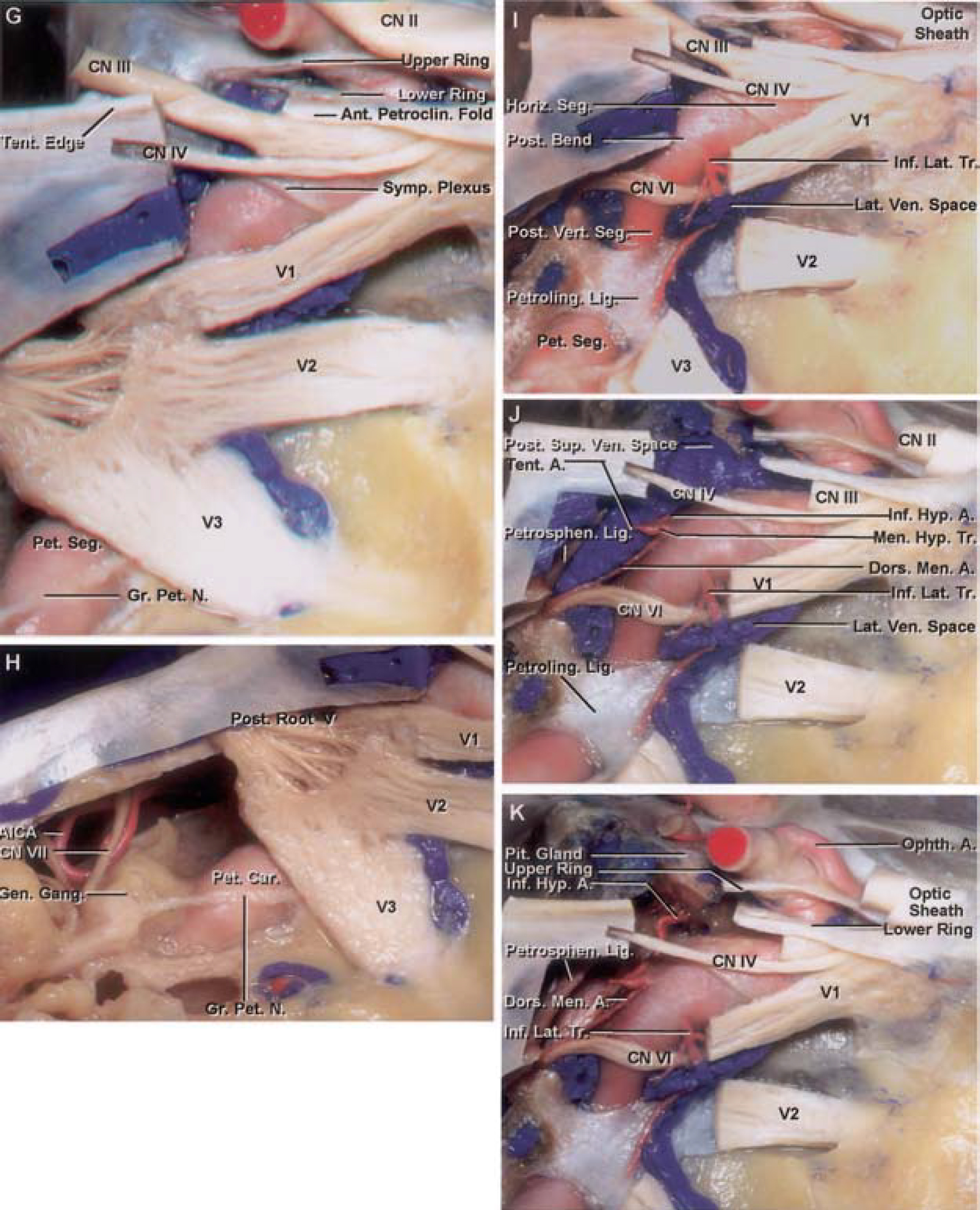
圖3 (G)- k).G,顯示頸動脈岩暴露於V的外側3.在岩大神經下麵。有時,頸動脈中窩底岩段末端沒有骨覆蓋,如本例所示。岩大神經在頸動脈岩部,交感神經叢的一個分支在頸動脈海綿內。H,視圖顯示內聲管的岩尖和頂部被切除,以暴露麵神經和小腦前下動脈。頸動脈岩暴露於V外側3.在岩大神經下麵。岩大神經起源於膝狀神經節。I,視圖顯示三叉神經節和後根被切除,盡管三個分區都被保留了。頸岩經過舌岩韌帶下方成為海綿內頸動脈,舌岩韌帶從蝶骨舌部延伸到岩尖。外展神經通過海綿內頸動脈後垂直段外側和V1內側到達眶上裂。下外側幹起於海綿內頸動脈的水平段並向外展神經的外側下行。海綿內頸動脈分為五個部分,由後向前依次為後垂段、後彎段、水平段、前彎段和前垂段。前彎段和前垂直段非常短,對應於斜向段。J,視圖顯示一段動眼神經被切除,部分後上靜脈間隙內的物質被抽出,以暴露腦膜-垂體幹的起源及其三個最常見的分支:下垂體、脊膜背側動脈和幕狀動脈。K,顯示後上靜脈間隙和內側靜脈間隙,以暴露下外側和腦膜垂體封閉幹。 The pituitary gland and the medial wall of the cavernous sinus are exposed between the intracavernous and supraclinoidal carotid. The abducens nerve passes below the petrosphenoid ligament (Gruber’s ligament) that forms the roof of Dorello’s canal and courses lateral to the posterior vertical segment of the intracavernous carotid and medial to the inferolateral trunk. A., artery; AICA, anteroinferior cerebellar artery; Ant., anterior; Clin., clinoid, clinoidal; CN, cranial nerve; Div., division; Dors., dorso; Fiss., fissure; For., foramen; Gang., ganglion; Gen., geniculate; Gr., greater; Horiz., horizontal; Hyp., hypophyseal; Inf., infero-, inferior; Infratroch., infratrochlear; Lat., lateral; N., nerve; Lig., ligament; Men., meningeal, meningo-; Oculom., oculomotor; Ophth., ophthalmic; Orb., orbital; Pericav., pericavernous; Pet., petrosal, petrous; Petroclin., petroclinoid; Petroling., petrolingual; Petrosphen., petrosphenoid; Pit., pituitary; Plex., plexus; Post., posterior, postero-; Seg., segment; Sup., superior; Supratroch., supratrochlear; Symp., sympathetic; Tent., tentorial; Triang., triangle; Tr., trunk; V, trigeminal; Ven., venous; Vert., vertical. (Images courtesy of AL Rhoton, Jr.)
前斜突上下表麵的硬腦膜向內側延伸形成上、下硬腦膜環,界定了頸內動脈斜突段的上下緣(圖2和圖3)。前斜突上表麵向內側延伸的硬腦膜形成硬腦膜上環的外側部分。硬腦膜向前和內側延伸,在視神經下方,排列在視神經支柱的上表麵形成硬腦膜上環的前部。最後,視支上表麵的硬腦膜向頸動脈內側和頸動脈溝後方延伸,形成硬腦膜上環的內側部分(圖2)。硬腦膜上環沒有後部,因為在硬腦膜上環的最後部,在前斜突尖端的水平,上環與下硬膜環連接,形成海綿竇頂部斜三角的頂點(圖2,H-J)。
硬腦膜位於前斜突下表麵並將斜突與動眼神經分開,硬腦膜向內側延伸環繞頸動脈。這種硬腦膜稱為頸動脈運動膜,形成下硬腦膜環(圖2,H-J)。它向內側向前延伸,襯著視神經支柱的下表麵,形成下環的前部。在內側,頸動脈運動膜與頸動脈溝的硬腦膜混合。這層膜向上旋轉,在頸動脈上下環之間形成一個頸圈,稱為頸動脈頸圈。在前斜突的後端,上硬膜環與下硬膜環連接形成斜突三角形的頂點(圖2,H-J,圖3,E-G)。
頸動脈頸圈是由下環硬膜向上旋轉,環繞在上下環之間的頸動脈段形成的(圖2,H-J)。頸動脈頸圈在到達硬腦膜上環之前不會與頸動脈壁緊密結合,在硬腦膜上環與頸動脈緊密結合。斜突靜脈叢是位於頸動脈頸圈和頸動脈斜突段外壁之間的一個小靜脈叢,與海綿竇前靜脈叢相通。因此,我們認為斜突段位於海綿體內。
硬腦膜形成上下環,斜向三角,頸動脈頸圈形成海綿竇頂部的前部。海綿竇頂部的後部由動眼肌三角形成,其邊界由硬腦膜結構確定。形成動眼三角邊界的硬膜結構為前、後岩斜突和斜突間硬膜褶皺(圖2)。前岩斜突硬膜褶皺從岩尖延伸至前斜突,後岩斜突硬膜褶皺從後斜突延伸至岩尖,前、後斜突硬膜間褶皺延伸至前、後斜突硬膜褶皺。動眼神經和滑車神經在動眼神經三角區穿過海綿竇頂部到達側竇壁(圖2)。因此,形成海綿竇頂部的硬腦膜可分為兩個三角形:斜向三角形(或海綿竇頂部的前部)和動眼神經三角(或海綿竇頂部的後部)。
海綿竇外側中窩襯裏的硬腦膜有一層內層與骨粘附,稱為內骨膜層,外層麵向大腦,稱為腦膜層(圖3、圖4)。在海綿竇外側下緣,兩層分離,腦膜層與內骨膜外側向上延伸,形成海綿竇外壁。而內骨膜層的內側向內側連續,形成內側竇壁的一部分。側竇壁解剖顯示,較厚的外層(腦膜層的延續)剝離,隻留下將神經包裹在側竇壁的薄內層(內骨膜層的延續)。側竇壁與覆蓋Meckel穴的硬腦膜融合(圖3,A-C)。海綿竇外側壁下緣與海綿竇內側壁在上頜神經上緣水平呈“龍骨狀”連接(圖3,C-G)。內層是內骨膜層的延伸,覆蓋在海綿竇外側壁內的神經。海綿竇外側壁的三角形是位於動眼神經和滑車神經之間的滑車上三角和位於滑車神經和三叉神經上緣之間的滑車下三角,也稱為帕金森三角(圖3C)。
![圖4。圖示通過海綿竇和腦垂體的冠狀切麵。圖A顯示硬腦膜分為腦膜層(橙色)和內膜層(綠色)。這兩層在中顱窩底部緊密相連,但在到達海綿竇的最下邊界,即第二三叉神經分區(v2)的上邊緣時,它們分離成兩層。腦膜層向上延伸,形成海綿竇外側壁和頂板的外層和鞍膈的上層。上頜神經上緣的骨內膜分為兩層。一層向上延伸構成海綿竇外側壁和頂板的內層,另一層粘附於蝶骨,覆蓋頸動脈溝和鞍底。從橫膈膜的自由邊緣,一層薄薄的硬腦膜向下延伸包裹,但很容易與腦下垂體分離。我們的解剖顯示,腦膜層形成海綿竇內側壁的鞍部,內骨膜層(綠色層)形成海綿竇內側壁的蝶部。硬腦膜和硬腦膜內膜在鞍底融合成一層。 B, diagram illustrating that it is easy to separate the menin-geal layer covering the inferior aspect of the pituitary gland from the endosteal layer covering the bony sellar floor. C, diagram illustrating an inferior intercavernous sinus that connects the paired cavernous sinuses. These intercavernous sinuses extend across the midline between the men-ingeal dural layer covering the inferior aspect of the pituitary gland and the endosteal layer covering the osseous sellar floor. A., artery; Car., carotid; CN, cranial nerve; Inf., inferior; Intercav., intercavernous; Pit., pituitary; Sphen., sphenoid (from, Yasuda A, Campero A, Martins C, Rhoton AL Jr, Ribas GC: The medial wall of the cavernous sinus: Micro-surgical anatomy. Neurosurgery 55:179–190, 2004 [50]). (Images courtesy of AL Rhoton, Jr.)](https://assets.neurosurgicalatlas.com/neuroanatomy/Rhoton_Pubs/Yasuda164Fig4.jpg)
圖4。圖示通過海綿竇和腦垂體的冠狀切麵。圖A顯示硬腦膜分為腦膜層(橙色)和內膜層(綠色)。這兩層在中顱窩底部緊密相連,但在到達三叉神經第二節(V2),即海綿竇的最下邊界,它們分為兩層。腦膜層向上延伸,形成海綿竇外側壁和頂板的外層和鞍膈的上層。上頜神經上緣的骨內膜分為兩層。一層向上延伸構成海綿竇外側壁和頂板的內層,另一層粘附於蝶骨,覆蓋頸動脈溝和鞍底。從橫膈膜的自由邊緣,一層薄薄的硬腦膜向下延伸包裹,但很容易與腦下垂體分離。我們的解剖顯示,腦膜層形成海綿竇內側壁的鞍部,內骨膜層(綠色層)形成海綿竇內側壁的蝶部。硬腦膜和硬腦膜內膜在鞍底融合成一層。B,圖顯示很容易將覆蓋腦垂體下側的腦膜層與覆蓋骨鞍底的骨內膜層分開。C,圖示連接成對海綿竇的下海綿竇。這些海綿間竇位於覆蓋腦垂體下側的腦膜硬膜層和覆蓋骨鞍底的骨內膜層之間的中線。 A., artery; Car., carotid; CN, cranial nerve; Inf., inferior; Intercav., intercavernous; Pit., pituitary; Sphen., sphenoid (from, Yasuda A, Campero A, Martins C, Rhoton AL Jr, Ribas GC: The medial wall of the cavernous sinus: Micro-surgical anatomy.神經外科55:179-190, 2004[50])。(圖片由AL Rhoton, Jr.提供)
內側竇壁的界限為前眶上裂,後鞍背,下上頜神經上緣水平與外側壁的交界處,上鞍膈肌(圖5)(50)。海綿竇內側壁分為鞍部和蝶部(圖5F)。在解剖解剖中,我們發現內側壁的鞍部是鞍膈肌的延續,鞍膈肌圍繞腦垂體前葉外側表麵向下折疊,由腦膜層構成(圖4和圖5,H-L)。內側壁鞍部與蝶部不連續,蝶部由覆蓋蝶骨體的骨內膜層形成,並沿鞍底向內側延伸。在我們的解剖中,我們發現鞍部,即腦膜層的延伸,沿垂體下表麵排列,很容易與蝶部分離,蝶部沿鞍底排列,是骨內膜層的延伸。海綿間竇位於兩層之間(圖5)。因此,蝶鞍的前、後、下表麵由兩層硬腦膜形成,腦垂體外側的壁由一層硬腦膜層形成。腦垂體後葉並不對著鞍部內側壁因為它位於鞍部內側壁後麵的鞍背凹處。我們的解剖解剖允許我們說,內側壁有一層,由鞍部的腦膜硬膜層和蝶部的骨內膜層(或顱內骨膜)構成。
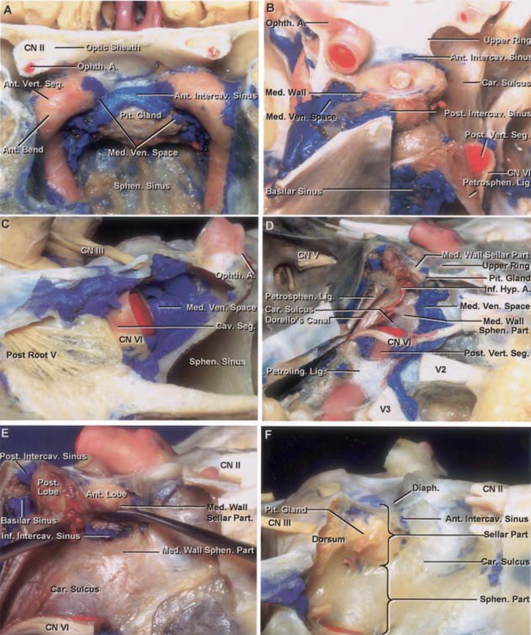
圖5 (A)- f).A-H,海綿竇內側壁的照片。A,前視圖顯示切除蝶竇壁後海綿竇。腦下垂體位於成對的海綿內頸動脈和海綿竇之間。內靜脈腔在腦垂體和動脈之間延伸。前海綿間竇穿過腦垂體前上表麵。眼動脈位於視神經下外側視神經管內。B,另一個標本的後上位視圖,顯示右側海綿內頸動脈、後斜突和鞍背鄰近部分被切除,暴露右側海綿竇內側壁。海綿竇的內側壁形成左側內側靜脈空間的內側邊界。前海綿間竇的前上段和後海綿間竇的後上段位於橫膈膜邊緣的垂體腺。基底竇是海綿竇中線上最大的交通線,位於背背部,通向兩個海綿竇的後部。 The petrosphenoid ligament, below which the abducens nerve passes to enter the cavernous sinus, extends from the petrous apex to the lower part of the lateral edge of the dorsum sellae. The abducens nerve passes lateral to the posterior vertical segment of the intracavernous carotid. C, lateral view showing the right cavernous sinus shown in B. A segment of the intracavernous carotid has been removed to expose the medial venous space located medial to the intracavernous carotid and in direct contact with the medial wall of the sinus. D, right lateral view showing another cavernous sinus. The intracavernous carotid has been removed and the medial venous space partially evacuated to expose the medial wall of the cavernous sinus. The medial wall has two parts: the sellar and sphenoidal. The sellar part is positioned lateral to the pituitary gland. The sphenoidal part lines the carotid sulcus on the body of the sphenoid bone. The sellar portion of the medial wall separates the lateral surface of the pituitary gland from the cavernous sinus. The sphenoidal part of the medial wall is formed by the dura lining the carotid sulcus on the body of the sphenoid bone. The petrous carotid passes below the petrolingual ligament to enter the cavernous sinus. The abducens nerve passes below the petrosphenoid ligament (Gruber’s ligament), which roofs Dorello’s canal, enters the cavernous sinus, and courses lateral to the posterior vertical segment of the intracavernous carotid. E, enlarged view showing that the dura lining the lower surface of the pituitary gland can be easily separated from the dura lining the sellar floor and that the inferior intercavernous sinus crosses between the two dural layers. The thin dural layer, which forms the sellar part of the medial wall of the cavernous sinus, separates the medial venous space from the pituitary gland. F, right lateral view of another cavernous sinus showing the nerves and intracavernous carotid removed to expose the medial wall of the cavernous, which has two parts: sellar and sphenoidal. The sellar part covers the lateral surface of the pituitary gland, and the sphenoidal part is formed by the dura lining the carotid sulcus. A., artery; Ant., anterior; Car., carotid; Carotidoculom., carotidoculomotor; Cav., cavernous; Clin., clinoid, clinoidal; CN, cranial nerve; Diaph., diaphragma; Gr., great; Hyp., hypophyseal; Inf., inferior; Intercav., intercavernous; Lig., ligament; Med., medial; Memb., membrane; Ophth., ophthalmic; PCoA, posterior communicating artery; Pet., petrosal; Petroclin., petroclinoid; Petroling., petrolingual; Petrosphen., petrosphenoid; Pit., pituitary; Port., portion; Post., posterior; Seg., segment; Sphen., sphenoid, sphenoidal; Ven., venous; Vert., vertical. (Images courtesy of AL Rhoton, Jr.)

圖5 (G)- l).繼續說。G,矢狀麵穿過蝶竇到海綿竇內側壁,顯示垂體位於蝶竇上方的蝶鞍中。前海綿間竇穿過腺體的前上側麵。基底竇是海綿竇之間最大的通道,穿過鞍背的背部,通向兩個海綿竇。H,放大圖顯示腦下垂體被切除,暴露右側海綿竇內側壁。前海綿間竇位於硬腦膜麵向腺體的腦膜層和襯於骨鞍壁的骨內膜層之間。圖示左海綿竇內側壁逐步暴露。I,圖示海綿竇的頂部和側壁暴露在外。通過切除前斜突暴露斜突間隙。形成海綿竇頂部和頸動脈頸圈前部的頸動脈運動膜向前折疊,露出頸動脈斜向段。 The oculomotor nerve enters the roof of the cavernous sinus through the oculomotor triangle located on the medial side of the anterior petroclinoid dural fold. A microdissector placed below the diaphragma sellae and lateral to the pituitary gland can be observed through the thin medial wall of the cavernous sinus. J, enlarged view showing the microdissector through the thin medial sinus wall that separates the cavernous sinus from the pituitary gland. K, view showing the intracavernous carotid and nerves removed to expose the medial wall of the cavernous sinus. The microdissector, placed below the diaphragma sellae and pituitary gland, can be observed through the thin semitransparent medial wall. L, view showing the medial wall of the cavernous sinus opened, with the leaves of the sellar portion of the medial wall folded outward to expose the lateral surface of the gland. The sphenoidal portion of the medial wall is exposed along the anterior and lower edges of the gland. A., artery; Ant., anterior; Car., carotid; Carotidoculom., carotidoculomotor; Cav., cavernous; Clin., clinoid, clinoidal; CN, cranial nerve; Diaph., diaphragma; Gr., great; Hyp., hypophyseal; Inf., inferior; Intercav., intercavernous; Lig., ligament; Med., medial; Memb., membrane; Ophth., ophthalmic; PCoA, posterior communicating artery; Pet., petrosal; Petroclin., petroclinoid; Petroling., petrolingual; Petrosphen., petrosphenoid; Pit., pituitary; Port., portion; Post., posterior; Seg., segment; Sphen., sphenoid, sphenoidal; Ven., venous; Vert., vertical. (Images courtesy of AL Rhoton, Jr.)
海綿竇後部位於鞍背外側,有一個大的靜脈彙合點,通向基底肌和岩上、岩下竇(圖6)。基底肌竇位於鞍背後表麵和上斜坡,是兩個海綿竇之間最大的連接。海綿竇後壁的下邊界位於岩蝶韌帶(Gruber’s韌帶)下方的岩斜裂縫的上邊緣,岩蝶韌帶位於岩尖和鞍背下外側邊緣之間,以頂頂Dorello’s管。第v腦神經穿過岩蝶韌帶下方斜坡硬腦膜,向上穿過多雷洛管到達海綿竇。海綿竇後壁的上邊界為岩斜後硬腦膜褶,外側邊界為三叉神經孔內側邊緣的硬腦膜。海綿竇後壁內側緣位於鞍背外側緣。
圖6。圖示海綿竇後壁逐步剝離的照片。A,後視圖顯示海綿竇後壁。海綿竇後壁位於三個點之間:後斜突,外展神經穿過斜坡硬腦膜的位置,以及三叉神經孔的內側。外展神經穿過斜坡硬腦膜,經多雷洛管後,呈向上運動。動眼神經穿過動眼三角中間的海綿竇頂部。岩上竇沿著岩脊在三叉神經後根之上。岩下竇沿岩斜裂隙向外展神經周圍延伸進入基底竇。在三叉神經孔下方的右側岩尖部分已被切除以暴露岩頸動脈。B,視圖顯示斜坡硬腦膜打開露出基底竇,這是海綿竇之間最大的連接。岩蝶韌帶(Gruber’s韌帶)位於Dorello’s管的頂部,從岩尖延伸到鞍背外側邊緣的下部。 The lateral limit of the posterior wall of the cavernous sinus is the medial aspect of the trigeminal porus. C, view showing part of the basilar sinus evacuated to demonstrate the upward course of the abducens nerve after piercing the clival dura. A., artery; Car., carotid; Cav., cavernous; Clin., clinoid; CN, cranial nerve; Hyp., hypophyseal; Inf., inferior; Lig., ligament; Sup., superior; Pet., petrosal; Petrosphen., petrosphenoid; Pit., pituitary; Post., posterior; Seg., segment; Triang., triangle. (Images courtesy of AL Rhoton, Jr.)
與海綿竇相關的神經包括頸動脈海綿內周圍的動眼神經、滑車神經、眼神經、展神經和交感神經叢(圖2、3和5)。動眼神經和滑車神經穿過海綿竇的動眼三角區。這個三角形的角位於岩尖和前、後斜突,三角形的邊緣是由連接三個結構的硬腦膜皺褶形成的。動眼神經穿過海綿竇頂部的一個短池,即動眼池,直到它到達前斜突的下緣,即池的末端,才與外側壁合並。動眼神經沿前斜突下緣進入眶上裂。硬腦膜排列於斜突下表麵,將斜突神經和動眼神經分開,向內側延伸形成頸動脈運動膜,環繞頸動脈形成硬腦膜下環。動眼神經經過視神經支柱外側和眶上裂後,穿過津環。它在眶上裂近端分為下段和上段,支配6個眼外肌中的4個(上斜肌和外側直肌除外)和瞳孔收縮肌(圖3D)。
滑車神經進入海綿竇頂部位於動眼三角的後外側,動眼神經入口後8.12±2.32 mm(範圍,4.52-13.1 mm),後斜突後外側13.82±2.39 mm(範圍,10.14-20.1 mm)(圖2、3和5)。在岩石斜突前、後硬膜皺襞交界處穿過海綿竇頂部後,滑車神經位於動眼神經下方的海綿竇外側壁。在前斜突水平,滑車神經從外側向內側穿過動眼神經的上表麵和前斜突和視神經支柱下緣的硬腦膜之間。滑車神經穿過眶上裂後,穿過提肌起點到達眶內側,支配上斜肌。
眼神經(三叉神經第一分支)與動眼神經和滑車神經一起嵌在海綿竇外側壁的內層內(圖2、3和5)。眼神經在滑車神經下方經過,到達眶上裂,在那裏分為淚支、額支和鼻纖毛支。隻有Meckel穴內側壁的上部和胃神經節的上三分之一位於海綿竇的正外側。上頜神經(三叉神經第二分支)在海綿竇下,不屬於海綿竇外側壁。海綿竇在上頜神經上緣的上方結束,海綿竇的內側壁和外側壁在這裏連接成龍骨狀。外展神經和頸動脈海綿內周圍的交感神經叢是僅有的完全在海綿內的神經。
外展神經穿過斜坡硬腦膜,有一個短的向上路徑,並通過位於岩蝶韌帶(Gruber’s韌帶)下方的Dorello’s管穿透海綿竇。它經過頸動脈海綿內動脈的後垂直段外側穿過海綿竇的側靜脈空間在頸動脈海綿內動脈水平段外側和下方和眼神經內側到達眶上裂。在海綿內頸動脈彎曲的情況下,展神經有時會在前下靜脈空間內運動。外展神經通常以一束形式穿過斜坡硬腦膜,但在前額神經池也可分成兩束;然而,它可能在海綿竇內分裂成多達五個束。海綿內頸動脈周圍的交感神經叢(圖3G)向外展神經發出分支;這些交感神經從展神經出發,到達眼部,然後到達支配虹膜瞳孔擴張纖維的長睫狀神經。
海綿竇包含頸內動脈的海綿內段及其分支。海綿內段開始於頸動脈管的顱內端,位於裂孔上方,後斜突外側,在那裏頸內動脈的岩段進入海綿竇。頸內動脈的岩段在下方的軟骨撕裂孔和上方的岩舌韌帶之間形成海綿腔內。岩舌韌帶從蝶骨舌突延伸至岩尖。海綿內段沿頸動脈溝向上向前延伸至視神經支柱後方和前斜突內側,穿過硬腦膜從前斜突上表麵向內側延伸出海綿竇(圖1)。
海綿內頸動脈有五個部分:1)後垂直段、2)後彎曲段、3)水平段、4)前彎曲段和5)前垂直段(圖1、D、E、3、5)。後垂直段開始於動脈從上方舌岩韌帶和下方撕裂孔之間的空間出口。它上升並在動脈前轉處結束,後斜突下外側,形成後彎。後彎有時會向上隆起並使海綿竇頂硬膜變形,就在後斜突外側。後彎止於水平段,它向前延伸到蝶骨的頸動脈溝。水平段轉向並結束於海綿竇頂部的前部,在那裏向上轉向形成前彎,前彎靠在視神經支柱的凹後表麵上,並與位於前斜突內側的前垂直段混合。前垂直段,也稱為斜突段,很短,隻有去除前斜突才能顯露。它被頸動脈頸圈和頸動脈頸圈內的斜向靜脈叢所包圍並受到上下硬膜環的限製。
海綿內頸動脈有兩個主要分支。第一個是腦膜垂體幹,起源於後彎。第二個是下外側幹,也稱為海綿竇下動脈,起源於水平段(圖3)。腦膜垂體幹通常起源於海綿內頸動脈的後彎,有三個分支:1)腦膜背側動脈,2)垂體下動脈,3)腦膜幕動脈(Bernasconi-Cassinari動脈)(圖3)。腦膜背側動脈向Dorello 's管方向向後通過,供給上斜坡硬腦膜。垂體下動脈向內側走行,支配垂體後囊和垂體葉。幕狀動脈首先沿竇外側壁向前,然後在幕狀動脈內向後轉。幕狀動脈分支到動眼神經和滑車神經。
腦膜垂體幹有完全和不完全兩種類型。完整型產生所有三個常見的腦膜垂體分支。不完全型產生一個或兩個通常的分支,其他分支直接產生於海綿內頸動脈。Inoue等人(18)報道了70%的完全型和30%的不完全型。腦膜垂體幹的所有三個常見分支可能很少直接起源於海綿內頸動脈。
下外側幹,也稱為下海綿竇動脈,通常起源於腦膜垂體幹起點遠端約5 - 8mm的水平段下表麵或外側表麵的中間三分之一。它幾乎總是經過外展神經上方,然後向下在外展神經和眼神經之間,供應海綿竇下外側壁硬腦膜和圓孔和卵圓孔周圍的鄰近區域(圖3)(17,18,30)。它很少起源於腦膜垂體幹。如果腦膜垂體幹沒有產生幕狀動脈,則通常有邊緣幕狀動脈起源於下外側幹。
其他可以起源於頸動脈海綿內動脈,但比腦膜垂體幹和下外側幹少得多的動脈有:1)麥康奈爾包膜動脈(占頸動脈的8%),起源於頸動脈海綿內內側,供應腦垂體包膜;2)眼動脈(占頸動脈的8%);3)頑固性三叉動脈,很少起源於海綿內頸動脈後彎的中央三分之一,向後穿過Dorello 's管外側海綿竇後壁,並在小腦上動脈和前下動脈之間與基底動脈吻合(18)。
海綿竇呈船狀,前部靠近眶上裂處最窄,後部與基底竇、岩上竇和岩下竇交界處最寬。海綿竇有四個靜脈空間(內側、前下、後上和外側),根據它們與海綿頸內動脈的位置(圖3)來定義。內側靜脈空間位於海綿頸內動脈和腦下垂體之間。如果海綿內頸動脈呈彎曲狀並膨入海綿竇內側壁,則可以不可見(圖5)。前下靜脈空間位於海綿內頸動脈後彎的前下側。眼上靜脈和眼下靜脈或它們的總幹通常進入前下靜脈空間。後上靜脈位於頸動脈海綿內動脈和海綿竇頂部後部之間,是海綿竇與基底竇連接的位置。外側靜脈腔位於海綿內頸動脈和眼神經之間,狹窄。外展神經在這個空間內走眼神經的內側,但如果海綿內頸動脈有一個曲折的路徑,它也可以走在前下靜脈空間。
與海綿竇相通的主要靜脈通道來自眼眶、大腦半球、後窩和對側海綿竇。兩個海綿竇之間的通信通過前、下、後海綿竇和基底竇(圖5和圖6)。海綿竇前通道位於前上,海綿竇後通道位於後上,海綿竇下通道位於腦垂體下方。這些鼻竇可以同時出現,也可以單獨出現。有時,前、後海綿間竇連同兩個海綿竇在橫膈膜鞍肌周圍相通,在橫膈膜周圍形成靜脈圈,稱為圓形竇。海綿竇前排入海綿竇後上靜脈腔,靠近前斜突尖端。海綿竇後腔流入海綿竇後上靜脈腔的後部。基底竇位於鞍背和上斜坡的後麵,與兩個海綿竇在鞍背側緣相通(圖5和圖6)。
海綿竇的另一個小靜脈組成部分是位於頸內動脈斜突段和頸動脈頸圈之間的斜突靜脈間隙。該空間內狹窄的靜脈通道與海綿竇頂板前部相通,並通過前斜突表麵的小孔和視支與眶頂雙曲靜脈相連。
我們選擇了一些臨床病例來說明成功的手術策略來接近海綿竇病理發現,並使用經海綿竇入路來接近海綿竇周圍的病理發現。這些入路由一位資深作者(EdO)執行,圖中附有屍體解剖,以說明執行入路時的重要解剖學考慮。海綿竇可通過其頂部或側壁進入。根據病理發現的部位不同,通過屋頂入路可以隻打開前部分,也可以同時打開前和後部分(圖7)。如果病理異常是斜旁動脈瘤,則隻打開海綿竇的前部分(圖8)。對於基底尖動脈瘤的經海綿體入路,打開頂板的前後部分(圖9)。Dolenc提出,通過外側壁入路用於外側壁結構引起的病變,如延伸至海綿竇的三叉神經瘤和垂體腺瘤(9)。
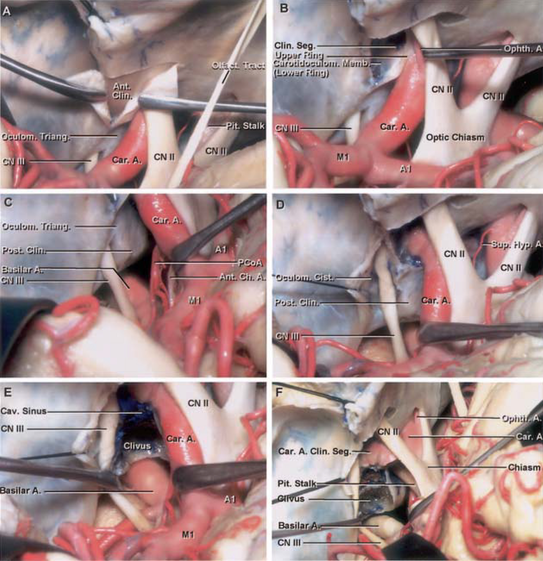
圖7。圖示左側海綿竇硬膜內入路。A,完整的額-眶-顴開顱術和顳前入路到海綿竇的視圖,顯示廣泛的sylvian裂剝離。頸動脈位於前斜突的內側,視神經位於頸內動脈的超內側。海綿竇入路首先是切除前斜突,露出竇頂的前部。前斜突上方硬腦膜已打開。動眼神經通過動眼三角進入海綿竇的頂部,動眼三角形成了海綿竇頂部的後部。B,顯示前斜突被移除。連續衝洗是必要的,以避免熱損傷視神經和斜突段時,使用鑽頭去除斜突。切除前床突暴露出床突間隙。硬腦膜從前斜突上表麵向內側延伸形成上環。 The carotidoculomotor membrane lines the lower surface of the anterior clinoid and extends medially to form the lower dural ring and carotid collar. C, view showing the carotid artery elevated to expose the posterior communicating and anterior choroidal arteries. The oculomotor nerve passes lateral to the posterior clinoid process and penetrates the roof of the cavernous sinus by passing through the oculomotor triangle. D, view showing the opening of the posterior portion of the roof, which begins by opening the oculomotor cistern. The incision follows the third nerve forward to the posterior edge of the clinoidal space. The posterior clinoid is exposed medial to the oculomotor nerve. E, view showing the roof of the cavernous sinus opened on the medial side of the oculomotor cistern. Gentle packing with Surgicel controls the bleeding. The posterior clinoid and adjacent part of the dorsum and upper clivus have been removed. The basilar trunk has been exposed behind the dorsum sellae. The supraclinoid carotid artery bifurcates below the anterior perforate substance in the A1 segment of the anterior cerebral artery and M1 segment of the middle cerebral artery. F, view showing a small segment of the supraclinoid carotid removed to expose the pituitary stalk. The pituitary gland can be reached between the initial supraclinoid segment of the carotid and the horizontal segment of the intracavernous carotid. A., artery; A1, A1 segment of the anterior cerebral artery; Ant., anterior; Car., carotid; Carotidoculom., carotidoculomotor; Ch., choroidal; Clin., clinoid, clinoidal; Cist., cistern; CN, cranial nerve; Hyp., hypophyseal; M1, M1 segment of the middle cerebral artery; Memb., membrane; Oculom., oculomotor; Olfact., olfactory; Ophth., ophthalmic; PCoA, posterior communicating artery; Pit., pituitary; Post., posterior; Seg., segment; Sup., superior; Triang., triangle. (Images courtesy of AL Rhoton, Jr.)
海綿竇的頂部有前部和後部。前部分有“上層”和“下層”(圖7-9),上層是前斜突的上表麵,下層是硬腦膜,硬腦膜排列在前斜突的下表麵,形成斜突三角形的底部。硬腦膜內襯前斜突下表麵形成的頸動脈運動膜,在去除斜突時暴露出來,形成竇靜脈間隙頂板的前部,向內側繞頸動脈延伸形成硬腦膜下環和頸動脈頸圈。頸動脈的斜突段位於視支柱的後方和前斜突的內側下方。硬腦膜從前斜突上表麵向內側延伸形成硬腦膜上環,它界定了頸動脈斜突段的上邊緣。

圖8 (A-F)。A-F,照片說明經海綿體入路向上導向的斜旁動脈瘤。G-L,照片說明經海綿體入路向下導向的斜旁動脈瘤。A,術前斜向血管造影顯示左側頸內動脈上有一個大的向上的斜旁動脈瘤。B,完成左側眶顴骨開顱術和顳前入路的視圖,顯示側裂打開,前斜突移除,海綿竇頂部前部暴露。硬膜外和硬膜內間隙暴露。大的向上導向的斜旁動脈瘤抬高視神經和額葉的鄰近部分。C,視圖顯示上環和視神經鞘打開,以幫助暴露動脈瘤頸。D,解剖解剖顯示暴露的結構。E,顯示下一階段動脈瘤頸夾層的中心插入片已經覆蓋在b的相應區域上。上環和視神經鞘已經打開,動脈瘤和眼動脈已經暴露。 F, view showing the aneurysm neck isolated and clipped using three straight clips. A., artery; A1, A1 segment of the anterior cerebral artery; Car., carotid; Cav., cavernous; CN, cranial nerve; Clin., clinoidal; Fr., frontal; M1, M1 segment of the middle cerebral artery; Op., operative; Ophth., ophthalmic; Seg., segment; Sphen., sphenoid; Temp., temporal. (Images courtesy of AL Rhoton, Jr.)
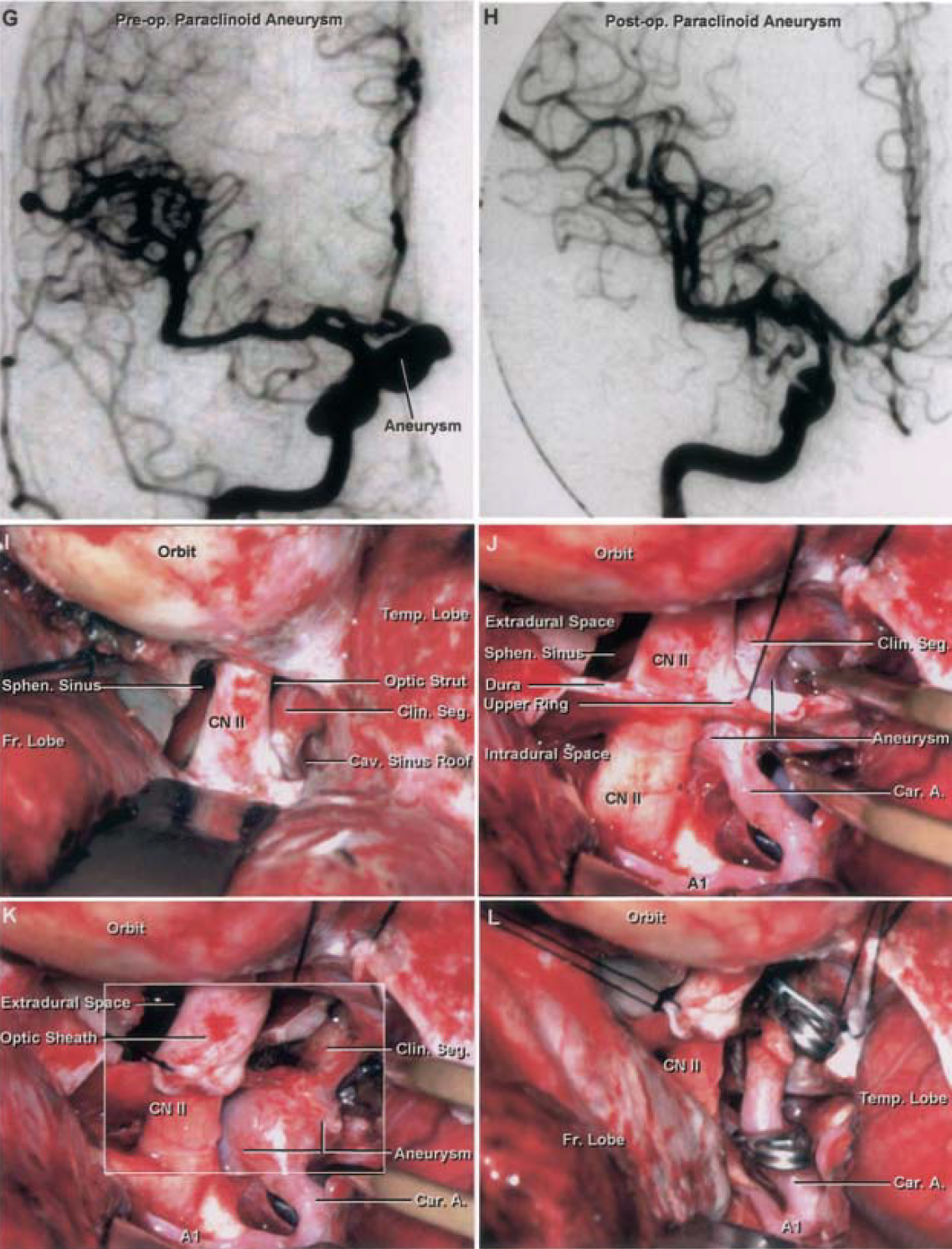
圖8 (G-L)。繼續說。G-H,照片顯示向下指向的右側斜旁動脈瘤。G,術前血管造影顯示動脈瘤。H,術後血管造影顯示動脈瘤被成功切除。I,顯示完成的右眶顴骨開顱術和前斜突硬膜外切除術。蝶竇暴露在視神經的內側。前斜突充氣。通過切除前斜突,暴露海綿竇頂部的前部。斜向三角形的結構,從前到後依次為視神經支柱、頸動脈斜向段和海綿竇頂部的前部。 J, view showing opened dura. The upper tip of the bipolar forceps is in the extradural space, and the lower tip is in the intradural space. The aneurysm can be observed below the supraclinoidal segment and above the clinoidal segment of the carotid. K, view showing upper ring and optic sheath opened to expose the aneurysm. L, view of clipped aneurysm using fenestrated right-angled clips. A., artery; A1, A1 segment of the anterior cerebral artery; Car., carotid; Cav., cavernous; CN, cranial nerve; Clin., clinoidal; Fr., frontal; M1, M1 segment of the middle cerebral artery; Op., operative; Ophth., ophthalmic; Seg., segment; Sphen., sphenoid; Temp., temporal. (Images courtesy of AL Rhoton, Jr.)

圖9 (A)- f).經海綿體入路治療基底尖動脈瘤的照片。術前血管造影側位圖顯示鞍背後方有一個巨大的基底端動脈瘤。B,術後血管造影顯示動脈瘤被成功切除。C,術中視圖顯示右側眶顴骨開顱術和顳前入路至海綿竇區。前斜突被切除以暴露斜突間隙和竇頂前部。頸動脈斜向段、視神經支柱和海綿竇頂部前部在斜向腔內暴露。這個間隙遠端由上(或遠端)硬膜環定義,近端由下(或近端)硬膜環定義。動眼神經穿過後斜突外側的動眼三角,穿過海綿竇頂部後部。頸鎖上動脈在大腦前動脈A1段和大腦中動脈M1段的前穿孔物質下麵分叉。在大腦中動脈顳早期分支的起始處可見一個小動脈瘤。 D, view showing carotid artery retracted medially to expose the space between the internal carotid artery and the oculomotor nerve, called the carotidoculomotor interval, through which the basilar artery can be approached. The posterior clinoid process blocks the approach to the basilar artery. E, view showing posterior portion of the roof of the cavernous sinus formed by the oculomotor triangle, which has been opened to expose the posterior clinoid process. Removing the dura of the posterior portion of the cavernous sinus exposes the pituitary gland anterior to the posterior clinoid process and dorsum sellae. F, anatomic dissection showing the same structures demonstrated in E. A., artery; A1, A1 segment of the anterior cerebral artery; Aneur., aneurysm; Arach., arachnoid; Ant., anterior; Bas., basilar; Br., branch; Car., carotid; Cav., cavernous; Clin., clinoid, clinoidal; CN, cranial nerve; M1, M1 segment of the middle cerebral artery; Op., operative; Petroclin., petroclinoid; Pit., pituitary; Post., posterior; Seg., segment; Temp., temporal. (Images courtesy of AL Rhoton, Jr.)

圖9 (G)- - - - - -J)。繼續說。G,顯示後斜突和部分鞍背切除以暴露硬腦膜後窩。H,硬腦膜內襯斜突和背側相鄰部分打開,露出覆蓋斜突的蛛網膜。I,打開蛛網膜,露出基底幹及其分叉和動脈瘤。比較去除斜突前的D與去除斜突後的I。J,解剖解剖,顯示I. A.動脈暴露的結構;A1,大腦前動脈A1段;Aneur。動脈瘤;Arach。, arachnoid; Ant., anterior; Bas., basilar; Br., branch; Car., carotid; Cav., cavernous; Clin., clinoid, clinoidal; CN, cranial nerve; M1, M1 segment of the middle cerebral artery; Op., operative; Petroclin., petroclinoid; Pit., pituitary; Post., posterior; Seg., segment; Temp., temporal. (Images courtesy of AL Rhoton, Jr.)
動眼三角形成海綿竇頂板後部,是動眼神經和滑車神經進入海綿竇頂板的位置(圖2和圖7)。通常隻有在去除前斜突暴露前部後,才會進入海綿竇頂板後部。使用90度顯微解剖器將硬腦膜抬舉到動眼神經上方,然後用鋒利的刀片沿著顯微解剖器的尖端切開硬腦膜。切口沿動眼神經延伸至海綿竇頂部的前部。然後將切口向後穿過海綿竇頂部的前後部分,到達後斜突。打開頂板後,可切除後斜突和上斜坡,為基底動脈提供額外通路(圖7和圖9)。
經海綿竇外側壁入路包括將海綿竇外側壁的硬腦膜外層與硬腦膜內層分離(圖10-12)。分離這些層可以使外側壁內層內的神經結構可視化。病變在離其最近的位置進入或侵入、膨出並使外側壁變形(圖11)。在圖11中,一個大的三叉神經瘤,其囊性部分延伸到後窩,已通過硬膜外入路完全切除。外側壁切口位置在腫瘤最突出部位上方,位於三叉神經第一、二節之間,注意不損傷三叉神經。腫瘤已經形成了自己通往後窩的路徑,這條通過擴大的梅克爾洞孔的路徑被用來切除延伸到後窩的腫瘤。了解這一區域的解剖結構有助於避免損傷隱藏在側壁或中窩底骨中的結構,如頸動脈的岩段。圖12顯示了經海綿體切除a垂體macroadenoma延伸到海綿竇。圖13和14顯示了合並入路治療海綿竇腦膜瘤。通過竇的側壁和頂部硬膜內和硬膜外入路。病灶已被移除,手術為診斷和鄰近組織減壓提供了組織,減少了輔助治療的病灶數量。

圖10。使用硬膜外入路逐步剝離右側海綿竇的照片。A,視圖顯示完成的顳前額-眶顴骨開顱術和顳前入路,顯露眶上裂外側邊緣的硬腦膜。B,視圖顯示,用鋒利的刀片在眶上裂外側邊緣切開硬腦膜帶後,海綿竇外層與內層分離。這個過程被稱為“剝去”中窩和海綿竇。當腦膜(外層)剝離時,外側壁內層的神經進入視野。前斜突已暴露。視神經和視神經管頂部位於前斜突內側。中窩剝落至V3後緣時,腦膜中動脈分裂。分離脊膜中動脈後,向後方和內側繼續剝離,在V3外側邊緣露出岩大神經。 The greater petrosal nerve usually courses above and serves as a good landmark for identifying the petrous carotid. The carotid artery may be exposed under the dura and the greater petrosal nerve at the lateral edge of the trigeminal nerve. The medial edge of the peeling of the middle fossa is at the anterior petroclinoid dural fold, and the posterior edge is at the petrous ridge. C, view showing anterior clinoid process removed extradurally using a high-speed drill with a diamond burr. Continuous irrigation is necessary to avoid heat spreading to the optic nerve and the clinoidal segment of the carotid. The drilling leaves a thin layer of bone over the optic nerve and the clinoidal segment that is removed with a microdissector. The anterior clinoid has attachments at its base to the sphenoid ridge, roof of the optic canal, and optic strut. The optic strut forms the floor of the optic canal and separates the superior orbital fissure from the optic canal. D, enlarged view showing removal the anterior clinoid exposes the carotidoculomotor membrane, lower ring, carotid collar, and clinoidal space or clinoidal triangle. The carotidoculomotor membrane lines the lower surface of the anterior clinoid and extends medially to form the lower dural ring and upward to form the carotid collar around the clinoidal segment. The “upper floor” of the anterior portion of the roof of the cavernous sinus is formed by the anterior clinoid process. The “lower floor” of the anterior portion of the roof of the cavernous sinus is the clinoidal space, which contains, from an anterior to posterior direction, the optic strut, clinoidal segment, and roof of the anterior part of the cavernous sinus. The venous spaces in the anterior part of the roof of the cavernous sinus are opened by incising the carotidoculomotor membrane. E, view showing the inner dural layer of the lateral sinus wall removed to expose the structures in this region. In surgery, the lesion is approached in the area at which it presents in the lateral wall. The middle fossa triangles exposed are the anteromedial triangle (between V1 and V2); the anterolateral triangle (between V2 and V3); the posterolateral triangle, also called Glasscock’s triangle, (between V3 and the greater petrosal nerve); and the posteromedial triangle, also called Kawase’s triangle (lateral to the trigeminal nerve and posterior to the greater petrosal nerve). The petrous carotid is exposed under the greater petrosal nerve. F, view showing some portions of the middle fossa floor and roof of the internal acoustic canal removed to expose the intraosseous segment of the greater petrosal nerve, the geniculate ganglion, and the contents of the fundus of the internal acoustic canal, including the facial and vestibulocochlear nerves. The tensor tympani muscle crosses below the middle fossa floor between the middle meningeal artery and the greater petrosal nerve. The cochlea is located at the angle formed by the greater petrosal and the facial nerves. A., artery; Ant., anterior; Car., carotid; Carotidoculom., carotidoculomotor; Clin., clinoid, clinoidal; CN, cranial nerve; Fiss., fissure; Fr., frontal; Gang., ganglion; Gen., geniculate; Gr., greater; Horiz., horizontal; Lat., lateral; M., muscle; Med., medial; Memb., membrane; Mid., middle; Men., meningeal; N., nerve; Orb., orbital; Pet., petrosal; Post., posterior, postero-; Seg., segment; Sup., superior; Temp., temporal; Tens., tensor; Tymp., tympani; Triang., triangle; Vert., vertical. (Images courtesy of AL Rhoton, Jr.)

圖11。計算機斷層掃描及照片顯示右三叉神經神經鞘瘤硬膜外切除。A,術前計算機斷層掃描和對比顯示一個大的三叉神經神經鞘瘤,囊性部分延伸到後窩。B,術後計算機斷層掃描和對比顯示,經海綿竇側壁硬膜外“剝離”入路,三叉神經神經鞘瘤全部切除。C,照片顯示完成的眶顴骨開顱術和顳前入路。硬腦膜從中窩底抬高,前斜突被移除,暴露海綿竇頂部前部的斜竇間隙,而不進入靜脈間隙。三叉神經神經鞘瘤在三叉神經第一節和第二節之間外側隆起(折線)。D,解剖解剖照片,顯示與c.e相同的結構,照片顯示三叉神經第一和第二節之間的切口在神經鞘瘤最突出的隆起處。腫瘤,包括其延伸到後窩,已被切除。腫瘤擴大了梅克爾洞的孔洞,並開辟了一條通往後窩的路。 F, photograph of the operation showing preservation of the trigeminal divisions and total resection of the lesion. The enlarged Meckel’s cave is exposed. Clin., clinoidal; CN, cranial nerve; Fr., frontal; Op., operative; Seg., segment; Temp., temporal. (Images courtesy of AL Rhoton, Jr.)

圖12。磁共振成像掃描和照片說明經海綿體切除垂體macroadenoma一例16歲女性患者,經三次蝶竇入路後,本病擴展至右側海綿竇及鞍上區。A,術前磁共振成像掃描顯示垂體腺瘤延伸至右側海綿竇和鞍上區。B,術後磁共振成像掃描顯示病灶完全切除。脂肪堆積在海綿竇內。病人用激素治療。C,照片顯示完成的眶顴開顱術和硬膜外顳前入路,抬高(剝離)硬腦窩,去除前斜突,暴露海綿竇頂部前部,而不打開靜脈空間。D,解剖照片顯示與c.e相同的結構,照片顯示使用硬膜外入路通過海綿竇側壁和頂部切除病變。壓下滑車神經和三叉神經第一節暴露出海綿內頸動脈後垂直段外側的外展神經。一個,動脈;騎兵。, cavernous; Clin., clinoid, clinoidal; CN, cranial nerve; Men., meningeal; Mid., middle; Op., operative; Pit., pituitary; Post., posterior; Seg., segment. (Images courtesy of AL Rhoton, Jr.)

圖13。磁共振成像掃描和照片顯示經海綿狀竇切除左側岩尖海綿竇腦膜瘤。A,術前磁共振掃描顯示岩尖海綿竇腦膜瘤壓迫腦幹。B,術後磁共振成像掃描顯示病灶切除。C,照片顯示完成的眶顴開顱術和顳前入路,硬膜外中窩“剝落”,去除前斜突,露出海綿竇頂部前部。D,解剖照片顯示與c.e相同的結構,照片顯示硬膜外和硬膜內聯合入路通過頂部和側壁切除病變。一個令人滿意的切除已經完成,並減壓腦幹。一個,動脈;的車。頸動脈; Clin., clinoid, clinoidal; CN, cranial nerve; Fr., frontal; Op., operative; Ophth., ophthalmic; PCA, posterior cerebral artery; Post., posterior; SCA, superior cerebellar artery; Seg., segment. (Images courtesy of AL Rhoton, Jr.)

圖14。磁共振成像掃描和照片說明手術切除右側海綿竇腦膜瘤。A,術前磁共振成像掃描顯示右側海綿竇腦膜瘤,海綿內頸動脈被腫瘤阻塞。B,術後磁共振成像掃描顯示海綿竇內病變部分被切除。C,照片顯示完成的眶顴開顱術和顳前入路,中窩“剝落”,前斜突移除,暴露斜竇間隙和海綿竇頂部前部。病變可觀察到向海綿竇側壁和頂部凸起並變形(折線)。頸動脈岩暴露在中窩底部就在三叉神經第三分支的後方和外側。D,解剖照片顯示c - E中暴露的結構,照片顯示海綿竇使用硬膜外和硬膜內聯合入路切除病變。術前檢查發現頸岩動脈被腫瘤阻塞,故用夾子切除頸岩動脈。騎兵。, cavernous; Clin., clinoidal; CN, cranial nerve; Fr., frontal; Gr., greater; N., nerve; Op., operative; Pet., petrosal; Seg., segment. (Images courtesy of AL Rhoton, Jr.)
海綿竇區位於顱底,與基底池接壤,周圍有重要的神經血管結構。自從Browder(4)和Parkinson(27)開創性地引入海綿竇手術以來,許多不同的方法被用於處理該區域及其周圍的病理發現。血管性、腫瘤性和炎性疾病影響海綿竇區。海綿竇入路包括硬膜內或硬膜外經顱和經基底入路(7 - 11,14,16,18,27,28,31 - 33,35,37,44,45)和經蝶入路(1,37)。盡管該區域的解剖結構已被廣泛描述(15,17,18,20,23,26,27,29,30,34,40,43,46,48),但關於不同類型病變的最佳治療和方法仍存在爭議(2,35,38),如海綿竇腦膜瘤(6,38)。人們一致認為,手術治療非腦膜腫瘤比手術治療腦膜瘤更安全,而且更經常導致完全切除(2,5,9,13)。直接入路到海綿竇的主要風險包括過度出血和海綿內頸動脈和顱神經的損傷。治療海綿竇病變的替代方法已經出現。在過去的十年中,放射外科單獨或作為部分切除的輔助手術發揮了突出的作用(3,12,19,24,25,39,47)。一些作者認為放射手術是海綿竇腦膜瘤的首選治療方法,因為其發病率和死亡率低,生長控製率高(12,19,25,42)。 However, radiosurgery is not completely absent of complications. Spiegelmann et al. (43) reported an incidence of 4.7% of new trigeminal neuropathy, a 2.8% incidence of new visual field defects, shunt-dependent hydrocephalus in 2 of 42 patients, and 1 patient with temporal lobe edema requiring surgical intervention. Cavernous sinus surgery can offer the possibility of tissue diagnosis and optic nerve decompression and can be used as a route to basilar artery aneurysms and extension of pituitary tumors. Conversely, excellent results have been achieved with surgical excision of meningiomas in this region (6, 33, 35). Microneurosurgery and radiosurgery have also been used for other types of tumors, such as pituitary adenomas (22), as well as for vascular lesions, such as hemangiomas (24). Another factor to consider is that new methods of treatment, such as radiosurgery and endovascular neurosurgery, are not available in all parts of the world; thus, neurosurgeons working in these parts of the world must rely on microsurgical technique combined with anatomic knowledge to deal with cavernous sinus pathological findings.
當計劃海綿竇手術時,術前評估是最重要的(31,33,35),外科醫生必須準備重建頸內動脈和與該區域相關的神經(32)。在進入海綿竇之前,應實現對頸內動脈血流的近端和遠端控製。根據Glasscock的指示(15),近端控製可通過暴露頸部的頸內動脈和顱底的頸岩管水平來實現。在硬膜外或硬膜內行前斜突切除術後,頸動脈的遠端控製得到(7,10,31)。重建海綿內頸動脈的主要技術是使用隱靜脈移植物在頸動脈或頸岩動脈和頸動脈之間搭橋(32,36,41)。建立近端對照的決定取決於海綿竇病理表現的類型和位置。
除了接近固有疾病外,海綿竇還可以作為接觸其他病變的途徑,如基底尖、眼頸動脈、旁楔動脈瘤以及鞍部和斜坡腫瘤(圖8和圖9)(8,9,11,21,33,37)。病變通常不在海綿竇內,而是在海綿竇周圍。因此,解剖學上熟悉該區域對於進入和保護海綿竇內及周圍的神經血管結構很重要,這些神經血管結構可能被竇壁或中窩骨所隱藏。
海綿竇入路通過頂或側壁(圖7和10)(7 - 11、14、16、18、27、28、31、33、35、37、44、45)。Umansky和Nathan(46)描述了海綿竇外側壁的兩層組成,這使得硬腦膜外層從海綿竇硬膜外入路的內層地板剝離。在這種方法中,在海綿竇外側壁的半透明內層中穿行的神經可以暴露出來,而無需直接進入海綿竇(7)。
經海綿竇頂部進入海綿竇的途徑結合了硬膜外和硬膜內的途徑(圖7-9)。他們需要進行顳前額眶顴骨開顱術,並在硬膜內或硬膜外切除前斜突以暴露竇頂的前部,之後可打開竇頂後部的動眼三角(7,8,13,30,33,35,37)。前部可單獨打開,也可與後部聯合打開。
海綿竇頂部的前部常用於入路治療斜突旁動脈瘤或頸動脈-眼動脈瘤(圖8)。如Dolenc(8)所述,可以硬膜內或通過硬膜內和硬膜外聯合入路打開海綿竇頂部(8)。前部,即斜竇間隙或斜竇三角形,比後部的排列更複雜。前斜突位於海綿竇頂部前部的上層。使用高速鑽頭去除前斜突,暴露出形成頂板前部下部的斜向空間或斜向三角形。切除前斜突可暴露頸動脈斜突段、頸動脈運動膜、視神經支架、眶上裂和視神經管,但不能打開海綿竇的靜脈空間。充分暴露海綿竇頂部的前部是接近旁斜動脈瘤的基礎。動脈瘤有時發生在頸動脈斜向間隙(或斜向三角形)內,累及頸內動脈斜向段。在接近這些動脈瘤時,熟悉海綿竇頂部的前部是至關重要的(圖8)。
在高速鑽和金剛石鑽頭的幫助下,在硬膜外或硬膜內切除前斜突。當前斜突被移除時,必須小心去除視神經管頂部。持續衝洗是必要的,以避免加熱損傷視神經。可以在視神經和頸動脈上留下一層薄骨,以保護它們不被鑽穿,之後使用微型解剖器將最後一層薄骨移除。在前斜突底部鑽孔並將其整體移除可能是危險的,特別是如果前斜突和中斜突之間存在骨橋,在頸動脈周圍形成頸突突孔。在這種情況下,切除前斜突會導致骨橋斷裂並損傷頸動脈的斜突段。
要充分暴露海綿竇的頂部,需要打開側裂並切除前斜突。此外,應將顳葉從其內側-基底表麵的蛛網膜附著物中解放出來,允許顳葉的收縮,以充分暴露海綿竇的頂部。在切除前斜突時,我們在硬膜外切除部分蝶小翼,然後打開硬腦膜完成切除。前斜突上方的硬腦膜在硬膜內暴露,切開四道切口:第一個向前延伸整個平麵附近開始內側視神經管的極限,第二個開始靠近床形的提示和蝶骨脊平行向前延伸,第三個加入前的第一個削減和延伸軌道上方的屋頂加入第二年底前削減,和第四個加入後的第一次和第二次削減通過鐮狀韌帶和前床形的過程。前斜突連著顱底的三個位置第一個是小蝶翼;第二,通過形成視神經管頂部的前根;第三,通過其後根或視神經支柱,形成視神經管的底部(圖1)。在硬膜外鑽蝶小翼,將斜突與小翼分離,通過打開視神經管頂部和鑽視神經支柱釋放其他附著物。
在這一步驟中必須小心,以避免損傷頸動脈的斜突段,它沿前斜突的內下表麵行進,並位於視支柱的後表麵。隻有從斜突下表麵向內側延伸的薄頸動脈運動膜將海綿竇靜脈叢與前斜突分開。前斜突切除術暴露海綿竇頂部的前部和斜突三角,而不打開靜脈叢。通過打開視神經外側的視鞘和頸內動脈周圍的遠端硬膜環,繼續暴露海綿竇頂部的前部。當暴露海綿竇頂部前部時,鼻竇出血是常見的,但用止血產品輕輕包裹很容易控製。打開遠端環後,可以活動頸內動脈的斜向段和鞘上段。重要的是要認識到硬膜遠端環是緊密地粘附在頸內動脈。這個環的開口必須小心地進行,留下一個硬膜環的袖口,它沒有與動脈分離。暴露海綿竇頂部的前部通常為接近旁斜肌和一些眼動脈瘤提供足夠的暴露(圖8)(8)。屋頂的後部可以與前部一起打開,以進入基底尖動脈瘤和內在海綿竇腫瘤。
海綿竇頂部的後部通過動眼三角接近,動眼神經通過動眼三角穿過海綿竇頂部(圖7和圖9)。神經不進入靜脈空間,而是在下降到動眼三角水平以下後穿過一個短池,動眼池。這個池終止於前斜突的頂端或下方,神經在這裏與竇外側壁的內層結合。海綿竇頂部後部的開口是通過在神經上方的眼動池插入一個90度微型解剖器,打開眼動池的上壁,小心地將硬腦膜抬高並切開,直到夾層開始。切口向前延伸至第三腦神經到神經與海綿竇外側壁結合的地方。解剖的下一步是打開第三腦神經斜向三角區的頸動脈運動膜。動眼神經和海綿內頸動脈水平段之間的空間沒有任何重要的神經血管結構。當頸動脈運動膜被打開時,下方的鼻竇會出現明顯出血。然而,用可吸收止血劑(如Surgicel;Ethicon, Inc., Somerville, NJ)有效地控製出血。打開上環時,膜部分打開。 The posterior edge of the upper ring at the tip of the anterior clinoid process fuses with the carotidoculomotor membrane (Fig. 2, C, J, and K). The last step in opening the posterior part of the roof of the cavernous sinus is an incision directed posteriorly through the oculomotor triangle toward the posterior clinoid process. Both parts of the roof of the cavernous sinus have now been opened, and the lateral, posterosuperior, and medial venous spaces have been exposed. The posterior bend, the horizontal segment, the anterior bend of the intracavernous carotid artery, and the pituitary gland between the horizontal intracavernous and supraclinoidal segments of the internal carotid artery may be exposed. The posterior clinoid and upper clivus can be removed to access the interpeduncular and prepontine cisterns for basilar tip aneurysms (11, 37) or the upper clivus area for tumors (Fig. 9) (33).
經海綿竇側壁入路首先從中窩底剝離硬腦膜(圖10-12)。剝離開始於蝶大翼,並向眶上裂前進,此處顱內骨膜與眶周連續。在眶上裂外側緣的硬腦膜上有一個淺切口,可以使硬腦膜從中窩底沿竇壁向內側分離。外層(腦膜硬膜)從內層(骨內膜層)剝離,露出顱神經III、IV、V1、V2和V3和胃神經節。有三個點的剝皮必須要小心。一個是從V2中分離層,另一個是從V3中分離層,最後一個是在帕金森三角形處分離層。兩層在V2和V3水平緊密粘附,帕金森三角內層較寬,容易損傷。從硬腦膜內膜剝離腦膜硬腦膜,暴露海綿竇的外側壁以及中窩底的四個三角形(30):V1和V2之間的前內側三角,V2和V3之間的前外側三角,岩上大神經前三叉神經外側的後外側三角或Glasscock三角,岩大神經後三叉神經外側的後內側三角或Kawase三角(圖10E)。側壁入路可根據病理異常部位進行調整(圖11-14)。 The “peeling of the dura of the middle fossa floor” away from the lateral wall while preserving the inner layer aids in visualization of the structures within the lateral wall because of the semitransparent nature of the inner layer, as reported by Dolenc (9, 10), who used the extradural approach for intracavernous carotid artery aneurysms (10) and for pituitary tumors extending beyond the sella (9). One of the senior authors (EdO) has used this approach to pituitary adenomas extending beyond the sella with good results (Fig. 12). As Dolenc (9) reports, this last approach is complementary to the transsphenoidal approach, but if the tumor has a parasellar extension, it offers a great chance of total removal through a single operation (9).
海綿竇區解剖結構複雜,在其硬腦膜壁內有高密度的神經血管結構。海綿竇區的病理表現多樣,包括內在病變和外在病變。海綿竇疾病的適當治療是有爭議的。所有我們做過海綿竇手術的病人都經曆過短暫性腦神經麻痹,但都完全康複了。放射手術治療某些類型的腫瘤(如腦膜瘤)已經提供了極好的結果(19,25,49)。不幸的是,這一技術進步在許多國家是不可用的,並且不能在具有不尋常的放射學特征的病例中提供組織診斷。此外,放射手術可能受到靠近視神經的限製,需要在放射手術治療前打開海綿竇來減輕腫瘤體積。事實上,像放射外科這樣的技術進步在許多國家都沒有,也不適用於治療靠近交叉的腫瘤,以及在該區域有經驗的外科醫生有可接受的結果,這意味著海綿竇手術在神經外科中繼續占有一席之地。準確了解該區域的解剖結構可以將海綿竇從一個不可接近的手術部位(47)轉變為一個可接近的手術部位。這些知識,加上外科醫生的經驗,是在或通過這個“解剖珠寶盒”進行令人滿意的手術的唯一途徑,正如帕金森(29)所描述的那樣。
貢獻者:A. Yasuda, A. Campero, C. Martins, A. L. Rhoton, Jr, E. de Oliveira, G. C. Ribas
內容來自安田A,坎佩羅A,馬丁斯C,羅頓AL, Jr,德奧利維拉E,裏巴斯FC。海綿竇顯微外科解剖及入路。神經外科外科(黑格斯敦)2005; 56:4-27。doi.org/10.1227/01.NEU.0000144208.42171.02.經牛津大學出版社許可,代表神經外科醫生協會。©神經外科醫生協會。
神經外科188bet手机app圖集很榮幸能夠保持Albert L. Rhoton, Jr, MD的遺產。
請登錄發表評論。
請務必在社交媒體上關注我們,獲取令人興奮的內容,並保持更新生活科恩醫生的會議,關於手術技術的問題,以及更多!


您必須登錄才能查看此資料。
的188bet手机app幾乎完全取決於你的捐款。
如果沒有你們的大量捐贈我們無法繼續製作《地圖集》。
請承諾每年至少向Atlas捐贈250美元。如果沒有這種承諾,Atlas將很快需要付費訂閱,世界各地的許多外科醫生將無法使用它,他們的病人的護理依賴於它。
請立即捐款!
如果沒有你們的大量捐贈我們無法繼續製作《地圖集》。請承諾每年至少向Atlas捐贈250美元。
如果沒有這個承諾,Atlas將很快需要付費訂閱世界各地的許多外科醫生都無法使用它,他們的病人的護理都依賴於它。請立即捐款!