


你可以有所作為。
的188bet手机app這幾乎完全取決於你的捐款。
如果沒有你們的大量捐贈,我們就無法繼續開展地圖集。
請承諾每年至少捐贈250美元給Atlas。如果沒有這種承諾,Atlas將很快需要付費訂閱,世界各地的許多外科醫生將無法獲得它,他們的病人的護理依賴於它。
現在請捐!

最後更新:2021年4月25日
側腦室和第三腦室的手術入路具有挑戰性,因為它們位於靠近顱內空間中心的深部,完全包裹在神經組織中,大腦內彎曲,不同腦葉形狀和大小可變,狹窄的通信孔容易阻塞,膨脹的性質使它們成為腫塊病灶,壁包含重要的運動、感覺和視覺通路以及重要的自主和內分泌中樞。側腦室為第三腦室和基底腦池提供了可接近的深腔。在本章中,在描述單個手術入路之前,回顧神經和血管的關係,為優化腦室手術獲得的結果提供了基礎。許多構成側腦室壁一部分的結構也可見於第三腦室。側腦室和第三腦室都與深靜脈係統密切相關,許多動脈供應側腦室和第三腦室的壁。
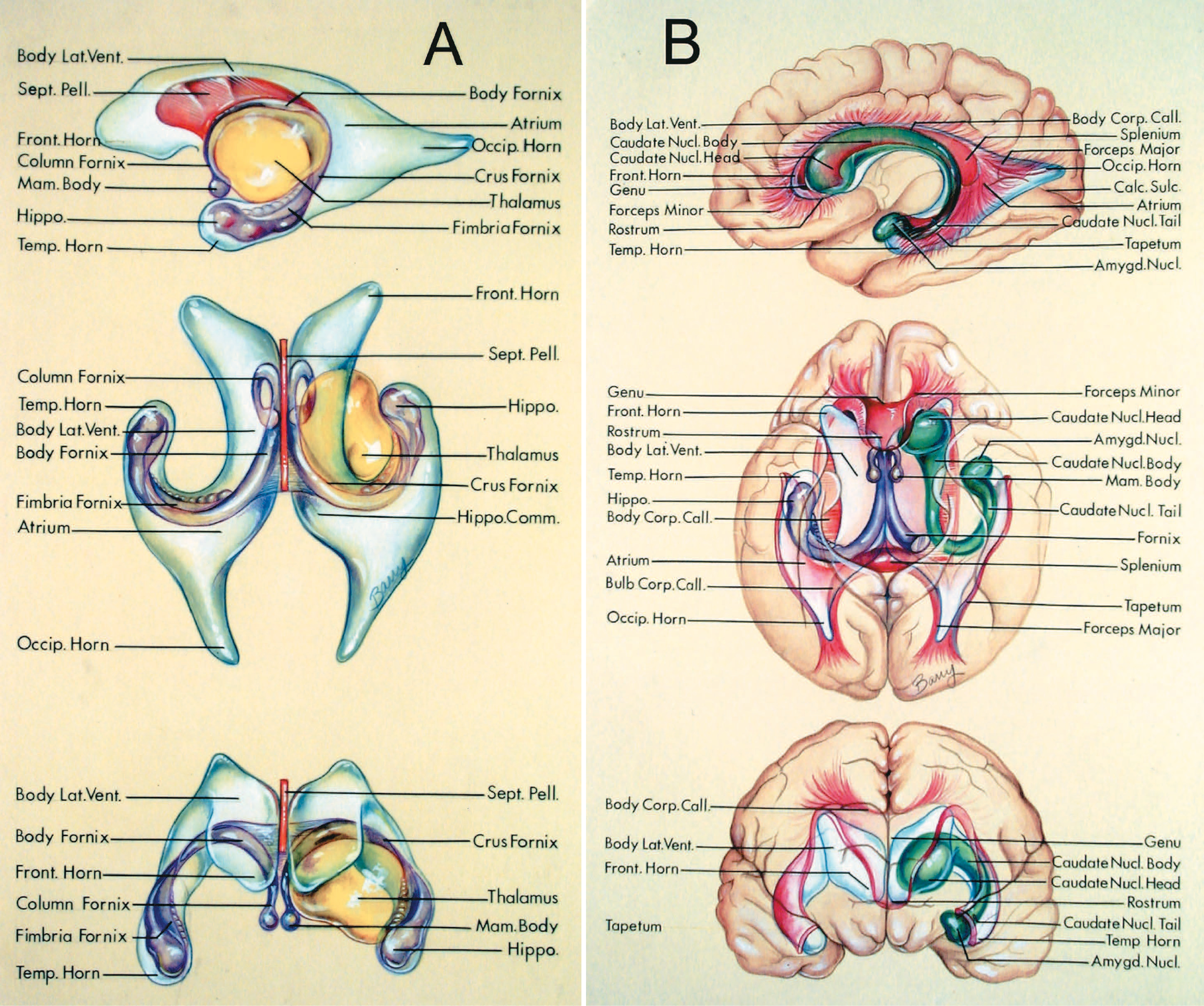
每個側腦室都是一個c形腔,環繞著丘腦,位於大腦深處(圖5.1)。每個側腦室有五個部分:額角、顳角和枕角、身體和心房。這五個部分都有內側壁、屋頂和地板。此外,額角和顳角以及心房都有前壁。這些壁主要由丘腦、透明隔、腦深部白質、胼胝體和兩個c形結構形成,尾狀核和穹窿包圍著丘腦。
丘腦位於側腦室的中央。每個側腦室包裹著丘腦的上、下、後表麵(圖5.1A)。側腦室體位於丘腦上方,心房和枕角位於丘腦後方,顳角位於丘腦下外側。丘腦上表麵形成身體的底板,丘腦枕部後表麵形成心房的前壁,丘腦下表麵位於顳角頂板的內側邊緣。
尾狀核是一個拱形的c形細胞團塊,包裹著丘腦,構成側腦室壁的重要組成部分(圖5.1B)。它有頭、身體和尾巴。頭部凸出進入額角的側壁和側腦室體。體形成心房外側壁的一部分,尾從心房延伸至顳角頂部,與顳角前尖附近的杏仁核連續。在側腦室中,尾狀核位於丘腦的上外側;在心房中,它位於丘腦的後外側;在顳角,它位於丘腦的下外側。終紋是一條與丘腦紋靜脈平行並深入的纖維束,發源於杏仁核,沿著尾狀核和丘腦之間的邊界在腦室壁從顳角延伸到身體。
穹窿是另一種c形結構,在腦室壁上包裹著丘腦(圖5.1A)。穹窿主要由海馬體、顳葉下丘腦和齒狀回的海馬乳頭束纖維組成。毛膜起於海馬組腦室表麵的顳角底部,向後方延伸成為穹窿的小腿。小腿包裹著丘腦枕部的後表麵,向胼胝體脾的下表麵超內側拱起。在心房和側腦室體的連接處,成對的腳彙合形成穹窿體,穹窿體在側腦室體的內側壁沿著丘腦的超內側邊界向前延伸。穹窿體將第三腦室的頂部和側腦室體的底部分開。在丘腦的前緣,穹窿體分離成兩列,沿蒙羅孔的上緣和前緣拱向乳頭體。在脾下方的區域,一薄層纖維連接著腳內側邊緣,形成海馬連合。在側腦室體中,穹窿體在內側壁的下部;在心房,穹窿的小腿在前壁的內側; and in the temporal horn, the fimbria of the fornix is in the medial part of the floor.
穹窿體大約在丘腦上表麵的內側和外側邊緣中間穿過丘腦。位於穹窿體側側的丘腦部分構成側腦室體的底板,位於穹窿內側的部分構成腹膜間質和第三腦室的部分側壁。穹窿的小腿穿過枕骨,大約在枕骨的內側和外側邊緣中間。穹窿小腿外側的枕部構成了心房前壁的一部分,穹窿內側的部分構成了四頭池前壁的一部分。穹窿的絲膜穿過丘腦下外側部分的下方就在內側和外側膝狀體的外側。丘腦中膜內側的部分形成了周圍池的頂部。
胼胝體構成了腦室壁的最大部分,構成了側腦室的五個部分(圖5.1B)。胼胝體有兩個前半部分,即喙部和膝,一個中心部分,即身體,和一個後部部分,即脾。前庭位於下方,形成前庭角的地板。膝有一大束纖維,小鉗,形成額角的前壁,當它斜向前和向外側掃過,連接額葉。膝和胼胝體構成了額角和側腦室的頂部。脾包含一個大纖維束,即大鉗,在心房和枕角內側壁的上部形成一個突出物,稱為球泡,因為它向後掃至連接枕葉。另一個纖維束,絨氈層,起源於身體的後部和胼胝體的脾,向外側和下方蔓延,形成心房的頂部和側壁,以及顳角和枕角。絨氈層將光輻射纖維與顳角分開。
透明隔由成對的薄片組成,在中線將側腦室的前角和側腦室體分開(圖5.1A)。在額角中,透明隔連接下麵的胼胝體,前麵的膝和上麵的身體。在側腦室體中,隔膜與上麵的胼胝體和下麵的穹窿體相連。透明隔前高後短,消失於身體和穹窿底端交界處,在那裏底端和海馬連合與胼胝體下表麵融合。透明隔的前後長度從28毫米到50毫米不等。在透明隔層之間的中線可能有一個空腔,即透明隔腔。
圖5.1。神經的關係。A,透明隔(橙色),丘腦(黃色),海馬形成和穹窿(紫色)與側腦室的關係。頂部,側麵圖;中間,優越的觀點;底,前視圖。每個側腦室環繞著丘腦。額角位於丘腦前方,身體位於丘腦上方,中庭和枕角位於丘腦後方,顳角位於丘腦下方和外側。透明隔位於額角和側腦室體的內側壁。海馬體在顳角底部。 The fornix arises in the hippocampal formation and wraps around the thalamus in the medial part of the temporal horn, atrium, and body. The fimbria of the fornix arises on the surface of the hippocampal formation in the temporal horn. The crus of the fornix is posterior to the thalamus in the wall of the atrium. The body of the fornix passes above the thalamus in the lower part of the medial wall of the body. The columns of the fornix are formed at the level of the foramen of Monro and pass inferior to the mamillary bodies. The crura of the fornix are connected across the midline in the roof of the third ventricle by the hippocampal commissure. The septum pellucidum, which separates the frontal horns in the midline, does not extend to the anterior tip of the frontal horn in the lateral view because the frontal horn is directed forward and laterally from the anterior margin of the septum pellucidum. B, relationship of the corpus callosum (red), caudate nucleus (green), and fornix and hippocampal formation (purple) to the lateral ventricles. Top, view through medial surface of the hemisphere; middle, view through inferior surface of the hemisphere; bottom, view through the anterior surface of the hemisphere. The head and body of the caudate nucleus form the lateral wall of the frontal horn and body of the lateral ventricle. The tail of the caudate nucleus extends into the anterior part of the lateral wall of the atrium and into the medial part of the roof of the temporal horn to the level of the amygdaloid nucleus, which is in the anterior wall of the temporal horn. The corpus callosum is made up of the rostrum (which is in the floor of the frontal horn), the genu (which forms the anterior wall and roof of the frontal horn), the body (which forms the roof of the body of the lateral ventricle), and the splenium (which gives rise to the fiber bundles making up the forceps major, which forms a prominence in the medial wall of the atrium called the bulb of the corpus callosum). The genu of the corpus callosum gives rise to a fiber bundle called the forceps minor, which forms the anterior wall of the frontal horn. The body and splenium give rise to a fiber bundle called the tapetum, which sweeps downward to form the roof and lateral wall of the atrium and temporal horn. The relationship of the hippocampal formation, fornix, and mamillary bodies to these structures is shown in the middle figure. A prominence in the medial wall of the atrium, called the calcar avis, overlies the calcarine sulcus. Amygd., amygdaloid; Calc., calcarine; Comm., commissure; Corp., corpus; Front., frontal; Hippo., hippocampal, hippocampus; Lat., lateral; Mam., mamillary; Nucl., nucleus; Occip., occipital; Pell., pellucidum; Sept., septum; Sulc., sulcus; Temp., temporal; Vent., ventricle.
在規劃腦室手術入路時,內囊與額角外側壁和側腦室體的密切關係常常被遺忘(圖5.2和5.3)。位於尾狀核和慢狀核之間的內囊前肢被尾狀核的頭部與額角隔開,位於丘腦和慢狀核之間的後肢被丘腦和尾狀核的體與側腦室體隔開。然而,內包膜的膝直接到達腦室表麵,並與側腦室壁接觸,緊挨著Monro孔的外側,在尾狀核和丘腦之間。
圖5.2。內囊與右側腦室的關係。內囊的前肢由尾狀核與側腦室隔開,後肢由丘腦與腦室隔開。膝直接到達腦室表麵,位於尾狀核和丘腦之間的Monro孔外側區域。摘除穹窿的右半部分露出第三腦室頂部的大腦內靜脈。

點擊這裏查看此圖像的交互模塊和相關內容。
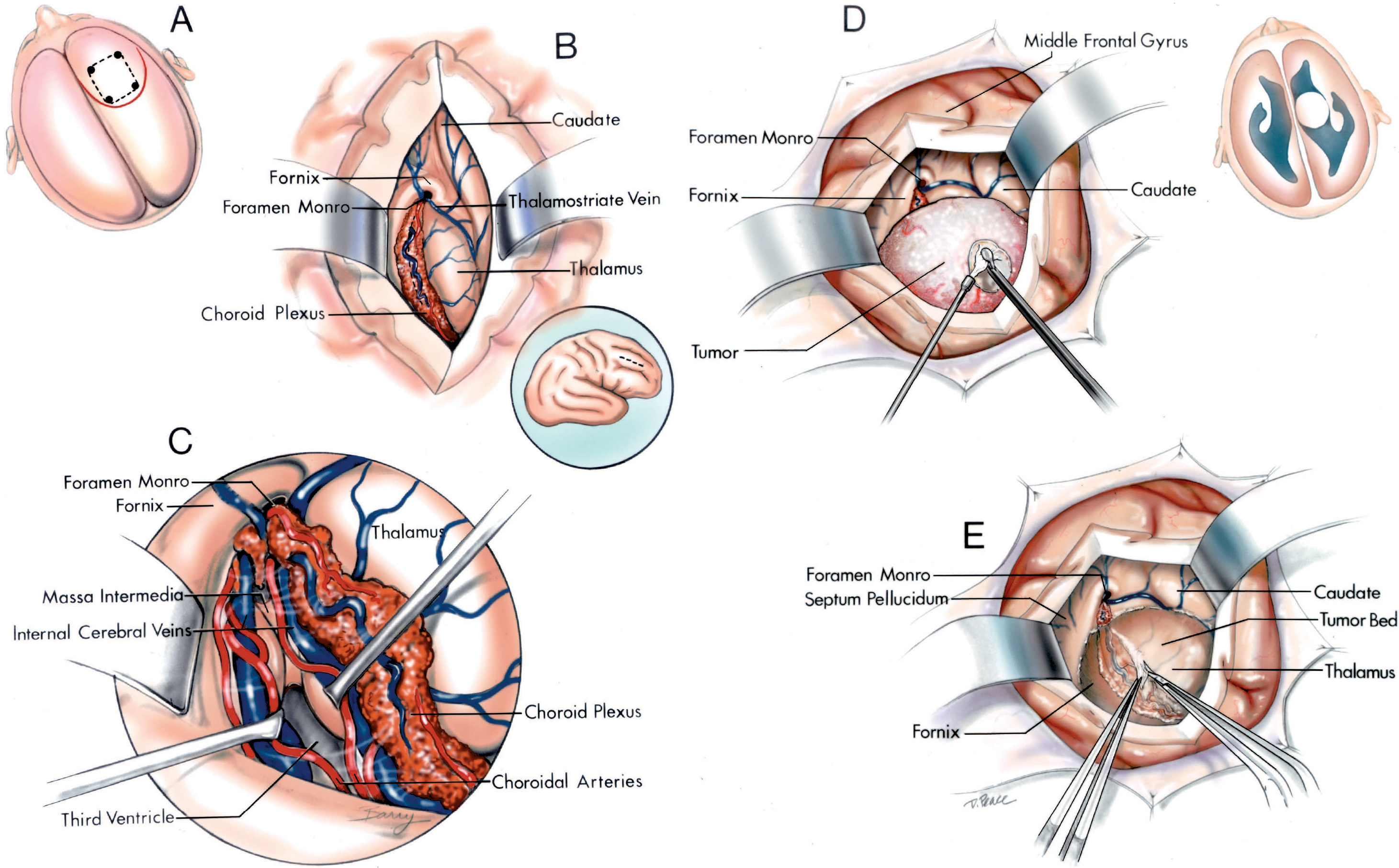
圖5.3模擬。在我們的顯微外科手術過程中使用的逐步剝離術來暴露側腦室和第三腦室以及脈絡膜裂。首先,解剖通過檢查經胼胝體前路進入第三腦室的關係開始。位於額葉中、後橋靜脈之間的右額葉已從鐮處縮回,露出位於胼胝體上表麵的大腦前動脈。插圖顯示了與冠狀線的關係。通常在冠狀線前方有一個區域,相對缺乏進入上矢狀竇的橋靜脈。經胼胝體入路的骨瓣三分之二放在冠狀線的前麵,三分之一放在冠狀線的後麵。B,放大圖。鐮和額葉已被收回,露出胼胝體上方的大腦前動脈。引流半球內側表麵的靜脈常與外側表麵的靜脈彙合,形成大的橋靜脈,流入矢狀竇。 C, the corpus callosum has been opened to expose the fornix coursing anterior and superior to the foramen of Monro. The transcallosal opening has been completed without sacrificing a bridging vein. D, enlarged view. The anterior caudate and superior choroidal veins join the anterior end of the thalamostriate vein. The column of the fornix passes anterior and superior to the foramen of Monro. The choroidal fissure begins at the posterior edge of the foramen of Monro where the choroid plexus is attached by the tenia fimbria and tenia thalami to the fornix and thalamus. The floor of the frontal horn is formed by the rostrum of the corpus callosum, the medial wall by the septum pellucidum, and the lateral wall by the caudate nucleus.

點擊這裏查看此圖像的交互模塊和相關內容。
圖5.3 E-J。E,半球的側麵視圖。下一步,檢查外側表麵的腦溝和腦回(圖1.1)。中央溝在中央前回和中央後回之間上升。中央前回位於腦蓋部後麵。中央後回位於邊緣上回前部的前麵。為了暴露心室以便在實驗室進行解剖,在側裂長軸(折線)後端上方1cm處完成穿過腦半球的軸向切割。F,切除動脈和靜脈後的同一個半球。暴露腦室的切口(斷線)穿過額下回、中央溝下部和邊緣上回。G,側腦室上方。 The caudate nucleus forms the lateral wall and the septum pellucidum forms the medial wall of the frontal horn and body of the lateral ventricle. The rostrum of the corpus callosum forms the floor of the frontal horn. The thalamus is in the floor of the body of the lateral ventricle. The third ventricle is located below the body of the fornix. The choroid plexus is attached along the choroidal fissure located between the fornix and thalamus. H, the frontoparietal operculum has been removed to expose the insula lateral to the frontal horn and body of the lateral ventricle. Branches of the middle cerebral artery cross the insula and the plana temporale and polare. I, superolateral view. The middle cerebral artery enters the operculoinsular compartment of the sylvian fissure by crossing the limen insula at the anteroinferior margin of the insula. The anterior part of the circular sulcus is separated from the frontal horn by the anterior isthmus of the central core of the hemisphere, and the posterior part of the circular sulcus is separated from the atrium by the posterior isthmus. J, enlarged view of the middle cerebral branches coursing along the insula. The upper temporal surface is formed posteriorly by the planum temporale where the transverse temporal gyri are located and anteriorly by the planum polare, an area free of gyri, which contains a shallow trough along which the middle cerebral artery courses. The lower part of the circular sulcus is located medial to the planum polare and temporale above the roof of the temporal horn.

點擊這裏查看此圖像的交互模塊和相關內容。
圖5.3 kp)。K,穿過腦半球的初始切口暴露了額角和側腦室體。然後完成三個切口,兩個冠狀切口和一個水平切口,以暴露心房和顳角的後部。後冠狀切口(第1號)沿心房內側壁斜向前。第二冠狀切口(2號)穿過心房前部的半球,位於枕骨後方。水平切口(3號)位於中庭樓層的水平位置。這三道切口暴露了從枕骨到內側壁的心房。L, K. M所示切口獲得的上外側切麵,兩次切口暴露顳角。一個(編號1)是通過圓形溝的下緣直達顳角,第二個是位於顳角底部水平的橫向切口(編號2)。切除兩個切口之間的組織塊暴露了顳骨角。 The collateral eminence overlying the deep end of the collateral sulcus is well seen, but it is difficult to see the hippocampus because it is located further medially below the insula and lentiform nucleus. N, a sagittal cut medial to the insula exposes the lentiform nucleus. The incision extends through the lentiform nucleus and amygdala. The full length of the choroidal fissure from the foramen of Monro to the inferior choroidal point, located behind the head of the hippocampus, is exposed. The bulb of the corpus callosum overlying the forceps major and the calcar avis overlying the deep end of the calcarine sulcus are exposed in the medial wall of the atrium. O, enlarged view of the foramen of Monro. The columns of the fornix pass around the superior and anterior margins of the foramen of Monro. The anterior nucleus of the thalamus sits in the posterior margin of the foramen of Monro. The thalamostriate vein passes forward between the caudate nucleus and thalamus and through the posterior margin of the foramen of Monro. The choroidal fissure in the body of the lateral ventricle is located between the body of the fornix and the thalamus. A superior choroidal vein passes along the choroid plexus. P, the opening in the choroidal fissure is begun by dividing the tenia fornix, the delicate membrane that attaches the lateral margin of the fornix to the choroid plexus. Opening the tenia on the thalamic side, by opening the tenia thalami, carries greater risk of damaging the thalamostriate vein than opening the forniceal side of the fissure. The internal cerebral vein and medial posterior choroidal arteries are exposed in the roof of the third ventricle.

點擊這裏查看此圖像的交互模塊和相關內容。
圖5.3 q v。Q,脈絡膜裂的開口已經通過分隔穹窿腱延伸到後連合上方的區域。脈絡膜神經叢在脈絡膜裂的丘腦側沒有受到幹擾。脈絡膜內側後動脈的分支與大腦內靜脈相連。R,穹窿間入路,穹窿體在中線縱向分割,已經完成。暴露中massa,渡槽,後連合,鬆果體隱窩和鬆果體。S,解剖的上外側視圖。腹膜間質位於上下層之間,是大腦內靜脈和內側後脈絡膜動脈經過的部位。附著於丘腦髓紋的底端還沒有打開。雙腦內靜脈均暴露於Monro孔後。 If a vein at the foramen of Monro is to be sacrificed, it is preferable to sacrifice the anterior septal rather than the thalamostriate vein. T, the exposure has been extended back to the atrium where the choroid fissure has been opened by dividing the tenia fornix along the edge of the crus of the fornix. The medial posterior choroidal arteries pass along the side of the pineal and through the quadrigeminal cistern to reach the roof of the third ventricle. U, the opening in the choroidal fissure has been extended to the temporal horn. The choroidal fissure has been opened by dividing the tenia on the edge of the fimbria of the fornix to expose the posterior cerebral artery and basal veins. The choroid plexus remains attached to the thalamus. V, the choroid plexus in the right lateral ventricle has been removed. The medial atrial vein drains into the internal cerebral veins. The amygdala is exposed below the globus pallidus and just behind the middle cerebral artery coursing in the sylvian fissure. The amygdala forms the anterior wall and anterior part of the roof of the temporal horn and superiorly blends into the lower margin of the lentiform nucleus. The middle cerebral artery courses above the amygdala in the medial part of the sylvian fissure.

點擊這裏查看此圖像的交互模塊和相關內容。
圖5.3 W-Y。W,優越的觀點。在打開從Monro孔到位於海馬頭部後方的脈絡膜下點的脈絡膜裂隙後,切除了右側側腦室中的脈絡膜叢。右半球的軸向切麵穿過內囊。內囊膝直接到達心室表麵在Monro孔外側的區域。顳角底的外側部分由側枝隆起形成,心房底由側枝三角形成。側支隆起和三角區都覆蓋在側支溝的深端,側支溝沿海馬旁回和枕顳回之間的半球基底麵延伸。位於鈣質溝深端上的結石前庭和位於大鉗上的結石球暴露在心房的內側壁。X,顳角和枕角上視圖,半球上半部分切除。這部分延伸到鈣質溝的深處。 The cuneus, forming the upper lip of the calcarine sulcus, has been removed to expose the lingula, forming the lower lip of the fissure. The calcarine sulcus extends so deeply into the medial part of the hemisphere that it produces a prominence, the calcar avis, in the medial wall of the atrium and occipital horn. Y, inferior view of the calcar avis. The lingula, forming the lower lip of the calcarine sulcus, has been removed to expose the cuneus, forming the upper lip of the sulcus. The calcarine sulcus cuts so deeply into the hemisphere that it produces a prominence in the medial wall of the atrium. The lateral atrial veins cross the lateral atrial wall. The lower part of the temporal lobe has been removed to expose the roof of the temporal lobe. The choroid plexus is attached to the lower surface of the thalamus. The anterior and lateral posterior choroidal arteries course along the medial edge of the choroid plexus. The anterior calcarine vein drains the depths of the calcarine sulcus. A.C.A., anterior cerebral artery; A.Ch.A., anterior choroidal artery; Ant., anterior; Atr., atrial; Calc., calcarine; Call., callosum; Cap., capsule; Caud., caudate; Cent., central; Cer., cerebral; Ch., choroidal; Chor., choroid, choroidal; Circ., circular; Cist., cistern; Col., column; Coll., collateral; Corp., corpus; Emin., eminence; Fiss., fissure; For., foramen; Front., frontal; Glob., globus; Hippo., hippocampal; Int., intermedia, internal; Lat., lateral; Lent., lenticular; M.C.A., middle cerebral artery; Med., medial; Mid., middle; M.P.Ch.A., medial posterior choroidal artery; Nucl., nucleus; Operc., opercularis; Pall., pallidus; P.C.A., posterior cerebral artery; Pell., pellucidum; Plex., plexus; Post., posterior; Postcent., postcentral; Precent., precentral; Quad., quadrigeminal; Rec., recess; Sag., sagittal; Sept., septal, septum; Sup., superior; Supramarg., supramarginal; Temp., temporal, temporale; Thal. Str., thalamostriate; Triang., triangularis; Trig., trigone; V., vein.
額角是側腦室位於Monro孔前方的部分,有透明隔形成的內側壁,胼胝體膝形成的前壁和頂,尾狀核頭部組成的外側壁,胼胝體喙部形成的狹窄底(圖5.3-5.5)。穹窿柱,當它們經過門羅孔前時,位於內側壁的後下方。
中庭和枕角一起形成一個大致三角形的腔體,其尖位於枕葉後方,底位於枕葉前部(圖5.3-5.5)。心房在丘腦上方的前部通向身體,在丘腦下方的前部通向顳角,在後側通向枕角。心房的屋頂由體、脾和胼胝體的絨氈層組成。內側壁由兩個大致水平的突起組成,它們一個位於另一個之上。上麵的突起被稱為胼胝體球,它覆蓋在被稱為大鉗的一束纖維上,並由其構成;下麵的突起被稱為胼胝體高突,覆蓋在鈣質溝的最深處。外側壁有前半部分,由尾狀核纏繞在枕部外側緣形成,後半部分,由絨氈層纖維沿腦室外側緣向前下掃形成。前壁的內側部分由穹窿的腳組成,因為它包裹著枕部的後部,外側部分由丘腦的枕部組成。底是由側枝三角區形成的,這是一個三角形區域,在側枝溝的後端向上凸起。在心房中,脈絡膜叢有一簇突出的血管球。
枕角從心房向後延伸至枕葉。它的大小不一,有的消失,有的延伸到枕葉後方很遠的地方,大小也可能左右不一。它的內側壁由胼胝體球和距距形成,頂部和側壁由絨氈層形成,底部由側副三角區形成。
顳角從枕葉下方的心房向前延伸,進入顳葉內側,盲目地終止於杏仁核後的前壁(圖5.3-5.5)。顳角的底部在內側由海馬體形成,平滑的凸起覆蓋在海馬體上,在外側由側支隆起形成,側支隆起覆蓋在側支溝上,側支溝在顳葉下表麵將海馬旁和枕顳回分開。屋頂的內側部分由丘腦的下表麵和尾狀核的尾部組成,它們被紋狀丘腦溝隔開。頂板的外側部分是由胼胝體的絨氈層形成的,它也向下延伸形成顳角的外側壁。絨氈層將顳角與視神經輻射分開。內側壁的唯一結構是狹窄的裂隙,即脈膜裂,位於丘腦的下外側部分和穹窿的絲膜之間。
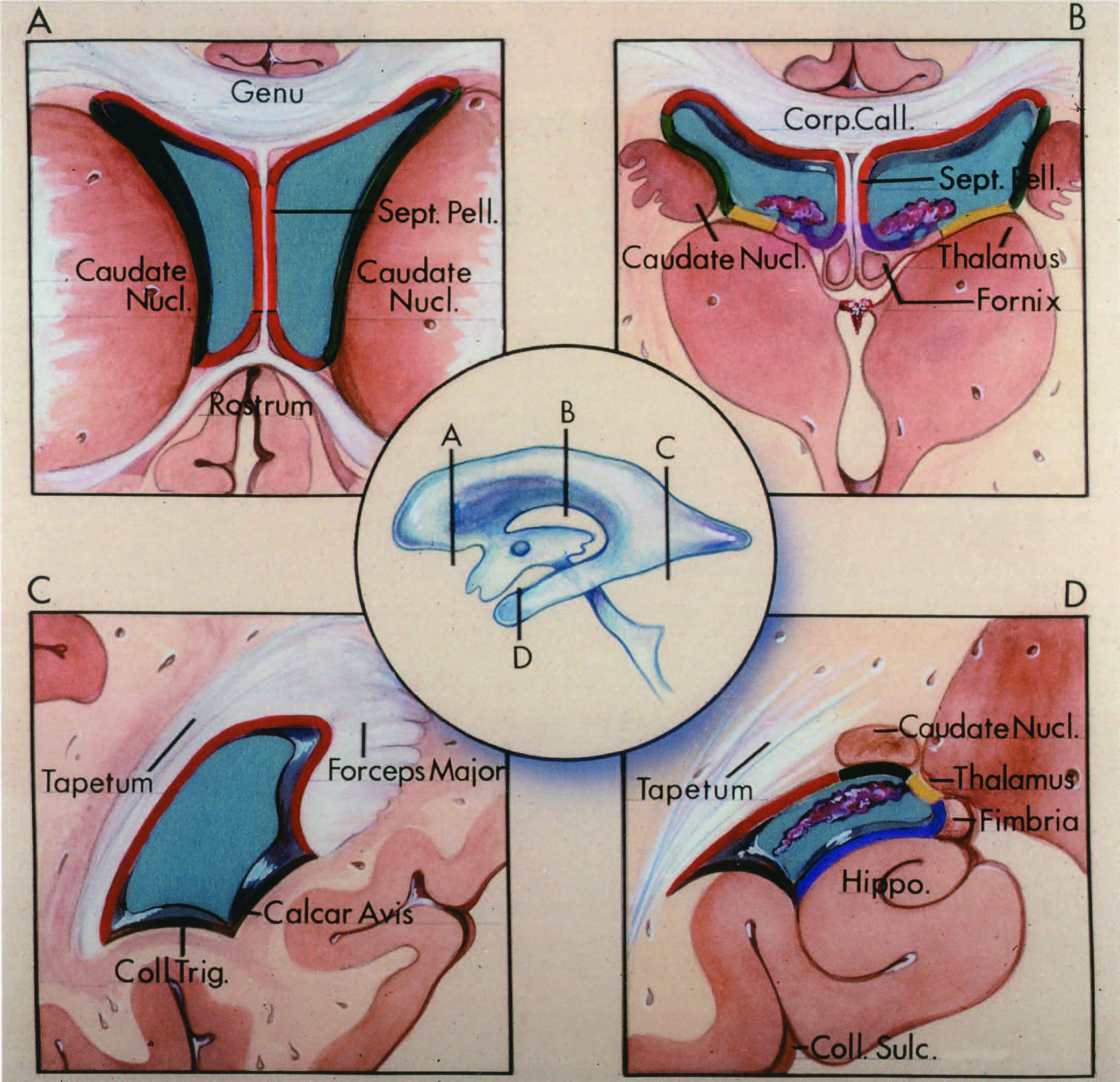
圖5.4。側腦室壁的結構。中心圖顯示的是通過額角(A)、體(B)、心房(C)和顳角(D)的橫斷麵水平。不同結構形成的心室表麵用不同的顏色表示:胼胝體,紅色;丘腦、黃色;穹窿和海馬形成,紫色;尾狀核、綠色;透明隔,橙色;覆蓋著側支和鈣質溝的突起,棕色。額葉角。胼胝體膝在頂,尾狀核在側壁,胼胝體的端部在底,透明隔在內側壁。 B, body of the lateral ventricle. The body of the corpus callosum is in the roof, the caudate nucleus is in the lateral wall, the thalamus is in the floor, and the septum pellucidum and fornix are in the medial wall. The choroidal fissure, the site of the attachment of the choroid plexus in the lateral ventricle, is situated between the fornix and the thalamus. C, atrium. The lateral wall and roof are formed by the tapetum of the corpus callosum, and the floor is formed by the collateral trigone, which overlies the collateral sulcus. The inferior part of the medial wall is formed by the calcar avis, the prominence that overlies the deep end of the calcarine sulcus, and the superior part of the medial wall is formed by the bulb of the corpus callosum, which overlies the forceps major. D, temporal horn. The medial part of the floor of the temporal horn is formed by the prominence overlying the hippocampal formation, and the lateral part of the floor is formed by the prominence called the collateral eminence, which overlies the deep end of the collateral sulcus. The roof is formed by the caudate nucleus and the tapetum of the corpus callosum, the lateral wall is formed by the tapetum of the corpus callosum, and the medial wall of the temporal horn is little more than the cleft between the fimbria of the fornix and the inferolateral aspect of the thalamus. Call., callosum; Coll., collateral; Corp., corpus; Hippo., hippocampus; Nucl., nucleus; Pell., pellucidum; Sept., septum; Sulc., sulcus; Trig., trigone.
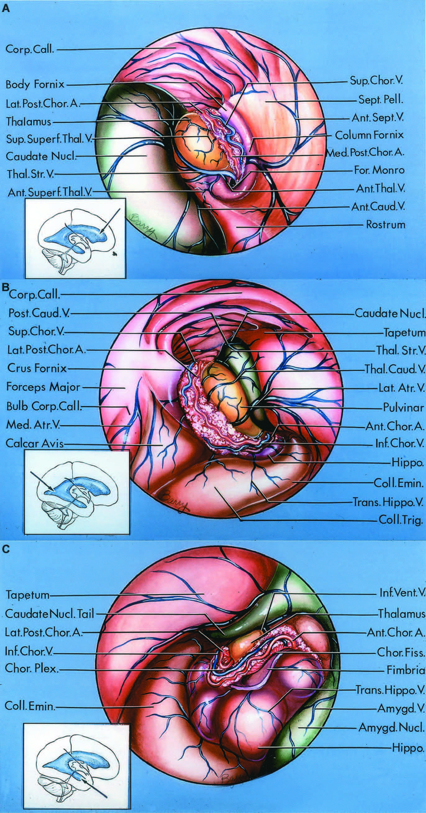
圖5.5。側腦室的視圖。腦室壁的結構用不同的顏色表示:黃色的是丘腦;尾狀核和杏仁核,綠色;胼胝體、紅;穹窿和海馬形成,紫色;透明隔,橙色;鈣質和側溝上方的突起是棕色的。A,前視圖,沿著圖中的箭頭,進入額角和側腦室體。額角位於Monro孔前方,內側壁有透明隔,頂部有胼胝體的膝和體,外側壁有尾狀核,前壁有胼胝體膝,底有胼胝體的端部。 The body of the lateral ventricle has the thalamus in its floor, the caudate nucleus in the lateral wall, the body of the fornix and septum pellucidum in the medial wall, and the corpus callosum in the roof. The choroid plexus is attached along the choroidal fissure, the cleft between the fornix and thalamus. The superior choroidal vein and branches of the lateral and medial posterior choroidal arteries course on the surface of the choroid plexus. The anterior and posterior septal veins cross the roof and the medial wall of the frontal horn and body. The anterior and posterior caudate veins cross the lateral wall of the frontal horn and body and join the thalamostriate vein, which passes through the foramen of Monro. A superior superficial thalamic vein courses on the thalamus. B, posterior view, along the arrow in the inset, into the atrium. The atrium has the tapetum of the corpus callosum in the roof, the bulb of the corpus callosum and the calcar avis in its medial wall, the collateral trigone in the floor, the caudate nucleus and tapetum in the lateral wall, and the crus of the fornix, pulvinar, and choroid plexus in the anterior wall. The temporal horn has the hippocampal formation and collateral eminence in the floor and the thalamus, tail of the caudate nucleus, and tapetum in the roof and the lateral wall. Branches of the anterior and lateral posterior choroidal arteries course on the surface of the choroid plexus. A thalamocaudate vein drains the part of the lateral wall of the body behind the area drained by the thalamostriate vein. The inferior choroidal vein courses on the choroid plexus in the temporal horn. The lateral and medial atrial veins cross the medial and lateral walls of the atrium. Transverse hippocampal veins cross the floor of the atrium and temporal horn. C, anterior view, along the arrow in the inset, into the temporal horn. The floor of the temporal horn is formed by the collateral eminence and the hippocampal formation. The roof and lateral wall are formed, from medial to lateral, by the thalamus, the tail of the caudate nucleus, and the tapetum of the corpus callosum. The medial wall is little more than the cleft between the thalamus and the fimbria, called the choroidal fissure, along which the choroid plexus is attached. The amygdaloid nucleus bulges into the anteromedial part of the temporal horn. The fimbria of the fornix arises on the surface of the hippocampal formation. Branches of the anterior and lateral posterior choroidal arteries course on the surface of the choroid plexus. The inferior ventricular vein drains the roof of the temporal horn and receives the amygdalar vein from the ventricular surface of the amygdaloid nucleus. The inferior choroidal vein joins the inferior ventricular vein. The transverse hippocampal veins drain the floor of the temporal horn. A., artery; Amygd., amygdaloid; Ant., anterior; Atr., atrial; Call., callosum; Caud., caudate; Chor., choroid, choroidal; Coll., collateral; Corp., corpus; Emin., eminence; For., foramen; Hippo., hippocampal, hippocampus; Inf., inferior; Lat., lateral; Med., medial; Nucl., nucleus; Pell., pellucidum; Plex., plexus; Post., posterior; Sept., septal, septum; Sup., superior; Superf., superficial; Thal., thalamic; Thal.Str., thalamostriate; Trans., transverse; Trig., trigone; V., vein.
脈絡膜裂是位於穹窿和丘腦之間的狹窄的c形裂縫,脈絡膜叢沿此附著(圖5.3-5.6)。當側腦室脈絡膜叢被撕裂時,裂縫被視為位於身體內側、心房和顳角的狹窄裂縫。穹窿形成了裂隙的外緣,丘腦形成了內緣。脈連膜裂在腦室內部受到上穹窿體和下丘腦體的限製,在心房受到後穹窿小腿和前枕骨的限製,在顳角受到下麵穹窿的絲膜和上麵的終紋和丘腦的限製。脈絡膜裂呈c型弧線,從蒙羅孔繞著丘腦的上、下、後表麵延伸到它的下端,稱為脈絡膜下點,位於海馬體頭部後麵,膝狀體外側。丘腦的位置是這樣的,它的表麵在脈膜裂隙外側的部分形成了側腦室壁的一部分,而在裂隙內側的部分形成了第三腦室壁或基底池的部分。
每個側腦室的脈絡膜叢穿過Monro孔,並與位於第三腦室頂的兩條平行脈絡膜叢連續。在心房,脈絡膜叢形成一個突出的三角形簇稱為血管球。與脈絡膜裂隙相鄰的丘腦和穹窿的邊緣有小脊,稱為絨毛膜,沿著絨毛膜,脈絡膜叢的膜,附著在脈絡膜上。丘腦一側的肌腱絛蟲稱為丘腦肌腱絛蟲或脈絡膜肌腱絛蟲。在裂隙的穹窿側的腱蟲稱為穹窿腱蟲,除了在顳角處稱為纖維腱蟲。
脈膜裂形成於胚胎發育的大約8周時,形成第三腦室上皮頂的血管髓膜內陷到大腦半球的內側壁。室管膜和軟膜之間沿著形成脈膜裂的內陷處沒有神經組織發育,因此形成了側腦室壁最薄的位置。脈絡膜動脈供應脈絡膜叢,起源於頸內動脈和腦後動脈,通過脈絡膜裂進入心室。此外,在心室壁內的靜脈通過室管膜下脈絡膜裂隙的邊緣離開心室,到達腦內靜脈、基底靜脈或大靜脈。在顱內手術中,通過側腦室的裂口可以進入一些很難或不可能通過腦外通路暴露的結構。
脈絡膜裂分為體部、心房部和顳部。體部位於側腦室體內穹窿體和丘腦上表麵之間(圖5.5和5.7)。腦內靜脈通過的膜間質位於第三腦室頂部裂縫的身體內側。從腦室體穿過脈絡膜裂的開口將暴露出膜間質和第三腦室的頂部。脈絡膜裂和脈絡膜叢不延伸到額角;然而,一些進入脈絡膜裂上部的手術入路是通過額角和身體的鄰近部分。心房位於側腦室的心房中穹窿小腿和枕骨之間(圖5.5和5.8)。裂縫沒有延伸到枕角。通過從心房打開的裂隙,可以暴露四邊形池、鬆果體區和周圍池的後部。顳部位於穹窿絲膜和丘腦下外側表麵之間的顳角(圖5.5和5.9)。 Opening through the choroidal fissure in the temporal horn exposes the structures in the ambient and posterior part of the crural cisterns. The cisternal side of the temporal portion of the fissure is situated in the superolateral edge of the ambient cistern. The fissure is the thinnest site in the wall of the lateral ventricle bordering the basal cisterns and the roof of the third ventricle.

點擊這裏查看此圖像的交互模塊和相關內容。
圖5.6。模擬。逐步剝離脈絡膜裂隙。側腦室上方圖。脈絡膜裂是穹窿和丘腦之間的裂口脈絡膜叢沿著它連接。額角位於前側,腦室體位於門羅孔後。丘腦形成了側腦室體的底部和心房的前壁。B,放大圖。穹窿柱形成了門羅孔的前緣和上緣。體內脈絡膜叢穿過Monro孔後緣,並與第三腦室頂的脈絡膜叢連續。 The right thalamostriate vein passes through the posterior edge of the foramen of Monro and the left thalamostriate vein passes through the choroidal fissure behind the foramen. The floor of the frontal horn is formed by the rostrum, and the anterior wall is formed by the genu of the corpus callosum. The lateral wall is formed by the caudate nucleus. The septum pellucidum is attached to the upper edge of the body of the fornix. C, enlarged view of the foramen of Monro. The columns of the fornix form the anterior and superior margins of the foramen. An anterior septal vein passes backward along the septum pellucidum and crosses the column of the fornix. The thalamostriate vein passes forward between the caudate nucleus and thalamus and turns medially to pass through the posterior margin of the foramen of Monro to empty into the internal cerebral vein. The choroid plexus is attached medially by the tenia fornix to the body of the fornix and laterally by the tenia thalami to the thalamus. D, the transchoroidal exposure is begun by dividing the tenia fornix that attaches the choroid plexus to the margin of the fornix. The tenia thalami that attaches the choroid plexus to the thalamus is not opened.

點擊這裏查看此圖像的交互模塊和相關內容。
圖5.6。情況。E,脈絡膜裂的開口從Monro孔向後延伸,暴露出大腦內靜脈和位於胸膜間質內的內側後脈絡膜動脈。前隔靜脈穿過透明隔。脈絡膜下層,附著於丘腦髓紋深至腦內靜脈處,完好無損。F,形成膜間質底部的脈留膜下層已被打開,暴露出第三腦室內的中間塊和後連合。G,大腦內靜脈被分離出來暴露出第三腦室的前下部分。中腦的上端形成了第三腦室底部的後部。乳狀體位於基底的中部。乳頭體前麵和漏鬥窩後麵的底非常薄,通常在第三腦室造口術中打開。 The chiasmatic recess extends forward above the posterior edge of the optic chiasm and below the anterior commissure. H, enlarged view of the inner surface of the anterior wall of the third ventricle. The columns of the fornix extend downward behind the anterior commissure toward the mamillary bodies. The lamina terminalis, chiasmatic recess, posterior edge of the chiasm, and the infundibular recess are located along the anterior and lower wall of the third ventricle.

點擊這裏查看此圖像的交互模塊和相關內容。
圖5.6。i n。沿著脈絡膜裂的開口已經向後延伸了通過沿著身體邊緣和穹窿小腿打開穹窿肌腱。四叉神經池的上部,也就是大腦內靜脈與蓋倫靜脈彙合的地方,已經暴露出來。脈絡膜內側後動脈與大腦內靜脈相連。J,脈絡膜裂的開口沿著脈絡膜裂向下延伸到四叉神經池的中央部分,暴露出大腦基底靜脈和內部靜脈、鬆果體和上丘。內側後脈絡膜動脈的分支在鬆果體旁邊。K,放大視圖。鬆果體的尖端向後突出於上丘的上方和大腦內靜脈的末端之間。L,剝離已經沿著脈膜裂向前延伸到顳角,通過在穹窿毛膜邊緣分裂肌腱,暴露基底靜脈,大腦後動脈和滑車神經在丘腦下部周圍池的後部。 M, the choroidal fissure in the temporal horn has been opened by dividing the tenia fimbria. The choroid plexus attachment to the thalamus has not been disturbed. The posterior cerebral artery and basal vein course through the ambient cistern on the medial side of the temporal portion of the choroidal fissure. N, the exposure has been extended through the amygdala anterior to the choroidal fissure to expose the oculomotor nerve and origin of the posterior cerebral artery. The posterior cerebral artery passes above the oculomotor nerve. A., artery; Ant., anterior; Bas., basilar; Call., callosum; Caud., caudate; Cer., cerebral; Ch., choroidal; Chiasm., chiasmatic; Chor., choroid; Col., column; Coll., colliculus; Comm., commissure; Corp., corpus; CN, cranial nerve; For., foramen; Front., frontal; Gen., geniculate; Infund., infundibular; Int., intermedia, internal; Lam., lamina; Lat., lateral; Mam., mamillary; M.P.Ch.A., medial posterior choroidal artery; Nucl., nucleus; P.C.A., posterior cerebral artery; Pell., pellucidum; Plex., plexus; Rec., recess; Sept., septal, septum; Tent., tentorial; Term., terminalis; Thal. Str., thalamostriate; V., vein; Vent., ventricle.
![圖5.7。經脈絡膜入路,通過胼胝體的開口穿過脈絡膜裂的體部。頭皮切口和骨瓣的位置如圖所示。A,手術暴露額角和右側腦室體。脈絡膜裂位於脈絡膜叢深處。側腦室壁的結構包括丘腦、尾狀穹窿核、門羅孔、透明隔和胼胝體的端部。聚集在脈絡膜裂隙上的血管結構包括脈絡膜內側和外側後動脈、前和後間隔、前和後尾狀體、上脈絡膜靜脈和丘腦靜脈。B,脈絡膜裂已經沿著穹窿腱切開。通過分離腦內靜脈,打開第三腦室頂部的脈絡膜層,暴露第三腦室中間塊及內部和底部。內側後脈絡膜動脈環繞大腦內靜脈。 A., artery; Ant., anterior; Call., callosum; Caud., caudate; Cer., cerebral; Chor., choroid, choroidal; Fiss., fissure; For., foramen; Front., frontal; Int., intermedia, internal; Lat., lateral; Med., medial; Nucl., nucleus; Pell., pellucidum; Plex., plexus; Post., posterior; Sept., septal, septum; Sup., superior; Thal. Str., thalamostriate; V., vein; Vent., ventricle. (From, Nagata S, Rhoton AL Jr, Barry M: Microsurgical anatomy of the choroidal fissure. Surg Neurol 30:3–59, 1988 [15].)](https://assets.neurosurgicalatlas.com/neuroanatomy/Rhoton_-_Missed_Images/Lateral_and_third_ventricles_5.7.jpg)
圖5.7。經脈絡膜入路,通過胼胝體的開口穿過脈絡膜裂的體部。頭皮切口和骨瓣的位置如圖所示。A,手術暴露額角和右側腦室體。脈絡膜裂位於脈絡膜叢深處。側腦室壁的結構包括丘腦、尾狀穹窿核、門羅孔、透明隔和胼胝體的端部。聚集在脈絡膜裂隙上的血管結構包括脈絡膜內側和外側後動脈、前和後間隔、前和後尾狀體、上脈絡膜靜脈和丘腦靜脈。B,脈絡膜裂已經沿著穹窿腱切開。通過分離腦內靜脈,打開第三腦室頂部的脈絡膜層,暴露第三腦室中間塊及內部和底部。內側後脈絡膜動脈環繞大腦內靜脈。 A., artery; Ant., anterior; Call., callosum; Caud., caudate; Cer., cerebral; Chor., choroid, choroidal; Fiss., fissure; For., foramen; Front., frontal; Int., intermedia, internal; Lat., lateral; Med., medial; Nucl., nucleus; Pell., pellucidum; Plex., plexus; Post., posterior; Sept., septal, septum; Sup., superior; Thal. Str., thalamostriate; V., vein; Vent., ventricle. (From, Nagata S, Rhoton AL Jr, Barry M: Microsurgical anatomy of the choroidal fissure. Surg Neurol 30:3–59, 1988 [15].)
![圖5.8。經脈絡膜入路通過脈絡膜裂隙的心房部分,使用頂葉上皮質切口。頭皮切口、骨瓣和皮質切口的位置如圖所示。A,脈絡膜叢沿著脈絡膜裂相連。脈絡膜裂隙的心房部分位於穹窿窩和枕骨之間。心房壁、體壁和側腦室顳角的結構包括枕狀區、穹窿區、尾狀核、胼胝體絨氈層和球狀區、距距區、海馬形成區、側隆起區和三角區。彙聚於脈絡膜裂隙的血管結構包括脈絡膜前動脈和後動脈、房側動脈和內側動脈、尾狀後動脈、上脈絡膜動脈和下脈絡膜靜脈以及海馬橫靜脈。B,通過切開穹窿肌腱,向後縮回穹窿底部,打開脈絡膜裂隙,暴露四頭池,大腦後動脈和內側脈絡膜後動脈,鬆果體,大腦內靜脈,基底靜脈和大靜脈。一個,動脈;螞蟻。, anterior; Atr., atrial; Call., callosum; Caud., caudate; Cer., cerebral; Chor., choroid, choroidal; Cist., cistern; Coll., collateral; Corp., corpus; Emin., eminence; Fiss., fissure; Hippo., hippocampal, hippocampus; Inf., inferior; Int., internal; Lat., lateral; Med., medial; Nucl., nucleus; P.C.A., posterior cerebral artery; Plex., plexus; Post., posterior; Quad., quadrigeminal; Sup., superior; Temp., temporal; Trans., transverse; Trig., trigone; V., vein; Vent., ventricle. (From, Nagata S, Rhoton AL Jr, Barry M: Microsurgical anatomy of the choroidal fissure. Surg Neurol 30:3–59, 1988 [15].)](https://assets.neurosurgicalatlas.com/neuroanatomy/Rhoton_-_Missed_Images/Lateral_and_third_ventricles_5.8.jpg)
圖5.8。經脈絡膜入路通過脈絡膜裂隙的心房部分,使用頂葉上皮質切口。頭皮切口、骨瓣和皮質切口的位置如圖所示。A,脈絡膜叢沿著脈絡膜裂相連。脈絡膜裂隙的心房部分位於穹窿窩和枕骨之間。心房壁、體壁和側腦室顳角的結構包括枕狀區、穹窿區、尾狀核、胼胝體絨氈層和球狀區、距距區、海馬形成區、側隆起區和三角區。彙聚於脈絡膜裂隙的血管結構包括脈絡膜前動脈和後動脈、房側動脈和內側動脈、尾狀後動脈、上脈絡膜動脈和下脈絡膜靜脈以及海馬橫靜脈。B,通過切開穹窿肌腱,向後縮回穹窿底部,打開脈絡膜裂隙,暴露四頭池,大腦後動脈和內側脈絡膜後動脈,鬆果體,大腦內靜脈,基底靜脈和大靜脈。一個,動脈;螞蟻。, anterior; Atr., atrial; Call., callosum; Caud., caudate; Cer., cerebral; Chor., choroid, choroidal; Cist., cistern; Coll., collateral; Corp., corpus; Emin., eminence; Fiss., fissure; Hippo., hippocampal, hippocampus; Inf., inferior; Int., internal; Lat., lateral; Med., medial; Nucl., nucleus; P.C.A., posterior cerebral artery; Plex., plexus; Post., posterior; Quad., quadrigeminal; Sup., superior; Temp., temporal; Trans., transverse; Trig., trigone; V., vein; Vent., ventricle. (From, Nagata S, Rhoton AL Jr, Barry M: Microsurgical anatomy of the choroidal fissure. Surg Neurol 30:3–59, 1988 [15].)
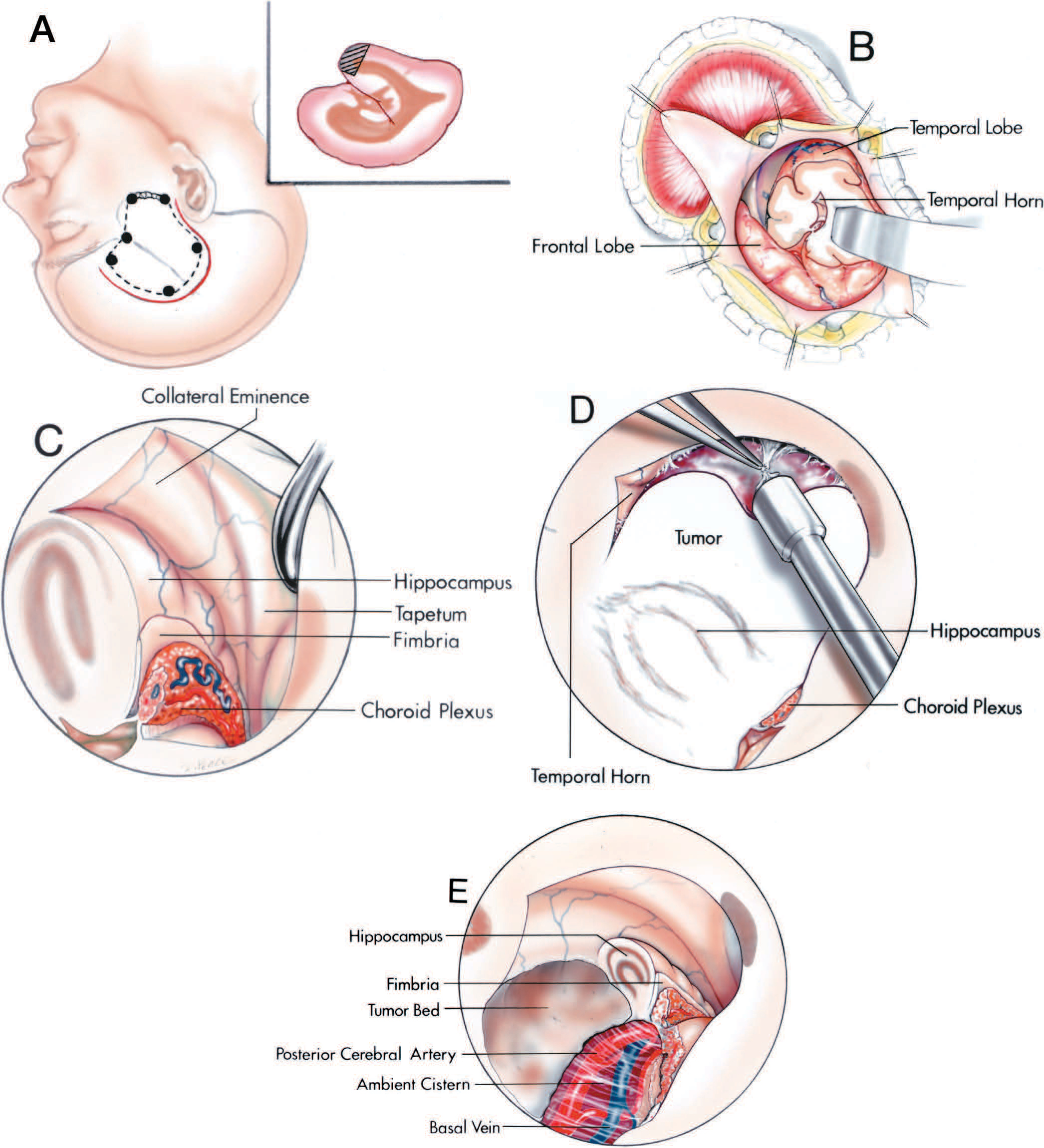
![圖5.9。經脈絡膜入路穿過脈絡膜裂的顳部。圖示頭皮切口和骨瓣的位置。第一,顳葉的下表麵已經打開,露出了顳角。脈絡膜叢沿脈絡膜裂附著。顳角壁的結構包括海馬形成,側支隆起,杏仁核和尾狀核,以及胼胝體的絨氈層。通過脈絡膜裂的血管結構包括前、後脈絡膜動脈和海馬橫靜脈、杏仁核、下脈絡膜靜脈和下心室靜脈。B,脈絡膜裂已經被打開了通過沿著纖維細肌切開並向上收回脈絡膜叢。這就暴露了周圍的腦池,大腦後動脈的分支和基底靜脈的支流。脈絡膜後內側動脈在大腦後動脈的內側。 A., artery; Amygd., amygdalar, amygdaloid; Ant., anterior; Caud., caudate; Chor., choroid, choroidal; Cist., cistern; Coll., collateral; Emin., eminence; Fiss., fissure; Hippo., hippocampal, hippocampus; Inf., inferior; Lat., lateral; Med., medial; Nucl., nucleus; P.C.A., posterior cerebral artery; Plex., plexus; Post., posterior; Temp., temporal; Trans., transverse; V., vein; Vent., ventricle. (From, Nagata S, Rhoton AL Jr, Barry M: Microsurgical anatomy of the choroidal fissure. Surg Neurol 30:3–59, 1988 [15].)](https://assets.neurosurgicalatlas.com/neuroanatomy/Rhoton_-_Missed_Images/Lateral_and_third_ventricles_5.9.jpg)
圖5.9。經脈絡膜入路穿過脈絡膜裂的顳部。圖示頭皮切口和骨瓣的位置。第一,顳葉的下表麵已經打開,露出了顳角。脈絡膜叢沿脈絡膜裂附著。顳角壁的結構包括海馬形成,側支隆起,杏仁核和尾狀核,以及胼胝體的絨氈層。通過脈絡膜裂的血管結構包括前、後脈絡膜動脈和海馬橫靜脈、杏仁核、下脈絡膜靜脈和下心室靜脈。B,脈絡膜裂已經被打開了通過沿著纖維細肌切開並向上收回脈絡膜叢。這就暴露了周圍的腦池,大腦後動脈的分支和基底靜脈的支流。脈絡膜後內側動脈在大腦後動脈的內側。 A., artery; Amygd., amygdalar, amygdaloid; Ant., anterior; Caud., caudate; Chor., choroid, choroidal; Cist., cistern; Coll., collateral; Emin., eminence; Fiss., fissure; Hippo., hippocampal, hippocampus; Inf., inferior; Lat., lateral; Med., medial; Nucl., nucleus; P.C.A., posterior cerebral artery; Plex., plexus; Post., posterior; Temp., temporal; Trans., transverse; V., vein; Vent., ventricle. (From, Nagata S, Rhoton AL Jr, Barry M: Microsurgical anatomy of the choroidal fissure. Surg Neurol 30:3–59, 1988 [15].)
第三腦室位於頭部中央,位於胼胝體和側腦室體下方,位於蝶鞍、垂體和中腦上方,位於大腦半球、丘腦兩半和下丘腦兩半之間(圖5.10和5.11)。它與威利斯及其分支的圈子和蓋倫及其支流的大脈密切相關。第三腦室區域的腫瘤是最難暴露和切除的。
操作第三腦室壁可導致下丘腦功能障礙,表現為意識、溫度控製、呼吸和垂體分泌紊亂,視交叉和束損傷導致視力喪失,第三腦室壁穹窿柱損傷導致記憶喪失(24,28,37)。

圖5.10。第三腦室的正中矢狀麵。底(藍色)從視交叉延伸到sylvius渡槽,包括視交叉下表麵,漏鬥,漏鬥隱窩,垂體腺,腫瘤塊莖,乳頭體,後穿孔物質,以及渡槽前的中腦部分。前壁(紅色)從視交叉延伸到門羅孔,包括視交叉上表麵、視隱窩、終板、前連合和門羅孔。頂部(綠色)由蒙羅孔延伸至棘突上隱窩,由穹窿和脈絡膜層構成,其間為腦內靜脈和脈絡膜後內側動脈。海馬連通性、胼胝體和透明隔位於屋頂之上。後壁從鬆果體上隱窩延伸到渡槽,包括鉤骨連合、鬆果體、鬆果體隱窩和後連合。動眼神經起源於中腦。下丘腦溝在第三腦室的丘腦和下丘腦表麵之間形成凹槽。螞蟻。, anterior; B., body; Call., callosum; Ch., chiasm; Cin., cinereum; Comm., commissure; Corp., corpus; For., foramen; Hab., habenular; Hippo., hippocampal; Hypothal., hypothalamic, hypothalamus; Infund., infundibular, infundibulum; Inter., intermedia; Lam., lamina; Mam., mamillary; N., nerve; O., optic; Pel., pellucidum; Perf., perforated; Pit., pituitary; Post., posterior; Sept., septum; Subst., substance; Sulc., sulcus; Ter., terminalis.

點擊這裏查看此圖像的交互模塊和相關內容。
圖5.11。模擬。第三腦室正中矢狀麵。A,第三腦室位於頭蓋骨的中心,在胼胝體、側腦室體和透明隔下方,在中腦和足間窩上方,在四叉神經池和蓋倫靜脈前方,在大腦前動脈後方。沿鐮側的半腦間裂是通向第三腦室的一條通道。第三腦室的後部也可以沿著鐮和幕的連接處靠近,與直竇相鄰。B,放大圖。透明隔隔開側腦室體和額角,並由前、後間隔靜脈交叉。大腦前動脈沿第三腦室前壁上升,基底分支位於地板下方,蓋倫靜脈阻斷了通向後壁的通路。C,第三腦室放大圖。 The anterior wall of the third ventricle is formed by the lamina terminalis and anterior commissure and blends above into the rostrum of the corpus callosum. The roof is formed by the body of the fornix and the velum interpositum through which the internal cerebral veins and medial posterior choroidal arteries course. The posterior wall, formed by the pineal and habenular and posterior commissures, is located anterior to the quadrigeminal cistern and the venous complex created by numerous veins converging on the vein of Galen. The floor is formed, from anterior to posterior, by the optic chiasm, tuber cinereum above the pituitary stalk, mamillary bodies, and upper midbrain. The section extends to the lateral side of the mamillary bodies. The velum interpositum is the space within the roof of the third ventricle along which the internal cerebral veins and medial posterior choroidal arteries pass. The body of the fornix is located above the velum interpositum. The upper wall of the velum interpositum is formed by the layer of tela choroidea attached to the lower margin of the fornix. The floor is formed by the layer of tela attached along the striae medullaris thalami. The internal cerebral veins and medial posterior choroidal arteries course between the two layers of tela. The choroid plexus in the roof of the third ventricle arises in the lower layer of tela. D, another third ventricle. This section extends just to the left of the midline through the column and body of the fornix. The body of the fornix forms the roof of the third ventricle. The columns pass anterior to the foramen of Monro and descend behind the anterior commissure to reach the mamillary bodies.

點擊這裏查看此圖像的交互模塊和相關內容。
圖5.11。情況。E,放大視圖。前壁由終板和前連合組成。視交叉、乳狀體和中腦都在眼底。F,放大視圖。交叉隱窩位於視交叉上方和終板後麵。漏鬥隱窩位於視交叉的下方和後麵。端板與胼胝體的端部融合。前連合位於胼胝體口和穹窿柱之間。 The thalamus and hypothalamus form the lateral wall of the third ventricle. G, enlarged view of the posterior part of the third ventricle. The posterior wall of the third ventricle is formed by the aqueduct, pineal, and habenular and posterior commissures. The pineal recess extends into the base of the pineal in the interval between the habenular and posterior commissures. H, lateral view of the third ventricle with the hippocampus and fornix preserved. The body of the fornix forms the roof of the third ventricle. The velum interpositum, through which the internal cerebral veins course, is located between the body of the fornix and the striae medullaris thalami. The quadrigeminal cistern and pineal region are located anteromedial to the crus of the fornix, and the ambient cistern and posterior cerebral artery are located medial to the temporal horn and the fimbria. Opening the choroidal fissure adjacent to the body of the fornix exposes the third ventricle. The medial posterior choroidal arteries turn forward beside the pineal to reach the velum interpositum. A., artery; A.C.A., anterior cerebral artery; Ant., anterior; Bas., basilar; Call., callosum; Car., carotid; Caud., caudate; Cer., cerebral; Chiasm., chiasmatic; Chor., choroid; CN, cranial nerve; Col., column; Coll., colliculus; Comm., commissure; Corp., corpus; For., foramen; Hab., habenular; Infund., infundibular; Int., intermedia, internal; Interpos., interpositum; Lam., lamina; Lat., lateral; Mam., mamillary; Med., medial; M.P.Ch.A., medial posterior choroidal artery; Nucl., nucleus; P.C.A., posterior cerebral artery; Pell., pellucidum; Pit., pituitary; Plex., plexus; Post., posterior; Rec., recess; Sag., sagittal; Sept., septal, septum; Str., straight; Sup., superior; Term., terminalis; Thal., thalami; V., vein; Vel., velum; Vent., ventricle.
第三腦室是一個狹窄的、漏鬥狀的、單眼的中線腔。它的前上緣通過Monro孔與每個側腦室相通,後緣通過sylvius渡槽與第四腦室相通。它有一個屋頂,一個地板,一個前壁,後壁和兩個側壁。
第三腦室的頂部形成一個平緩向上的拱形,從前麵的Monro孔延伸到後麵的棘突上隱窩(圖5.10-5.13)。屋頂有四層:一層由穹窿形成的神經層,兩層由脈絡膜組成的薄膜層,以及在脈絡膜層之間的一層血管。脈絡膜裂隙位於顱頂外側緣。第三腦室頂板前部的上層由穹窿體構成,頂板後部由腳和海馬連合構成。透明隔附著於穹窿體的上表麵。
在穹窿下麵的三層中有兩層是由脈絡膜組成的。背部脈絡膜由兩層薄薄的半渾濁的膜組成,膜起源於腦膜,由組織鬆散的小梁相互連接。頂部的最後一層是位於脈絡膜兩層之間的維管層。血管層由脈絡膜內側後動脈及其分支和腦內靜脈及其支流組成。平行的脈絡膜叢從脈絡膜下部向中線的兩側向下延伸到第三腦室上部。
腹膜間質是第三腦室頂兩層脈絡膜之間的空間。它位於脈膜裂隙體部內側,位於穹窿體下方第三腦室頂部,位於丘腦超內側表麵之間。脈絡膜的上層附著於穹窿和海馬連合的下表麵。下壁的前部與纖維束自由邊緣的小脊相連,稱為丘腦髓紋,它沿著丘腦的超內側邊界從門羅孔延伸到下丘腦連合。下壁的後部附著於鬆果體的上表麵。第三腦室的鬆果體上隱窩位於脈絡膜下層和鬆果體上表麵之間。第三腦室頂部的成對平行脈絡叢與脈絡膜下部相連。許多引流額角和身體的靜脈彙合於腹膜間質,形成腦內靜脈。大腦內靜脈起於膜間質前部,就在Monro孔後麵,它們從鬆果體上方的膜間質出進入四頭池並與大靜脈彙合。腹膜間質通常是一個封閉的空間,在Monro孔的後麵逐漸變細到一個狹窄的頂端,但它很少在脾和鬆果體之間有一個開口,與四頭池相連,形成腹膜間質池。 There also may be a space above the velum interpositum between the hippocampal commissure and splenium called the cavum vergae.

點擊這裏查看此圖像的交互模塊和相關內容。
圖5.12。模擬。第三腦室的頂部。優越的觀點。第一,大腦半球的上部被切除露出額角和側腦室體。脈絡膜叢沿著位於穹窿體和丘腦之間的脈絡膜裂隙相連。脈絡膜上靜脈沿著脈絡膜叢走行。丘腦紋靜脈穿過門羅孔的後緣。穹窿柱穿過門羅孔的前方和上方。穹窿體形成了第三腦室頂的上部。 B, the right lateral edge of the fornix has been removed to expose the upper layer of tela choroidea that spans the interval below the body of the fornix and forms the upper wall of the velum interpositum in the roof of the third ventricle. The velum is positioned between an upper layer of tela attached to the lower surface of the body of the fornix and a lower layer of tela attached below the internal cerebral veins to the striae medullaris thalami. The internal cerebral veins and medial posterior choroidal arteries course in the velum interpositum. C, the body of the fornix has been folded backward. The upper layer of tela that rests against the lower surface of the body of the fornix has been preserved. The tela is a thin, arachnoid-like membrane, through which the internal cerebral veins and the medial posterior choroidal arteries can be seen. The anterior septal veins pass above the foramen of Monro. D, the upper layer of tela has been removed to expose the internal cerebral veins and medial posterior choroidal arteries. The internal cerebral veins have been retracted laterally. The anterior septal veins course along the septum and join the internal cerebral veins near the foramen of Monro.

點擊這裏查看此圖像的交互模塊和相關內容。
圖5.12。E-F。E,打開網膜,暴露中massa,乳頭體和後連合。F,暴露在第三腦室的後部。導水管位於後縫和韁縫的下方。鬆果體隱窩向後延伸,在鉤骨和後密縫之間進入鬆果體基部。螞蟻。前;Cer。腦; Ch., choroidal; Chor., choroid; Col., column; Comm., commissure; For., foramen; Hab., habenular; Int., intermedia, internal; Mam., mamillary; M.P.Ch.A., medial posterior choroidal artery; Plex., plexus; Post., posterior; Rec., recess; Sept., septal; Sup., superior; Thal.Str., thalamostriate; V., vein.

點擊這裏查看此圖像的交互模塊和相關內容。
圖5.13。模擬。第三腦室的底部和頂部。A,第三腦室底部位於鉤椎和前穿孔物質的內側,在中腦上方。從前到後,底包括視交叉的下緣,被灰質塊莖包圍的垂體柄,乳頭體和中腦。踝間窩位於地板後部下方。視束的前部沿著地板的外側邊緣延伸,但在更遠的後方,視束向外側偏離地板,繞過腦梗的上邊緣。B,放大圖。癌塊莖位於垂體柄周圍。漏鬥隱窩延伸到莖的基部。 A third ventriculostomy is commonly performed by opening through the thin area (yellow arrow) in the floor just in front of the mamillary bodies. The oculomotor nerves arise behind the mamillary bodies below the posterior part of the floor of the third ventricle. C, another specimen showing the thin area in front of the mamillary bodies (yellow arrow) through which a third ventriculostomy is completed. The anterior perforated substance and optic tracts are positioned lateral to the anterior part of the floor of the third ventricle. The mamillary bodies and upper midbrain are positioned below the posterior part of the floor. D, view of another third ventricle from below with the vascular structure preserved. The internal carotid, posterior communicating, anterior choroidal, and posterior cerebral arteries all give rise to branches that reach the walls of the lateral and third ventricles. The thalamoperforating branches of the posterior cerebral artery supply some of the posterior part of the floor of the third ventricle.

點擊這裏查看此圖像的交互模塊和相關內容。
圖5.13。情況。E,下視圖,第三腦室底部切除,露出頂部。垂體柄向前反射,暴露出漏鬥隱窩和終板的腦室側。終肌板從交叉的上邊緣向上傾斜到前合角前麵的區域在那裏它與胼胝體的端部融合。穹窿柱在門羅孔上方和前方交叉,向下延伸至乳頭體。中間塊穿過第三心室的中部。腦膜間質位於第三腦室頂的丘腦之間,是大腦內靜脈和內側後脈絡膜動脈的通道。後連合暴露在鬆果體下方。蓋倫靜脈位於第三腦室的後麵,基底靜脈都流入蓋倫靜脈。 F, enlarged view. The infundibular recess is located below the optic chiasm in the base of the pituitary stalk, and the chiasmatic recess is located above the optic chiasm. The lamina terminalis forms the anterior wall of the chiasmatic recess. The anterior commissure crosses the anterior wall in front of the columns of the fornix. The foramina of Monro open upward into both lateral ventricles. The lower wall of the velum interpositum is formed by the layer of tela choroidea, in which the choroid plexus in the roof of the third ventricle arises, and which is attached laterally to the striae medullaris thalami. The internal cerebral veins can be seen through the layer of tela forming the lower wall of the velum interpositum. G, another specimen with the floor of the third ventricle removed. The posterior cerebral arteries, from which the lateral and medial posterior choroidal arteries arise, passes around the midbrain. The lamina terminalis is exposed above the optic chiasm and slopes upward toward the anterior commissure. The columns of the fornix pass along the anterior and superior margins of the foramen of Monro and behind the anterior commissure. The lower layer of tela choroidea in the velum interpositum has been removed to expose the vascular layer in the roof of the third ventricle formed by the internal cerebral veins and medial posterior choroidal arteries. Another layer of tela, which spans the interval above the internal cerebral veins and below the body of the fornix, separates the vascular layer from the body of the fornix. H, enlarged view. The upper layer of tela choroidea that spans the interval below the body of the fornix has been removed. The body of the fornix, exposed by removing the upper layer of tela, blends anteriorly into the columns of the fornix that pass along the anterior and superior margin of the foramen of Monro. The lamina terminalis has been opened in the interval between the optic chiasm and anterior commissure to expose the perforating branches of the anterior cerebral artery. A., artery; A.C.A., anterior cerebral artery; A.Ch.A., anterior choroidal artery; Ant., anterior; Calc., calcarine; Car., carotid; Cin., cinereum; CN, cranial nerve; Col., column; Comm., commissure; For., foramen; Gen., geniculate; Infund., infundibular; Int., intermedia, internal; Interped., interpeduncular; Interpos., interpositum; Lam., lamina; Lat., lateral; Mam., mamillary; M.C.A., middle cerebral artery; M.P.Ch.A., medial posterior choroidal artery; Olf., olfactory; P.C.A., posterior cerebral artery; P.Co.A., posterior communicating artery; Ped., peduncle; Perf., perforated; Pit., pituitary; Post., posterior; Rec., recess; Subst., substance; Term., terminalis; Thal.Perf., thalamoperforating; Tr., tract; V., vein; Vel., velum.
地板從前方視交叉延伸到後方sylvius渡槽口(圖5.10、5.13和5.14)。前半部地由間腦結構構成,後半部地由中腦結構構成。從下看,形成地板的結構包括,從前到後,視交叉,下丘腦漏鬥,灰質塊狀體,乳突體,後穿孔物質,和(最後方)位於腦梗內側上方的中腦被蓋部分。視交叉位於第三腦室底和前壁的交界處。交叉從它與視神經的連接處向後方和上方傾斜。交叉的下表麵形成地板的前部,上表麵形成前壁的下部。視束起於交叉後外側緣,並從底部向中腦外側緣傾斜延伸。漏鬥、灰質塊莖、乳狀體和後穿孔物質位於前方和外側受視交叉和束限製的空間,後方受腦梗限製。
下丘腦漏鬥是一個中空的漏鬥狀結構,位於視交叉和灰腺塊狀之間。垂體(垂體)附著於漏鬥,漏鬥中的軸突延伸到垂體的後葉。腫瘤塊狀是位於乳腺體前方的下丘腦灰質突出腫塊。癌塊莖在前方與漏鬥融合。在漏鬥基部周圍的灰質塊莖凸起形成一個突起,稱為中隆起。乳狀體形成成對的圓形突起,位於癌塊莖的後方。後穿孔物質是位於前側乳突體和後側腦梗內側表麵之間的灰質凹陷的點狀區域。地板的後部分延伸到腦梗內側的後上方和中腦被蓋的上方。
從第三腦室的上麵和裏麵看,視交叉在地板的前邊緣形成一個突出。漏鬥隱窩延伸到視交叉後的漏鬥。乳頭狀體在漏鬥窩後的地板內表麵形成成對的突起。在乳狀體和sylvius的導水管之間的地板部分具有從一側到另一側凹進去的光滑表麵。這個光滑的表麵位於前麵的後穿孔物質和後麵的腦梗內側部分和中腦被蓋的上方。

點擊這裏查看此圖像的交互模塊和相關內容。
圖5.14。地板和第三腦室下部的前視圖。第一,右丘腦被切除。第三腦室底的後部是由位於乳突體後麵的中腦上表麵形成的。天幕邊緣在位於引水渠後麵的四合院水池的天幕頂端連接。腦室體的脈絡膜裂位於穹窿體和丘腦上表麵之間。視交叉和乳頭體之間的基底位於交叉池的上方。第三腦室造口術最常見的位置是位於乳頭體的前麵。B,左丘腦前部被切除以暴露腦梗和第三腦室兩側的中腦上部。動眼神經起於第三腦室底後部的下方。 The infundibular recess is located behind the optic chiasm. The pons is exposed below the mamillary bodies and infundibular recess. C, both thalami have been removed. The third ventricular floor extends from the optic chiasm to the aqueduct. The choroidal fissure in the body of the ventricle is located between the body of the fornix and the thalamus, in the atrium it is between the crus of the fornix and the pulvinar, and in the temporal horn it is between the fimbria and lower surface of the thalamus. D, enlarged view. The upper midbrain and pons are located below the floor of the third ventricle. The oculomotor nerves exit the midbrain below the floor. The aqueduct and posterior commissure are positioned in the posterior wall of the third ventricle in front of the tentorial apex and quadrigeminal cistern. A., artery; Car., carotid; Chor., choroid; CN, cranial nerve; Comm., commissure; Infund., infundibular; Mam., mamillary; Parahippo., parahippocampal; Ped., peduncle; Plex., plexus; Post., posterior; Rec., recess; Tent., tentorial.
第三腦室的前緣從上麵的Monro孔延伸到下麵的視交叉(圖5.10、5.11和5.15)。在大腦的外表麵隻能看到前表麵的下三分之二;上三分之一隱藏在胼胝體的後部。在表麵可見的前壁部分是由視交叉和終板形成的。終椎板是一層薄薄的灰質和腦膜,附著在視交叉上表麵,向上延伸,填充視交叉和胼胝體頂之間的間隔。
當從內部觀察時,前壁的邊界由上到下由穹窿柱、門羅孔、前連合、終板、視隱窩和視交叉形成。兩側的Monro孔位於屋頂和前壁的交界處。孔是一種導管狀的通道,在穹窿和丘腦之間打開,進入側腦室,並在穹窿下方作為單一通道延伸到第三腦室。Monro孔的前麵是身體和穹窿柱的交界處,後麵是丘腦的前極。Monro孔的大小和形狀取決於腦室的大小:如果腦室很小,每個孔都是一個新月形的開口,前麵是穹窿的凹曲線,後麵是丘腦的凸前結節。隨著心室的擴大,兩側的孔變得更圓。穿過孔的結構是脈絡膜叢,內側後脈絡膜動脈的遠端分支,丘腦紋靜脈,上脈絡膜靜脈和間隔靜脈。
前連合是一束緊密的纖維,穿過穹窿柱前的中線。前連合前後徑變化範圍為1.5 ~ 6.0 mm(37)。在我們的標本中,從前連合後端到Monro孔前緣的距離為1.0 - 3.5 mm(平均2.2 mm),從視交叉上緣到前連合前緣的距離為8 - 12 mm(平均10 mm)。終板填充前連合和視交叉之間的間隙。視板附著在視交叉上表麵的中部,在視交叉的上半部分和視板之間留下一個小裂口,稱為視隱窩。

點擊這裏查看此圖像的交互模塊和相關內容。
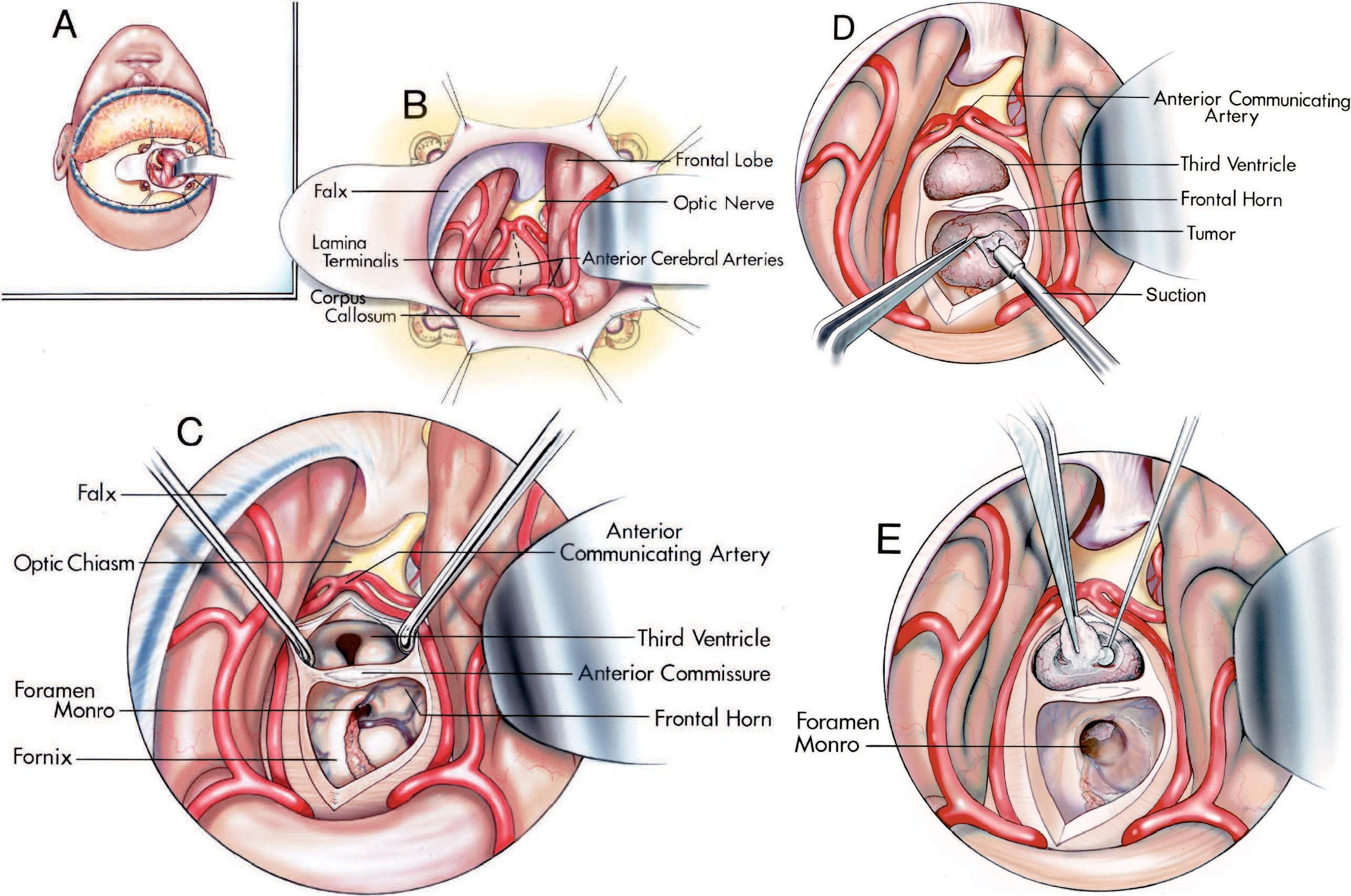
圖5.15。模擬。第三腦室前壁。第一,額葉和頸前動脈被抬高以暴露視交叉和終板。垂體柄從第三腦室底部向下延伸。視神經束沿第三腦室底側緣延伸。端板在上麵與胼胝體的端部融合。嗅覺束向後穿過視神經上方。B,打開終板,暴露交叉隱窩、乳頭體和導水管。垂體柄暴露在視交叉後的漏鬥隱窩下方,位於乳頭體的前方。 Superior hypophyseal arteries pass medially from the carotid artery. C, another third ventricle. The anterior communicating artery commonly passes in front of the lamina terminalis. Perforating arteries arise from a precallosal branch of the anterior communicating artery and penetrate the anterior wall of the third ventricle to reach the columns of the fornix. D, anterior view of a cross section through the anterior part of the third ventricle and body of the lateral ventricle. The lamina terminalis, which has been opened, extends upward in front of the anterior commissure and blends into the rostrum of the corpus callosum. The anterior cerebral arteries have been folded forward. The choroid plexus extends through the foramen of Monro into the roof of the third ventricle below and the body of the lateral ventricle above.

點擊這裏查看此圖像的交互模塊和相關內容。
圖5.15。情況。E,另一個第三腦室的橫切麵穿過前連合。穹窿位於心室的底部。穹窿柱繞過門羅孔的上緣和前緣,在前連合的後麵。端板從交叉向上延伸。F,放大視圖。椎板被打開了。交叉隱窩位於視交叉末端板下部和視交叉後部之間。基底核位於前連合外側的下方。 G, another third ventricle. The lamina terminalis extends upward from the optic chiasm and blends into the rostrum of the corpus callosum. H, the lamina terminalis has been opened. The posterior margin of the chiasm is exposed behind the anterior communicating artery. The anterior commissure is exposed behind the upper edge of the lamina terminalis. The incision has been extended upward through the rostrum of the corpus callosum between the columns of the fornix. This exposes the roof of the third ventricle above the anterior commissure. The choroid plexus hangs down from the tela into the roof of the third ventricle. The mamillary bodies are exposed in the floor. A., artery; A.C.A., anterior cerebral artery; A.Co.A., anterior communicating artery; Ant., anterior; Car., carotid; Chiasm., chiasmatic; Chor., choroid; CN, cranial nerve; Col., column; Fiss., fissure; For., foramen; Hyp., hypophyseal; Lam., lamina; Mam., mamillary; M.C.A., middle cerebral artery; Nucl., nucleus; Olf., olfactory; Pell., pellucidum; Perf., perforating; Pit., pituitary; Plex., plexus; Precall., precallosal; Rec., recess; Sept., septum; Sup., superior; Suprachiasm., suprachiasmatic; Term., terminalis; Thal. Str., thalamostriate; Tr., tract; V., vein; Vent., ventricle.
第三腦室後壁從上方的錐體上隱窩延伸至下方的sylvius導槽(圖5.10和5.11)。從前麵和第三腦室內部看,它由上至下由鬆果體上隱窩、下丘腦隱窩、鬆果體及其隱窩、後隱窩和sylvius導水管組成。鬆果體上隱窩向後突出,位於鬆果體上表麵和頂部的脈絡膜下層之間。鬆果體從其莖部向後延伸到四頭池。鬆果體的柄有一個上板和一個下板。鉤骨連合與鉤骨相連,在上椎板穿過中線,後連合在下椎板穿過。鬆果體隱窩在後伸入鬆果體,位於兩個椎板之間。水楊樹渡槽口的形狀為三角形;三角的底部位於後連合處,其他兩肢由中腦的中心灰質構成。當從後麵看,唯一的結構在後壁是鬆果體。 The pineal gland projects posteriorly into the quadrigeminal cisterns and is concealed by the splenium of the corpus callosum above, the thalamus laterally, and the quadrigeminal plate and the vermis of the cerebellum inferiorly.
側壁在大腦外表麵看不見,而是隱藏在大腦半球之間(圖5.10和5.11)。它們是由下丘腦和上丘腦形成的。側壁有一個輪廓,就像鳥的頭部和張開的喙的側麵輪廓。頭部由丘腦的卵形內側表麵形成;開口的喙,向前方和下方突出,由下丘腦的凹處表示:尖尖的上喙由視隱窩形成,下喙由漏鬥隱窩形成。下丘腦和丘腦表麵由下丘腦溝分開,這是一個通常定義不清的溝槽,從Monro孔延伸到sylvius渡槽。第三腦室丘腦表麵的上界限以狹窄隆起的脊為標誌,稱為丘腦髓紋。這些條紋從枕骨向前延伸,沿著丘腦的超內側表麵,靠近脈絡膜下部的附著處。鞍窩是位於丘腦背內側表麵鬆果體前麵的小突起。在鬆果體的吻端柄的中線上,由韁骨連合連接著韁骨。 The massa intermedia projects into the upper half of the third ventricle and often connects the opposing surfaces of the thalamus. It is present in approximately 75% of brains, being located 2.5 to 6.0 mm (average, 3.9 mm) posterior to the foramen of Monro. The columns of the fornix form distinct prominences in the lateral walls of the third ventricle just below the foramina of Monro, but inferiorly they sink below the surface.
側腦室和第三腦室位於幕內切肌上方,位於幕內遊離邊緣和鞍背之間的三角形空間(圖5.16)(18,23,27)。切骨的頂端位於鬆果體後的中腦背側,基部位於鞍背上。中腦位於門骨的中央。中腦與遊離邊緣之間的區域分為(a)位於腦幹前方的前切骨空間;(b)位於中腦外側的成對中切口間隙;和(c)位於中腦後方的後切口空間。前角位於前切口上方;側腦室的主體位於門骨中央的正上方,它們位於門骨中央,並被丘腦與門骨中央分開;心房位於後切口上方;顳角位於中切口空間的上外側。 The three incisural spaces contain some of the basal cisterns and are so intimately related to the lateral ventricles that some operative approaches to the basal cisterns situated within the incisura are directed through the lateral ventricles and choroidal fissure.
前切骨間隙位於中腦前方,沿第三腦室前壁斜向上延伸,繞視交叉延伸至胼胝體口下方和額角底部。這個空間包括位於腦梗之間的椎間池和位於視交叉下方的交叉池。交叉池與腦池終板在視交叉周圍相通,後者位於終板的前方,位於額角底以下的區域。
中切骨間隙,位於顳葉和中腦之間,與顳角和脈絡膜裂的顳部密切相關,因此一些進入該空間的手術途徑是通過顳角。顳角延伸至顳葉內側,位於中切口間隙外側,終止於距顳葉前極約3cm處。這個空間是腳和周圍蓄水池的位置。底腦池位於腦梗和鉤椎之間,由視道頂起,向後方打開進入周圍腦池。
周圍池是一個狹窄的交流通道,內側由中腦劃分,上方由枕部劃分,外側由海馬旁回、齒狀回和穹窿的絲膜劃分。脈絡膜裂顳部的池側位於周圍池的上外側,位於脈絡膜和下丘腦表麵之間。腳池不能通過脈絡膜裂到達,因為裂隙的終點就在背帶後麵,而腦池在脈絡膜下點。從脈絡膜下點通過杏仁核向前延伸的切口可以從顳角露出腳池。
後切口空間,四叉腦池的位置,位於心房的內側。這個水池包含了一個與鬆果體區域相對應的空間,有屋頂、地板、前壁和側壁。脈絡膜裂位於四頭池前壁和外壁的交界處。四隔池的側壁將池與心房隔開。每一側壁都有前後兩部分:前部分由穹窿小腿構成,後部分由位於脾下方的枕葉內側表麵部分構成。
池前壁有內側和外側兩部分。前壁的內側部分由四邊形板和鬆果體組成。第三腦室的鬆果體上隱窩向鬆果體上方的貯池凸起。池前壁的外側部分是由位於穹窿小腿內側的枕部組成的。在丘的下方,腦池延伸到中腦和小腦之間的裂縫,稱為小腦後腦裂。這個裂隙不能通過脈絡膜裂隙到達。滑車神經起於下丘下方,向外側繞過中腦和枕骨下方進入周圍池。
貯水池的頂部由脾的下表麵和包圍大靜脈及其支流的寬膜狀包膜組成。這一寬闊的蛛網膜組織包膜被應用於脾髒的下表麵,並與腹膜間質向前相連。正是在這個包膜內,在池的超內側部分,靜脈結構的密度最大。貯池內大靜脈的超內側位置與位於貯池下外側的大動脈位置形成對比。
四腔池在枕骨下方向前打開,進入周圍腔池。四叉腦池可與腹膜間質相通。另一個可能與四叉腦池相通的腔是淺腔,它位於海馬連通性和脾髒之間的腹膜間質正上方。由於海馬連通性通常與脾髒的下表麵融合,故疣腔不常出現。

點擊這裏查看此圖像的交互模塊和相關內容。
圖5.16。顯示側腦室與幕切肌的關係的上視圖。A,幕切肌分為位於腦幹前方的前切肌空間,位於中腦和幕邊緣之間的中切肌空間,以及位於幕尖和中腦後表麵之間的後切肌空間。前切口間隙包含交叉池和踝間池。中切口空間與周圍池和腳池相通。後切口空間包含四叉神經池。B,左腦和左半幕切除後的上視圖。額角位於前切口上方。丘腦位於中腦的正上方,位於腦幕切骨的中央。中切口位於中腦和腦幕邊緣之間。 The atrium faces the posterior incisural space and quadrigeminal cistern. C, another specimen. The axial section of the right hemisphere extends through the internal capsule. The frontal horn is located above the anterior incisural space. The thalamus is located above the midbrain in the center of the incisura and above the middle incisural spaces. The medial wall of the atrium forms the lateral wall of the quadrigeminal cistern and posterior incisural space. The internal capsule is situated above the lateral edge of the three incisural spaces.D, comparison of the relationships in the tentorial incisura (D-left) and temporal horn (D-right). The neural structures on the right have been removed except the temporal horn. The temporal lobe on the left was removed to expose the tentorial incisura. The choroidal fissure opens between the fimbria and the thalamus into the middle incisural space located lateral to the midbrain. The temporal horn is positioned lateral to the middle incisural space. The lower part of the medial wall of the atrium faces the posterior incisural space. A., artery; Ant., anterior; Car., carotid; Caud., caudate; CN, cranial nerve; Coll., collateral; Front., frontal; Incis., incisural; Lat., lateral; Lent., lenticular; Med., medial; Nucl., nucleus; Parahippo., parahippocampal; P.C.A., posterior cerebral artery; P.Co.A., posterior communicating artery; Post., posterior; S.C.A., superior cerebellar artery; Temp., temporal; V., vein; Vent., ventricle.
側腦室和第三腦室的每一部分都有重要的外科動脈關係。威利斯圓的所有動脈組成部分都位於側腦室額角和體下方的前切骨空間。頸內動脈在額角以下的區域分叉成大腦前動脈和大腦中動脈,形成脈絡膜前動脈,其分支通過脈絡膜裂隙到達脈絡膜叢。威利斯圓的後部和基底動脈的頂端位於丘腦、側腦室體、第三腦室底和顳角之間。大腦前動脈繞過第三腦室的前壁和前角的底部和前壁,到達前角和身體的頂部。大腦後動脈通過顳角和心房內側,形成脈絡膜後動脈,後動脈通過脈絡膜裂隙,為顳角、心房和身體的脈絡膜叢供血。大腦後動脈、胼胝體周動脈、小腦上動脈和脈絡膜動脈毗鄰後壁。大腦前動脈和後動脈都有分支進入大腦頂,大腦中動脈從額角下方穿過,到達腦裂,然後穿過腦島,在側腦室體的外側。內頸動脈、前脈絡膜動脈、前腦動脈和後腦動脈以及前後交通動脈形成穿孔分支,這些分支到達側腦室和第三腦室壁內或附近的結構(圖5.17和5.18)(8,9,20,21,29,30)。下麵將更詳細地介紹這些動脈和心室之間的關係。

點擊這裏查看此圖像的交互模塊和相關內容。
圖5.17。側腦室的動脈關係。側位(上),上位(中),前位(下)。內頸動脈及其分支用橙色表示,基底動脈及其分支用紅色表示。內頸動脈、基底動脈、前、中、後腦膜動脈、前、外側和內側後脈絡膜動脈都與額角、顳角和枕角以及側腦室的心房和體有重要關係。頸動脈在額角後部下方的區域分叉成它們的大腦前支和大腦中支。大腦中動脈的起源位於額角以下。大腦前動脈在額角的前內側下方穿過,形成胼胝體周支和胼胝體小支,它們繞著額角的前壁和頂部彎曲。脈絡膜前動脈進入顳角的前部。後交通動脈位於丘腦和側腦室體的下方。 The basilar artery bifurcates below the bodies of the lateral ventricles into the posterior cerebral arteries, which course below the thalami near the medial aspect of the temporal horns and atria. The medial posterior choroidal arteries arise from the proximal part of the posterior cerebral arteries, encircle the brainstem below the thalami, and pass forward in the roof of the third ventricle, where they give branches to the choroid plexus in the roof of the third ventricle and the bodies of the lateral ventricles. The lateral posterior choroidal branches of the posterior cerebral arteries pass laterally through the choroidal fissures to enter the temporal horns and atria of the lateral ventricles. The middle cerebral arteries course on the insulae in the area above the temporal horns and lateral to the bodies of the lateral ventricles. The posterior cerebral arteries bifurcate into the calcarine and parieto-occipital arteries in the area medial to the atria. A., artery; A.C.A., anterior cerebral artery; Ant., anterior; Bas., basilar; Cal. Marg., callosomarginal; Calc., calcarine; Car., carotid; Chor., choroidal; Comm., communicating; Front., frontal; Lat., lateral; M.C.A., middle cerebral artery; Occip., occipital; Par. Occip., parieto-occipital; P.C.A., posterior cerebral artery; Post., posterior; Temp., temporal; Vent., ventricle.
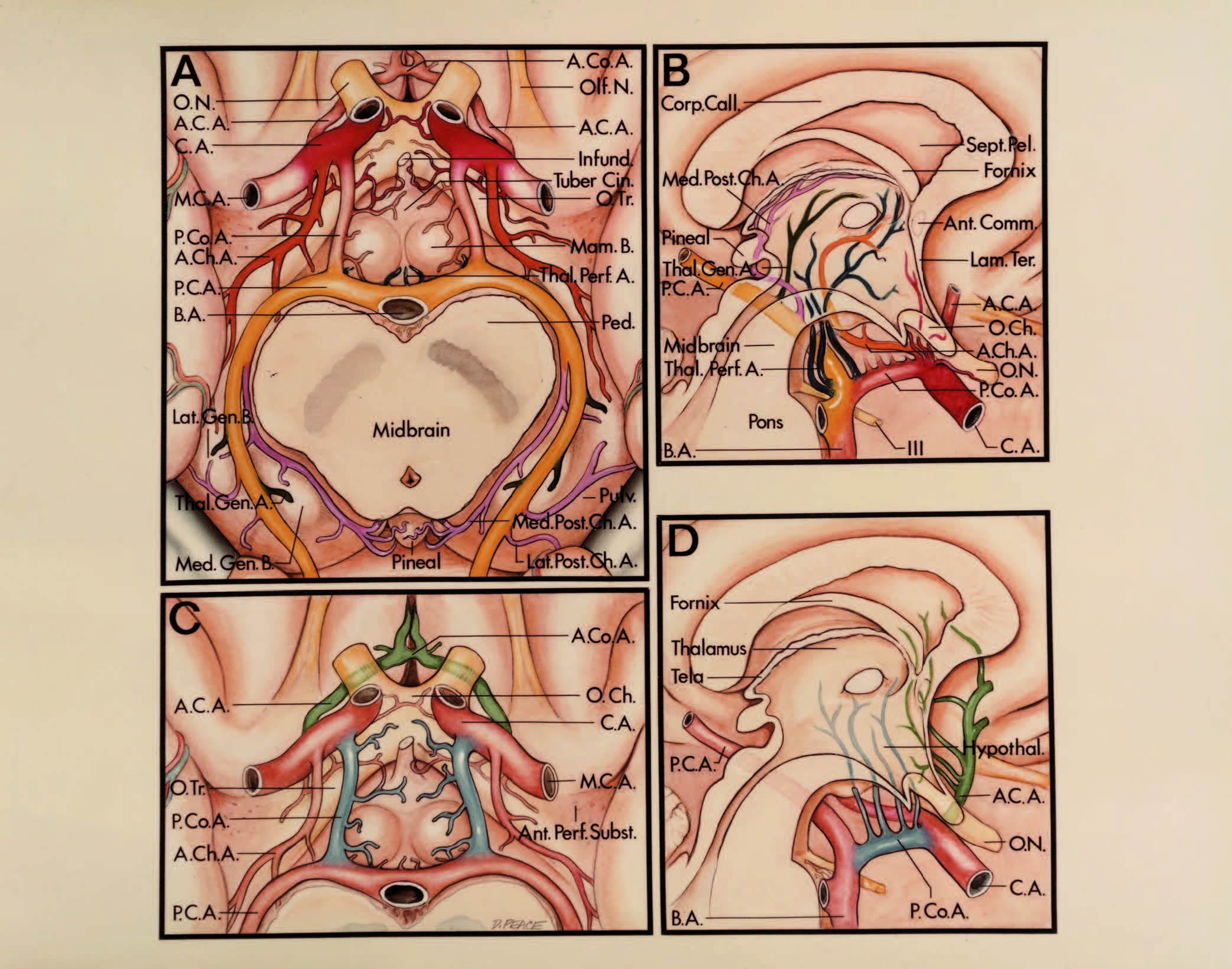
圖5.18。第三腦室的動脈關係。A和C是第三心室底部的下視圖,B和D是穿過第三心室的正中矢狀麵。A和B表示以下動脈到第三腦室的主幹和穿支的關係:頸內動脈(暗紅色)、脈絡膜前動脈(橙色)、基底動脈頂動脈(黃色)、腦後動脈(黃色)、內側脈絡膜後動脈(粉色)、外側脈絡膜後動脈(粉色)、丘腦開孔動脈(藍色)和丘腦膝狀動脈(深綠色)。C和D分別為大腦前動脈(淺綠色)、前通動脈(淺綠色)和後通動脈(藍色)到第三腦室的主幹和穿支關係。嗅覺和視神經位於第三腦室底的前方。底層的結構是視交叉、視束、漏鬥、灰質塊莖和乳狀體。中腦和腦梗低於後半地板。前穿孔物質位於視道外側。外側膝狀體和內側膝狀體附著於丘腦下緣靠近枕部,位於中腦外側。 The structures in the anterior wall of the third ventricle are the anterior commissure, lamina terminalis, and optic chiasm. The corpus callosum and septum pellucidum are above the roof of the third ventricle. The roof is formed of the two layers of tela choroidea, the fornix, and a vascular layer composed of the internal cerebral veins and the medial posterior choroidal arteries. The oculomotor nerve exits from the midbrain. A., artery; A.C.A., anterior cerebral artery; A.Ch.A., anterior choroidal artery; A.Co.A., anterior communicating artery; Ant., anterior; B., body; B.A., basilar artery; C.A., carotid artery; Call., callosum; Ch., chiasm, choroidal; Cin., cinereum; Comm., commissure; Corp., corpus; Gen., geniculate; Hypothal., hypothalamus; Lam., lamina; Lat., lateral; Mam., mamillary; M.C.A., middle cerebral artery; Med., medial; N., nerve; O., optic; Olf., olfactory; P.C.A., posterior cerebral artery; P.Co.A., posterior communicating artery; Ped., peduncle; Pell., pellucidum; Perf., perforated; Post., posterior; Pulv., pulvinar; Sept., septum; Subst., substance; Term., terminalis; Thal.Gen., thalamogeniculate; Thal.Perf., thalamoperforating; Tr., tract.
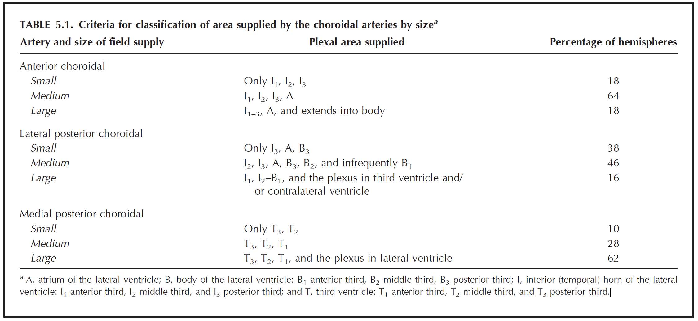
與側腦室和脈絡膜裂隙最密切相關的動脈是在側腦室和第三腦室供應脈絡膜叢的脈絡膜動脈。它們起源於基底池的頸內動脈和腦後動脈,通過脈絡膜裂隙到達脈絡膜叢(圖2.9、2.10、2.33和5.19;表5.1 - -5.3)。
側腦室的脈絡膜叢由前、後脈絡膜動脈供血(7,26)。脈絡膜後動脈分為外側和內側兩組稱為脈絡膜後外側動脈和內側動脈。在第二章中回顧了與這些動脈相關的插圖和文本。每條脈絡膜動脈沿著它的路線向神經結構分支。最常見的模式是脈絡膜前動脈供應部分脈絡膜叢在顳角和心房;脈絡膜後外側動脈供應部分脈絡膜叢在心房,身體和顳角的後部;還有內側後脈絡膜動脈供給第三腦室頂部的脈絡膜叢和側腦室的一部分。由前、後脈絡膜動脈供血的叢區大小成反比:隨著一條動脈供血麵積的增大,另一條動脈供血麵積的減小。脈絡膜後外側動脈和內側後動脈供血區域之間也有同樣的反向關係。一側的脈絡膜外側動脈和內側後動脈很少有分支到對麵側腦室的脈絡膜叢。
脈絡膜前動脈起於切膜前間隙的頸內動脈,向後延伸至切膜中間隙,在切口中間隙處通過臨近下脈絡膜點的脈絡膜裂隙,沿脈絡膜叢內側邊界向外延伸,與後脈絡膜外側動脈緊密相連。它沿著神經叢的後方和背側穿過,在幾個半球到達Monro孔。脈絡膜叢表麵的前、外側後脈絡膜動脈分支之間經常有吻合。
脈絡膜後外側動脈是由大腦後動脈或其皮層分支在周圍池和四頭池中形成的一組動脈。這些分支在脈絡膜前動脈分支後麵進入心室。它們在枕骨周圍橫向穿過脈絡膜裂,在穹窿的毛膜、小腿和體的水平到達顳角、心房和體的脈絡膜叢。如果脈絡膜前動脈供給顳角和心房的脈絡膜叢,則脈絡膜後外側動脈會沿脈絡膜裂的顳部和心房部的內側沿腦室外,穿過脈絡膜裂的體部到達脈絡膜叢。外側後脈絡膜動脈可從一側側腦室體通過Monro孔發出分支,或在穹窿和丘腦之間發出分支到第三腦室的脈絡膜叢,或通過Monro孔到達對側側腦室體的脈絡膜叢。這些分支在心室體和Monro孔處與內側後脈絡膜動脈的分支混合。
內側後脈絡膜動脈最常出現為一至三個分支,來自於腦後動脈近端在踝間和腳池的後內側。這些分支環繞大腦後動脈主幹內側的中腦,在鬆果體一側向前轉進入第三腦室頂,並在丘腦之間的膜間質中穿過,與大腦內靜脈和相對的內側後脈絡膜動脈相鄰。少數內側後脈絡膜動脈可能起源於大腦後動脈或其皮層分支的遠端,並從其起點向前或逆行延伸至第三腦室頂。內側後脈絡膜動脈供應第三腦室頂的脈絡膜叢,有時通過同側Monro孔或脈絡膜裂供應側腦室的脈絡膜叢。偶有分支通過對側Monro孔和脈絡膜裂供應對側側腦室的脈絡膜叢。它們可能沿其路徑發送微小的分支到腦梗,膝狀體,被蓋,丘,枕葉,鬆果體,後連合,下丘腦,丘腦髓紋,枕葉皮層和丘腦。
圖5.19。A.上視圖:上視圖,部分大腦半球、胼胝體和穹窿被切除,顯示側腦室、第三腦室和脈絡叢之間的關係。左半球顯示心室和脈絡膜叢的關係。側腦室脈絡膜叢從顳角延伸到心房和側腦室體。它不向後延伸到後角或向前延伸到前(額)角,但通過Monro孔,並在第三腦室頂部繼續向後延伸到鬆果體上方的鬆果體上隱窩。右半球顯示脈絡膜動脈和脈絡膜叢之間的關係。脈絡膜前動脈起源於頸動脈供應顳角叢和心房。脈絡膜後外側動脈起源於大腦後動脈或其分支,供應顳角、心房和側腦室體後部的神經叢。內側後脈絡膜動脈起源於大腦後動脈並供應第三叢,在很多情況下,還供應側腦室體。左下:脈絡叢的分類。 The portion of the choroid plexus within the temporal horn and body of the lateral ventricle and the third ventricle is subdivided into an anterior, middle, and posterior third. The subdivisions within the lateral and third ventricles are designated as follows: inferior (temporal) horn of the lateral ventricle—anterior third I1, middle third I2, and posterior third I3; atrium of the lateral ventricle—A; body—anterior third B1, middle third B2, and posterior third B3; and third ventricle—anterior third T1, middle third T2, and posterior third T3. The criteria used to divide the area of supply of each artery into small, medium, and large groups are listed in Table 5.1. Lower right: Schematic illustration of the choroid plexus showing the most common pattern of supply (22% of hemispheres). The anterior choroidal artery is shown in red, the lateral posterior choroidal artery in blue, the medial posterior choroidal artery in yellow, and the contralateral lateral posterior choroidal artery in green. The area of the field of supply of the choroidal arteries is as follows: anterior choroidal artery, medium; lateral posterior choroidal artery, small; and medial posterior choroidal artery, large. The medial posterior choroidal arteries are shown together in both hemispheres. B, schematic illustration of the choroid plexus showing size of the area supplied by the choroidal arteries. The criteria used to divide the area of supply of each artery into small, medium, and large groups are listed in Table 5.1. The second to seventh most common patterns are listed in Table 5.2.
表5.1
表5.2
頸內動脈沿前臥突內側表麵出海綿竇,在額角以下分岔(圖5.15-5.18)(9,29)。從眼段和通信段發出的分支經過視神經、交叉、道和第三腦室底,但從脈膜段發出的分支向上通過前穿孔物質供應側腦室和第三腦室壁內或附近的結構,包括內囊的膝和後肢、蒼白球的鄰近部分和丘腦。頸內動脈也由下垂體上動脈發出,下垂體上動脈在第三腦室底下方向內延伸,到達腫瘤塊莖並與對麵的腫瘤塊莖結合形成一個環繞漏鬥的血管環。
後交通動脈起於頸內動脈後壁,位於前切口空間額角下方,並在視束後內側下方和第三腦室底部與大腦後動脈相連(圖2.8、5.17和5.18)。它的分支穿過第三腦室底部,位於視交叉和頸動脈梗之間,到達下丘腦、丘腦、下丘腦和側腦室體下方的內囊。
大腦前動脈在端板和第三腦室前壁的前方上升,到達額角底部以下區域(圖5.11、5.15、5.17和5.18)(20,21)。然後,它穿過腦頂下方,繞著胼胝體膝,靠近腦底、前壁、額角頂和側腦室體頂。額角周圍曲線的緊度是側腦室大小的一個很好的指標。大腦前動脈的遠端部分不僅可能暴露在胼胝體上方,也可能暴露在胼胝體下方,因為胼胝體周圍動脈的末梢分支可能繞過脾髒,在第三腦室頂向前延伸,最遠可達Monro孔。胼胝體周圍分支穿過胼胝體到達透明隔和額角和身體內側壁的穹窿。
大腦前動脈和前交通動脈形成穿孔分支,終止於第三腦室的整個前壁,並到達下丘腦、穹窿、透明隔和紋狀體的鄰近部分。胼胝體前動脈可能起源於大腦前動脈或前交通動脈,向上穿過終板,並將分支送入前壁。
大腦前動脈的返支常在第三腦室前部和額角以下入路遇到。它和前交通動脈近端的大腦前動脈段向額角和身體外側壁附近的區域發送分支。這些分支供應部分膝和內囊、蒼白球的前肢,以及較少見的丘腦。
大腦中動脈位於額角下方(圖5.17和5.18)(8)。大腦中動脈的貫通分支供應額角外側區域和側腦室體的結構,稱為紋狀體動脈。它們進入側腦室額角和體側的深層結構,包括慢狀核、內囊的整個前後長度以及尾狀核的體和頭。
基底動脈與大腦後動脈的分叉位於第三腦室底後半部分下方和側腦室體下方(圖5.13、5.17和5.18)(30,38)。一個高的基底分岔可能使地板凹陷。它的分支到達側腦室的顳角、心房和體壁,以及第三腦室的底、頂和後側壁。
丘腦膝狀動脈和丘腦操作動脈是大腦後動脈的兩個較大的穿支。丘腦工作動脈通過後穿孔物質進入大腦,供應第三腦室底和側壁的結構,包括側腦室體底以下區域的丘腦前三分之二。它們也向腦梗、下丘腦、中腦和內囊發送分支。丘腦膝狀動脈起源於周圍環境,在膝狀體區域進入大腦,並向丘腦後外側部分發送分支,包括膝狀體和內囊的鄰近部分。
該動脈起源於基底動脈,在大腦後動脈下方環繞中腦,並穿過四頭神經池到達小腦上表麵(10)。在通向第三腦室後部的幕上和幕下手術入路中,四頭池的動脈段暴露出來,幕下入路暴露出其皮層分支。腦後動脈和小腦上動脈的穿支供應腦池壁。腦後動脈支配著上丘和下丘之間的溝以上的結構,小腦上動脈支配著這一層次以下的結構。
腦深部靜脈係統與側腦室、第三腦室和基底池的壁密切相關。在第4章的“深靜脈”一節中提供了與這些靜脈相關的插圖和更廣泛的文本(圖4.16、4.17和5.20)。這些靜脈對從側腦室到第三腦室的手術入路以及後壁、心房、鬆果體區和四頭池區域構成了巨大的障礙,在這些區域內腦內靜脈和羅森塔爾基底靜脈分別與蓋倫大靜脈彙合。
大腦的深靜脈係統彙集成通道,在室管膜下位置通過側腦室壁和第三腦室壁,並彙聚到腦內靜脈、基底靜脈和大靜脈(圖5.3、5.6、5.11、5.12和5.20)(14,17,19)。來自額角、側腦室體和周圍灰質和白質的靜脈流入腦內靜脈;顳角和鄰近心室周圍結構的靜脈流入基底靜脈;而引流心房和鄰近腦區的則流入基底靜脈,腦內靜脈或大靜脈。從室周白質和灰質收集血液的靜脈在側腦室壁彙合形成室管膜下通道。
在側腦室手術中,靜脈比動脈更常見地提供定位標誌,因為心室壁的動脈較小,不容易看到,但靜脈較大,很容易通過室管膜看到。當神經結構之間的正常角度消失時,這些靜脈標記對腦積水尤其有幫助。在腦血管造影中,這些靜脈可能比動脈更準確地估計病變的位置和大小,因為它們比動脈更緊密地附著在腦室管膜和腦膜表麵。
心室靜脈起源於引流基底神經節、丘腦、內被膜、胼胝體、透明隔、穹窿和深白質的支脈,沿著心室壁在室管膜下的位置向脈膜裂隙延伸。根據室性靜脈是否經過脈膜裂的丘腦側或穹窿側,將室性靜脈分為內側組和外側組:外側組通過脈膜裂的丘腦側或內側,內側組通過裂隙的外周或穹窿周。
外側組引流額角,顳角和枕角的外側壁,身體,心房,身體的底部,心房的前壁,和顳角的頂部。內側組引流額角和枕角的內側壁和頂部,身體,心房和顳角的底部。由內側和外側組組成的靜脈經常在脈絡膜裂附近彙合,形成一個共同的莖,然後終止於腹膜間質和基底池中的大靜脈。
額角的內側靜脈群由前間隔靜脈組成,外側靜脈群由前尾狀靜脈組成。體內的內側靜脈組由後間隔靜脈組成,外側靜脈組由丘腦紋靜脈、丘腦尾狀靜脈和尾狀後靜脈組成。心房和枕角內靜脈組由心房內靜脈組成,外側靜脈組由心房外靜脈組成。內側靜脈組位於顳角的底部,外側靜脈組位於顳角的頂部。頂部和側壁主要由下心室靜脈排出,底部由海馬橫靜脈排出。

點擊這裏查看此圖像的交互模塊和相關內容。
圖5.20。側腦室的靜脈關係。側視圖(上),前視圖(中),上視圖(下)。心室靜脈分為內側靜脈組和外側靜脈組。心室靜脈流入內腦靜脈、基底靜脈和大靜脈。外側組包括額角的尾狀前靜脈;身體內的丘腦紋靜脈,尾狀後靜脈和丘腦尾狀靜脈;心房和枕角的側心房靜脈;還有顳角的心室下靜脈和杏仁靜脈。內側組由額角的前隔靜脈構成; the posterior septal veins in the body; the medial atrial veins in the atrium; and the transverse hippocampal veins in the temporal horn. The transverse hippocampal veins drain into the anterior and posterior longitudinal hippocampal veins. The superior choroidal veins drain into the thalamostriate and internal cerebral veins, and the inferior choroidal vein drains into the inferior ventricular vein. The great vein drains into the straight sinus. Amygd., amygdalar; Ant., anterior; Atr., atrial; Caud., caudate; Cer., cerebral; Chor., choroidal; Front., frontal; Hippo., hippocampal; Inf., inferior; Int., internal; Lat., lateral; Long., longitudinal; Med., medial; Occip., occipital; Post., posterior; Sept., septal; Str., straight; Sup., superior; Temp., temporal; Thal.Caud., thalamocaudate; Thal.Str., thalamostriate; Trans., transverse; V., vein; Vent., ventricular, ventricle.
脈絡膜上靜脈和下靜脈是脈絡膜叢上最大的靜脈(圖5.3、圖5.6和圖5.12)(19)。脈絡膜上靜脈是最大的脈絡膜靜脈,在側腦室體的脈絡膜叢上向前延伸,終止於丘腦紋靜脈或腦內靜脈或其支流的Monro孔附近。脈絡膜下靜脈流經顳角和心房的脈絡膜叢。
在顱腔中,心房內側的四叉池內的靜脈關係最為複雜,因為腦內靜脈、基底靜脈和大靜脈及其許多支流都彙聚於此區域(圖5.3、5.6、5.11、5.12和5.20)。腦內靜脈從腹膜間質流出,基底靜脈從周圍池流出,到達四叉池,並在那裏連接形成蓋倫靜脈。
腦內靜脈起源於Monro孔處的多條支靜脈,後延於第三腦室頂,位於脈絡膜兩層之間的丘腦髓紋上方。它的前部與中線相鄰,從另一側靠近它的配偶。它沿鬆果體上外側表麵從中線發散。再往後,在胼胝體脾的下方,它在中線彙合,並從另一側與它的配偶結合形成蓋倫靜脈。
基底靜脈起源於前穿孔物質的表麵,由多條靜脈結合而成,穿過腳池和周圍池。它在鉤椎後正中上方延伸至腦梗的前部。在足梗前基靜脈的最內側點,向後外側轉彎到達腦梗的最外側點,然後向後內側轉彎繞過枕骨下、後側,與蓋倫靜脈或四頭池內的腦內靜脈彙合。
側腦室和第三腦室是大腦中手術最難到達的區域。自從Dandy的開創性工作(圖5.21和5.22)(3-5)以來,已經描述了許多進入心室的手術入路。到達側腦室和第三腦室的路徑是(a)從上方通過胼胝體或大腦皮層;(b)從前,通過前半球間裂、胼胝體和終板;(c)從下方,通過基底池、鞍上區,或通過顳葉或在顳葉下方;(d)從後側,穿過半腦間裂、四叉腦池、胼胝體和大腦皮層。最佳手術入路的選擇取決於病變與側腦室和第三腦室的關係、腦室的大小和所涉及的結構,包括門羅孔、sylvius渡槽、視神經和交叉、鬆果體、蝶鞍、垂體腺、穹窿、中腦、丘腦、胼胝體、半球間裂和基底池。在考慮具體的手術方法之前,先回顧一些一般原則。這些原則適用於本問題中討論的所有操作方法神經外科.

點擊這裏查看此圖像的交互模塊和相關內容。
圖5.21。側腦室的外科入路。每個入路的皮膚切口(實線)和骨瓣(虛線)的位置。側腦室前部可經胼胝體前入路、皮質前入路和額路入路到達。通向側腦室的後路有胼胝體後路、皮質後路和枕側後路。側腦室下部采用額顳部和顳部入路。螞蟻。前;職位。,後。

點擊這裏查看此圖像的交互模塊和相關內容。
圖5.22。頭部正中矢狀麵顯示手術進入第三腦室。沿著中線或靠近中線的入路用實線表示,遠離中線的入路用虛線表示。進入第三腦室前下方的中線或近中線入路是經蝶竇和額下入路。額下手術路徑分為四種不同入路:(a)經終板入路;(b)經頸動脈三角的頸動脈入路;(c)視神經間視交叉下方的視交叉下入路;(d)經蝶平麵和蝶竇的額-蝶入路。離中線的第三腦室底和前下部分的入路是顳下和額顳。進入第三腦室前上段Monro孔區域的入路有前胼胝體入路和前皮質入路。 The supratentorial approaches to the posterior part of the third ventricle are the posterior transcallosal, posterior transcortical, and occipital transtentorial. The infratentorial supracerebellar approach is directed below the tentorium cerebelli to the posterior part of the third ventricle.
開顱皮瓣的放置應盡量減少腦後縮的需要。用於到達側腦室壁和第三腦室壁的收縮部位包括額葉的眶麵,以到達交叉區;額葉和頂葉旁矢狀皮質經胼胝體入路;額葉下表麵和內側表麵用於額葉前入路;額葉的下表麵和顳葉的前部和下部用於額顳入路;顳葉下表麵用於顳下入路;枕骨葉的下表麵和內側用於枕骨入路;小腦上表麵用於幕下入路。為了盡量減少大腦回縮的需要,外科醫生應按以下方式進行開顱手術。對於矢狀旁入路,皮瓣應延伸至或越過中線。 For the occipital approach, the flap should reach the margins of the sagittal and transverse sinuses and the torcular herophili. For the anterior frontal approach, the flap should have its medial margin on the midline and, if needed, its anterior margin on the floor of the anterior fossa. For the subfrontal, subtemporal, and frontotemporal approaches, the flap should have its lower border on the floor of the anterior and / or middle fossa. For the posterior frontotemporal approach, the flap should be based on the floor of the frontal and temporal fossae and the lateral half of the sphenoid ridge should be removed. For the infratentorial approaches the opening should reach the margin of the transverse sinus and torcular herophili.
使用自持式牽開器,而不是手持式牽開器。如果存在腦積水,可通過腦室造口引流腦脊液,如果腦積水不明顯且暴露時可通過基底池引流腦脊液,如果沒有腦室梗阻,可通過腰椎引流,從而增加腦外空間,並進一步減少後撤的需要。
不打開某些神經結構是不可能到達側腦室和第三腦室的。側腦室和第三腦室的外科入路可能需要在額葉、頂葉或顳葉和胼胝體前後切開皮質切口,移位或切開穹窿,打開末梢板、脈膜裂、透明隔、第三腦室底,並將腫瘤從四頭肌板、視神經、交叉和束、垂體及其柄中分離出來,還有腦梗。大腦可能會被縮回以暴露第三腦室或側腦室的外壁,如胼胝體或終板,但隨後必須切開該壁才能到達腦室。到達側腦室後,需要打開脈絡膜裂或通過穹窿等部位的另一個神經切口,以暴露延伸到第三腦室或基底池的病變。通過腦室體的脈膜裂隙打開會暴露出膜間質和第三腦室的頂部,通過心房裂隙打開會暴露出四頭池和鬆果體區,在顳角打開它會暴露周圍的腦池。在打開脈絡膜裂隙時,通過絨毛膜肌腱層打開比通過絨毛膜肌腱層打開更好,因為通過絨毛膜肌腱層的動脈和靜脈較少(圖5.3、5.6 - 5.9和5.23)。
神經結構的切口和收縮到達側腦室和第三腦室,如嗅覺和動眼神經,視神經通路和四頭肌板,導致明確的缺損和對應的損傷區域。其他神經結構的犧牲產生了不同的結果:在一些情況下沒有缺損,在另一些情況下缺損是短暫的或永久性的,或導致生命的損失。犧牲的結構有不同的結果,包括胼胝體的前後部分和穹窿的不同部分。胼胝體切口導致了半球間信息轉移、視覺空間轉移、雙運動任務學習和記憶障礙,也導致了失讀症、失用症和對視識別等缺陷(15,24,28,33)。兩側穹窿分裂可能導致失憶。前、後經胼胝體入路和經心室手術入路的皮質切口需要大腦收縮,這導致了抽搐、偏癱、沉默、意識障礙和視野喪失。對延伸到第三腦室壁的病變的操作可能導致下丘腦功能障礙,表現為溫度控製、呼吸、意識和垂體分泌障礙;視交叉和視束損傷導致的視力喪失;以及由於身體和穹窿柱的損傷而導致的失憶。心房內側四邊形板區域的解剖可引起眼動障礙、瞳孔導水管水腫性關閉、丘或膝狀體水腫引起失明,以及由於神經核或腦幹中樞通路水腫引起眼外麻痹(5)。
打開脈絡膜裂隙有損傷穹窿的風險。然而,對穹窿的單側損傷不會產生缺陷,而對兩個半球穹窿纖維的損傷通常不會導致永久性記憶喪失(15,24,28,33)。打開脈絡膜裂的顳部有損傷毛膜和海馬形成的風險。大量的實驗和臨床證據表明,大量的雙側海馬結構損傷,會導致近期記憶的損傷。然而,海馬結構的單側損傷不會產生缺陷。位於脈絡膜裂顳部的終紋是杏仁核複合體到終紋核最顯著的傳出通路。然而,沒有證據表明終末紋或杏仁核的單側病變會引起情緒障礙。雙側杏仁核複合體的病變可使情緒興奮性降低。

點擊這裏查看此圖像的交互模塊和相關內容。
圖5.23。模擬。經脈絡膜入路,沿脈絡膜裂隙的穹窿側進入第三腦室。A,額角和側腦室體的上方視圖。穹窿體形成了第三腦室頂的上部。左側丘腦紋靜脈穿過Monro孔後緣右側丘腦紋靜脈穿過脈絡膜裂在孔後幾毫米處。前間隔靜脈和前尾狀靜脈穿過額角壁。後間隔靜脈和後尾狀靜脈穿過側腦室體壁。丘腦位於身體的底部。脈絡膜裂位於丘腦和穹窿之間,通過將將脈絡膜叢連接到穹窿外側邊緣的穹窿肌腱分隔開,使脈絡膜叢與丘腦的連接不受幹擾。 B, enlarged view. The columns of the fornix form the anterior and superior wall of the foramen of Monro. The massa intermedia is seen through the foramen. Anterior and posterior septal veins cross the septum pellucidum and fornix. C, the tenia fornix, which attaches the choroid plexus to the fornix, has been divided and the body of the fornix retracted medially to expose the internal cerebral vein and medial posterior choroidal arteries. The lower layer of tela, which attaches to the striae medullaris thalami and forms the floor of the velum interpositum, is intact. D, the separation of the fornix and choroid plexus has been extended posteriorly to the junction of the atrium and body of the ventricle. The lower layer of tela remains intact.

點擊這裏查看此圖像的交互模塊和相關內容。
圖5.23。情況。E,打開心室下層,露出中間塊、後連合和第三腦室底。覆蓋前間隔靜脈的室管膜已被打開,以便一小段靜脈可以活動。通過讓脈絡膜叢保持附著在丘腦和靜脈上表麵,減少了損傷丘腦紋靜脈的可能性。F-H, interforniceal方法。F,穹窿間入路通過在中線縱向切開穹窿完成。穹窿體的每半部分已向外側縮回,露出大腦內靜脈、內側後脈絡膜動脈和與丘腦髓紋相連的脈絡膜層。G,打開心室暴露第三腦室底和中間塊。H,視野已經指向後麵的導水管以及後和韁骨相通處。 The pineal recess extends into the base of the pineal between the habenular and posterior commissures. The pineal gland extends backward from the pineal recess. Ant., anterior; Caud., caudate; Cer., cerebral; Chor., choroid; Col., column; Comm., commissure; Fiss., fissure; For., foramen; Hab., habenular; Int., intermedia, internal; M.P.Ch.A., medial posterior choroidal artery; Nucl., nucleus; Plex., plexus; Post., posterior; Rec., recess; Sept., septal; Thal.Str., thalamostriate; V., vein; Vent., ventricle.
腦室內腫瘤和動靜脈畸形通常由脈絡膜動脈供血(圖5.19;表5.1 - -5.3)。脈絡膜動脈彙聚並穿過脈絡膜裂的事實有助於識別這個位於丘腦外圍的裂隙,通過它手術程序可以定向到第三腦室、鬆果體區、周圍池和四頭池。通過裂隙打開將暴露心室病變近端的動脈;從腦室體的裂隙打開,暴露出腹膜間質和第三腦室頂的內側後脈絡膜動脈;通過心房的裂隙打開會暴露四頭池和鬆果體區的內外側脈絡膜後動脈;在顳角的開口會暴露周圍池的前,內側和後外側脈絡膜動脈。
在切除側腦室和第三腦室腫瘤時也可能暴露的其他動脈是位於第三腦室前壁區域的前腦動脈和前交通動脈以及側腦室的額角和體;威利斯圓的後部,基底動脈的頂端,大腦後動脈的近端在第三心室底以下和顳角內側;腦後動脈的遠端,位於第三腦室後區和心房內側;大腦後動脈、胼胝體周動脈、小腦上動脈和毗鄰第三腦室後壁和心房內側的脈絡膜動脈;還有大腦前動脈和後動脈它們的分支進入側腦室的頂部。此外,頸內動脈、大腦前、中、後動脈以及前、後交通動脈形成了連通側腦室壁和第三腦室壁的穿支。隻有很少的情況下才應該犧牲其中的任何一個。阻斷這些動脈在威利斯環前段的穿支容易導致記憶和人格障礙,阻斷威利斯環後段的穿支更容易導致意識水平障礙,並常合並眼外運動障礙。在後交通動脈在顳下入路的穿支犧牲導致基底神經節梗死(32)。顳角內側池內丘腦操作動脈閉塞可引起昏迷和死亡。 Injuries to the superior cerebellar artery in approaches to the posterior part of the third ventricle may cause a cerebellar deficit.
表5.3
心室靜脈為在心室手術中引導外科醫生到達Monro孔和脈絡膜裂提供了有價值的標誌(圖5.3、5.6、5.11、5.12、5.20和5.23)。腦室腫瘤常發生腦積水,腦室壁神經結構之間的邊界隨著腦室擴張而變得不那麼明顯,如果腦積水則尤其如此。丘腦紋靜脈有助於劃分尾狀核和丘腦的交界處,因為它通常沿著分隔這些結構的溝走。
在接近心室病變時犧牲的靜脈數量應該保持在最小,因為失去它們會產生不良後果。深靜脈,包括腦大靜脈、基底靜脈和腦內靜脈及其支流,以及從大腦到硬腦竇的橋靜脈,在到達和切除腦室內或腦室附近的一些腫瘤時是不可避免的。在犧牲這些靜脈之前,外科醫生應該試著將它們置於中等甚至嚴重的拉伸下(接受它們可能被撕裂的事實),如果這樣做能讓人滿意的暴露,並產生保留靜脈的可能性。在犧牲基底靜脈、腦內靜脈和大靜脈之前,外科醫生應該嚐試繞過它們,或將它們移出手術路線,或嚐試隻分割它們的幾個小分支,這樣可以將主幹移出手術野。
淺靜脈係統和深靜脈係統的犧牲分支造成了不穩定的缺損。Dandy(5)注意到,在很多情況下,一條腦內靜脈被切除而沒有效果,在少數情況下,兩條靜脈甚至Galen大靜脈都被結紮恢複,而沒有任何明顯的功能障礙。另一方麵,該靜脈網絡的損傷可能導致間腦水腫、精神症狀、昏迷、高熱、心動過速、呼吸急促、瞳孔縮小、四肢僵硬和深肌腱反射過度(15,24,28,33)。丘腦和門羅孔處的其他靜脈阻塞可引起嗜睡、偏癱、沉默和基底神經節出血性梗塞。經胼胝體入路時,大腦與上矢狀竇前或後靜脈之間的靜脈閉塞,雖然通常不會造成缺損,但可伴有偏癱。犧牲枕內靜脈或從枕極到上矢狀竇或橫竇的橋靜脈可導致偏盲。小腦與幕間橋靜脈橫斷後小腦腫脹已有報道(15,24,28,33)。
在將包膜腦室腫瘤的包膜與鄰近組織分離之前,應先將包膜內的組織取出。如果腫瘤可能是囊性的,第一步是用針穿刺。如果腫瘤被包膜,則打開包膜,對腫瘤進行活檢,並完成囊內切除。在囊的內容物被取出後,囊從神經和血管結構中分離出來。腫瘤緊密粘附的最常見原因不是包膜和周圍結構之間的粘連;確切地說,它是囊內的殘餘腫瘤將腫瘤楔入原位。當囊內內容物被移除時,腫瘤就會塌陷,因此可以通過小範圍的暴露移除更多的腫瘤。腫瘤通常不會如此緊密地附著,以至於在囊內內容物被移除後,它們不容易被移除。如果在囊內內容物被移除後,腫瘤還不能輕易與神經組織分離,短暫的等待通常會讓大腦的搏動將腫瘤移到暴露的地方,然後就可以經常從囊內移除更多的腫瘤。在放大鏡下,可以用微型儀器將重要結構和腫瘤之間的個別粘連分開。 This technique has been especially helpful in removing適應證.通常可以切除顱咽管瘤和累及第三腦室的表皮樣腫瘤的包膜,但不能切除厭色腺瘤的包膜。憎色腺瘤的包膜是顱底硬腦膜,它向上延伸到腫瘤之上。在厭色腺瘤穹窿上拉伸的硬腦膜可被切除,但試圖將硬腦膜假包膜從附著於顱底的位置拉出可能導致嚴重的血管和神經損傷。如果腫瘤囊緊緊地附著在諸如視神經或視交叉、丘、丘腦或下丘腦等重要結構上,可能會留下腫瘤囊的殘餘。顱咽管瘤、恐色腺瘤、鬆果體瘤和一些膠質瘤對放射治療的反應足夠好,可以依賴放射治療來處理殘餘腫瘤。如果腫瘤是惡性或浸潤性的,切除通常僅限於活檢或內部減壓。
一個膠體囊腫首先用針從門羅孔吸入。通常可以通過Monro孔進行整個手術,特別是當Monro孔擴大時。然後用微吸法取出囊腫內的粘液,也許還需要用鑷子取出。對於非常大的膠質囊腫,通過脈絡膜裂入路比切開穹窿更可取。穿過腫瘤囊到神經組織的動脈應該被保留。任何位於包膜表麵以上的血管在開始時都應該像處理大腦供血的血管一樣處理。當腫瘤從囊內取出後,應嚐試使用小解剖器將血管移離腫瘤囊。
如果在手術結束時,腦脊液在Monro孔、sylvius渡槽、第三腦室或幕切肌的流動仍然受阻,則可能需要進行分流。如果最初的手術創造了一個從第三腦室穿過終板、第三腦室底或鬆果體區進入蛛網膜下腔的開口,這可能就足夠了。在乳狀體前麵的第三腦室底可以用內窺鏡技術打開。如果使用枕下暴露術來接近鬆果體區的腫瘤,可以從側腦室或從第三腦室後部的開口通向大池,從而形成Torkildsen分流。
側腦室手術入路分為前入路、後入路和下入路(圖5.21)。前入路通向額角、側腦室體和第三腦室前部。後入路通向心房和後第三腦室,下入路通向顳角和基底池。前入路包括胼胝體前路、皮質前路和額葉前路。後入路包括胼胝體後路、腦室後路、枕部幕前路和幕下小腦上路。外側入路包括翼點、額顳後部和顳下。經蝶竇和額下入路也可用於涉及第三腦室前壁和底的選定病變;然而,這些入路最常用於涉及蝶鞍的病變,將在第8章進行回顧。
這種經胼胝體的入路適用於到達位於側腦室額角、體和第三腦室前上段的病變(圖5.24)。經胼胝體前入路的另一選擇是經皮質前入路,經額中回。通過這個入路比通過前經皮質入路更容易將Monro孔的開口暴露到兩側側腦室。如果心室大小正常或最小擴大,經胼胝體入路比經心室入路更容易。
患者仰臥位,矢狀麵縫合在垂直平麵上,頭部抬高20 ~ 30度。另一種體位是右側朝下的側臥位,這樣重力會幫助右腦半球的內側表麵遠離鐮的右側。采用右額部馬蹄形、雙冠狀或s形皮膚切口。右額骨瓣延伸至矢狀竇外側緣或穿過矢狀竇,位於冠狀縫線前三分之二,後三分之一。硬腦膜與矢狀竇基部一起打開。冠狀縫線前的區域通常相對缺乏進入上矢狀竇的橋靜脈;有些(通常不多於一個)可能需要被分割,以允許右額葉內側表麵從鐮處回縮(圖5.3)。在鐮遊離邊緣處的蛛網膜被打開,露出胼胝體和大腦前動脈。來自胼胝體和額葉鄰近部分的流向下矢狀竇前端的小靜脈可能必須被切除。打開鐮下方的蛛網膜,可暴露大腦前動脈的分支,它們可穿過胼胝體上方的中線。 The right and left cingulate gyri, which face each other, are separated to expose the corpus callosum and the pericallosal arteries. The approach is best directed between the pericallosal arteries, although some of their branches may cross the midline above the corpus callosum. If both pericallosal arteries are retracted to one side, it may be necessary to divide some of the branches that run laterally from the pericallosal arteries to the corpus callosum and the cingulate gyrus to reach the corpus callosum.
胼胝體在Monro孔上方的部分在中線裂開。長度為2cm的切口可滿足進入兩側側腦室的要求。圖像引導有助於選擇打開胼胝體的位置。經胼胝體或經腦室入路打開心室後,沿著脈絡膜叢和丘腦靜脈向前延伸,直至它們在腦室孔處彙合,即可發現Monro孔。脈絡膜叢沿著穹窿和丘腦之間的脈絡膜裂隙相連,而丘腦紋靜脈在丘腦和尾狀核之間的凹槽中位於更側向的位置。額角的靜脈後向Monro孔引流,因為它們通過脈絡膜裂隙到達腦內靜脈,但並沒有延伸到額角。
收回側腦室壁時,應注意內囊膝與Monro孔的密切關係(圖5.2和5.3)。內囊膝在靠近丘腦前極的Monro孔外側接觸腦室壁。收回這一區域時應謹慎操作,因為薄的腦鏟很容易切入這一關鍵區域的心室壁,導致偏癱。
胼胝體開口可能暴露額角和身體在顱骨暴露的同側或相反側,但解剖學表明這一點很明顯。判斷左側腦室或右側腦室是否暴露的一個簡單規則是判斷丘腦靜脈是在脈絡膜叢的左側還是右側;如果丘腦靜脈比脈絡膜叢更靠近患者左側,則說明左側腦室已打開;如果丘腦靜脈比脈絡膜叢更靠近患者右側,則右側側腦室已進入。進入透明隔葉之間的空腔可能會引起混淆,直到外科醫生意識到沒有室內室結構存在。打開透明隔將提供通往對麵側腦室的通道和通往兩側側腦室的門羅孔的開口。
如果需要探查第三心室更深的部分,有幾種擴大Monro孔開口的方法(圖5.24B)。一種是在門羅孔的前上邊緣切開穹窿的同側柱。除了切開穹窿外,經腦膜入路是一個更好的選擇,該入路打開位於穹窿和丘腦之間的脈膜裂隙,從而使穹窿推到另一側,暴露第三腦室頂的結構(圖5.23和5.24)。裂是側腦室壁與第三腦室頂交界處最薄的部位。另一種可選方法是穹窿間入路,即穹窿主體在中線處,沿纖維方向切開,露出腹膜間質(2,13,35)。經腦膜和穹窿間入路的優點是,通過置換而不是橫切穹窿內的纖維,可進入Monro孔後的第三腦室中央部分(15,36)。這兩種入路均能較經胼胝體入路提供令人滿意的第三腦室視野。在Monro孔後緣切開丘腦紋靜脈也被認為是切開穹窿以擴大進入第三腦室的開口的另一種選擇;然而,這可能引起嗜睡、偏癱和沉默,Monro孔靜脈阻塞已引起基底神經節出血性梗死。
在經腦膜入路中,從Monro孔後緣開始沿著穹窿肌打開脈膜裂隙,並將穹窿移至另一側,第三腦室的頂部通過打開脈膜背層進入(圖5.23和5.24)。從Monro孔的後緣開始打開1厘米的脈膜裂隙,通常可以看到心室底回到導水管的景象。經耳膜入路經穹窿肌腱比經丘腦肌腱更安全,因為大靜脈,如丘腦紋靜脈,排出內囊和半球中央部分和脈絡膜動脈,通過丘腦肌腱而不是穿過穹窿肌腱。經脈絡膜入路特別適用於由脈絡膜動脈末端分支供血的病變。
第三腦室的頂部通常可以進入,而不犧牲任何大腦內靜脈的分支,如果進入之間的靜脈;然而,如果第三腦室位於靜脈與丘腦之間的腦內靜脈的外側,則通常需要犧牲一些腦內靜脈的分支(圖5.12和5.23)。內側和外側脈絡膜後動脈的小分支可能穿過腹膜間質中線,但犧牲這些細分支的風險是最小的。打開脈絡膜下部後,與第三腦室頂、中間塊和第三腦室底的脈絡膜叢相遇。
在早期的報道中,脈絡膜裂隙是通過切開丘腦腱來打開的。這種入路被稱為脈絡膜下入路,由於穿透脈絡膜肌腱,有損傷丘腦和穿過裂丘腦側的血管的風險(6,11,13)。為了與脈絡膜下入路區分,我們將經穹窿肌的入路稱為經脈絡膜或脈絡膜上入路。
打開脈絡膜裂隙有損傷穹窿的風險。然而,對穹窿的單側損傷不會產生缺陷,而對兩個半球穹窿纖維的損傷通常不會導致記憶喪失。有證據表明,小腿和海馬連合處的損傷比身體或柱狀部位的損傷對記憶的危害更大。在打開脈絡膜裂的身體部分時,有損傷丘腦背內側核的風險。該核的傳入纖維主要來自杏仁核複合體和顳葉新皮層,傳出纖維直接來自前額葉皮層。背內側核的病變產生的情緒變化類似於眼窩前額皮質消融所產生的情緒變化(15,28)。穹窿間入路,像經腦室入路一樣,也可用於暴露位於第三腦室頂以下和Monro孔後的病變。穹窿間切口沿著穹窿體的中線向後延伸。穹窿體的左右半部分分別移至同側,穹窿下方的第三腦室頂部打開。第三腦室病變的穹窿間入路具有雙側穹窿損傷的潛在風險,但使用該入路導致的記憶缺陷通常是短暫的(1,2)。

點擊這裏查看此圖像的交互模塊和相關內容。
圖5.24。得了。經胼胝體入路至側腦室和第三腦室。A-C,正常心室解剖。A,通過胼胝體前部的切口暴露出右側腦室體和額角。左上角的插圖顯示了頭部位置、頭皮切口(實線)和骨瓣(虛線)。骨瓣穿過上矢狀竇。另一種方法是使用Souttar切口,讓骨瓣隻延伸到上矢狀竇的外側邊緣。B,用於到達第三腦室病變的切口位置:(1)Monro孔可通過在Monro孔的前上緣切開穹窿同側柱來擴大;(2)經穹窿入路通過在穹窿體中線處的切口完成; and (No. 3) the transchoroidal approach is completed by opening the choroidal fissure by incising along the tenia fornicis. C, the transchoroidal approach is completed by incision along the tenia fornicis rather than the tenia choroidea, also referred to as the tenia thalami, because more veins and arteries pass through the tenia choroidea than the tenia fornicis. The internal cerebral veins course in the roof of the third ventricle.

點擊這裏查看此圖像的交互模塊和相關內容。
圖5.24。d。D和F,去除一個大膠體囊腫通過經類固醇入路。D,一個阻塞Monro孔的膠質囊腫正在用針抽吸。E,通過沿著脈絡膜叢與穹窿的連接處打開脈絡膜裂隙,暴露出阻塞Monro孔的膠質囊腫。在Monro孔後暴露大腦內靜脈和內側後脈絡膜動脈。囊腫的內容物正在用吸力吸出。F,囊腫附著在脈絡膜叢上的最後殘餘正在凝固。G,穹窿柱已被切開以擴大Monro孔,囊腫內的半膠狀物質正在用杯形鉗和吸力取出。

點擊這裏查看此圖像的交互模塊和相關內容。
圖5.24。設定h。H,透明隔已經打開,露出兩個側腦室的前角和體。穹窿柱位於門羅孔開口的前方和上方,進入兩側側腦室。穹窿體是第三腦室頂的一部分。第一,穹窿主體在中線處裂開露出第三腦室。大腦內靜脈和內側後脈絡膜動脈位於第三腦室的頂部。這種經穹窿入路適用於暴露位於Monro孔後第三腦室的病變。
該入路通過半腦間裂和胼胝體前部,適用於側腦室前部和第三腦室前上方的病變,特別是腫瘤主要位於入路一側的側腦室時(圖5.25)。經皮質入路比經胼胝體入路更難暴露側腦室前段。如果側腦室擴大,經皮質入路更容易。
患者取仰臥位,頭部輕微旋轉至額葉對麵,通過額葉接近心室。頭皮和骨瓣位於非支配半球額中回的中央部分。隻有當腫瘤向顯性腦半球側腦室大麵積擴展時,才選擇顯性腦半球。如果是通過顯性腦半球入路,要注意將皮層切口置於額下回表達語言中心的上方和前方,並置於中央前運動帶的前方。擴張的額角通過位於額中回長軸的皮質小切口到達。心室內的標記在經胼胝體入路的章節中有描述。可能有必要通過打開脈絡膜裂隙來擴大進入第三腦室的開口。經腦室開口比經皮質入路的穹窿間開口能更好地觀察到第三腦室的頂部。對於大多數病變,打開脈絡膜裂隙優於穹窿間入路或切除同側穹窿柱。打開透明隔將提供進入前角和身體的通道。
圖5.25。經皮質入路至側腦室和第三腦室。A,頭皮切口(實線)和骨瓣(虛線)位於額中回的中心。B和C,正常心室解剖。B,皮質開口暴露右側腦室。右下插圖顯示皮質切口的位置。進入右側腦室的開口暴露尾狀核、穹窿、門羅孔、丘腦和丘腦靜脈。C,第三腦室通過沿著脈絡膜叢與穹窿的連接處打開脈絡膜裂暴露出來。可見第三腦室頂的大腦內靜脈和內側後脈絡膜動脈。D,經皮質入路暴露脈絡膜叢乳頭狀瘤。 The inset on the upper right shows the site of the tumor and the position of the head for the operation. The tumor is being removed using a small cup forceps and suction. E, the last remnant of tumor is being removed from its attachment to the choroid plexus. The tumor has compressed the structures in the floor at frontal horn.
對於涉及形成額角前下壁和第三腦室鄰近部分結構的病變,很少考慮采用額前入路(圖5.15和5.26)(28)。這種入路足以暴露額角前壁底部和下部的病變,或從胼胝體端部延伸到端板後第三腦室的病變。這種入路不適合於到達額角後部靠近Monro孔區域、側腦室體底或Monro孔後第三腦室上部區域的病變。
患者仰臥位,麵部直視上方。使用Souttar頭皮切口。病變一側的單側骨瓣向上延伸至上矢狀竇邊緣。根據病變的位置,骨瓣位於眶上脊和冠狀線之間的間隙。如果病變位於額脊和端板的鄰近部分,則皮瓣的下緣將與眶上脊相鄰,但如果病變位於胼胝體膝的下部,則皮瓣的下緣將位於前額上方。硬腦膜與上矢狀竇的基底一起打開。抬高額葉眶麵可使視神經交叉前的蛛網膜和沿腦裂內側的蛛網膜打開,暴露出頸內動脈的脊上段、大腦前動脈和大腦中動脈的起始段以及前交通動脈和循環動脈。一條嗅覺神經可能需要在篩狀板上方分開,以暴露視交叉上方的區域。進一步的回縮將暴露視交叉上方的終板,可能還會暴露磨床的下部。額葉的內側表麵從鐮的前部縮回,露出胼胝體的喙部和膝區域的半球間裂的前部。 One or two small cortical veins entering the lateral margin of the superior sagittal sinus may need to be sacrificed to retract the frontal pole away from the falx. Small veins entering the anterior end of the inferior sagittal sinus may need to be obliterated and divided before the arachnoid in the depths of the interhemispheric fissure is opened. Opening the arachnoid will expose the A2 segments of the anterior cerebral arteries and their bifurcation into the pericallosal and callosomarginal arteries. The surgeon may need to push the anterior cerebral arteries toward their respective hemispheres or to one side to reach the rostrum part of the corpus callosum. Care should be taken to avoid occluding the perforating branches of the anterior communicating artery, which extend into the walls of the third ventricle to supply columns of the fornix. A vertical incision beginning in the lamina terminalis and extending upward into the rostrum of the corpus callosum will expose a lesion in the floor of the frontal horn formed by the anteroinferior part of the corpus callosum and straddling the lateral and third ventricular sides of the foramen of Monro.
圖5.26。經額側入路進入側腦室和第三腦室的前部。A,頭皮和骨瓣部位。B,右額葉已從鐮部縮回以暴露視神經,端板,胼胝體的端部,和大腦前動脈。C,正常心室解剖。打開胼胝體端板和端部,露出第三腦室和額角。穹窿的前連合和柱被保留。D,腫瘤跨越Monro孔,並延伸到額角和第三腦室。額角內的部分腫瘤正在用吸盤和鑷子切除。E,第三腦室內的腫瘤部分正在用精細解剖器切除。
適合於側腦室和第三腦室後部病變的入路為皮質後路、胼胝體後路、枕部幕前路和幕下小腦上路(圖5.21和5.22)。經皮質後入路和經胼胝體入路最適合於心房病變,但也可用於涉及心房內側壁並延伸至四頭池和後第三心室的特定病變。枕部幕後和幕下小腦上入路最適合於第三腦室後和四腦池的病變。
完全位於心房和側腦室體後部的病變最好使用後經皮質入路暴露。經胼胝體後入路或枕部半球間入路可暴露部分病變。如果病變累及胼胝體脾,並從心房頂或心房內側壁上端延伸至側腦室,可考慮經胼胝體入路。如果累及心房內側壁的病變延伸至第三腦室和四頭池內側壁,則應采用沿枕極和半球間裂的枕入路。
鬆果體腫瘤已被通過心房內側壁的後經皮質入路切除。然而,該區域非常狹窄,血管化嚴重,很難通過這條路徑接近鬆果體腫瘤。在暴露鬆果體腫瘤時,最常用的入路是枕部幕後入路和幕下小腦上入路。
該入路通過頂葉上皮質切口,暴露了心房內部和身體後部,對於完全位於心房內的病變,或起源於脈絡膜叢血管球的病變,或累及心房、後第三腦室和四邊形腦池的病變,可能是首選入路(圖5.27)。
患者取四分之三俯臥位,麵部朝向地麵,以便將待手術的頂骨區置於最上方。一個高的頂骨後瓣,位於上頂骨小葉上方,位於中央後回後方。皮層切入頂葉上小葉的長軸,位於中央後回後麵的區域,最好是在穿過小葉的溝中。這個皮質切口避開了穿過頂葉的視覺通路以及頂葉和顳葉交界處的言語區。側腦室進入身體與心房交界處上方,身體與穹窿小腿上方(圖5.3、5.6和5.8)。脈絡膜叢提供了一個定位的標誌。它形成一個邊緣,附著在丘腦的上表麵和後表麵,沿著身體的外側邊緣和穹窿的小腿。在這個區域,穹窿體和穹窿底端填充脈絡膜叢和胼胝體下表麵之間的間隔。這種入路將暴露內側壁的胼胝體的距和球部,前側壁的枕部和底側的副側三角區。海馬連通性位於穹窿小腿內側。
進入心房後,位於心房內側的四頭池、鬆果體區和第三腦室後部可通過以下三種路徑之一到達:打開脈膜裂、沿著穹窿腳開口、或打開形成心房內側壁的枕葉。脈絡膜裂是通過將脈絡膜叢的球球向外側縮回並沿著穹窿肌腱開口而打開的。通過小腿或脈絡膜裂的開口會暴露出腹膜間質中的腦內靜脈的後端或四頭池中的基底,腦內和大靜脈。向內側和後方收小腿暴露四頭池和周圍池的尾部。應小心地應用回縮,因為它可能會損害距距和潛在的視覺皮層。為了防止主導腦半球的語言和言語障礙,應盡量減少枕狀核的收縮,因為枕狀核是丘腦纖維的主要來源,這些纖維連接著聯係皮層,以及與語言和視覺有關的頂葉、顳葉和枕葉的連接處。另一種方法是沿著小腿纖維的方向使用弧形切口,穿過由小腿穹窿形成的心房薄薄的內側壁。這個切口進入四叉腦池並提供一條通往第三腦室頂部的通路。這個切口應該保留對側穹窿的一半。然而,在心房中避開穹窿對側的一半比在心室體中更容易,因為腳在心房水平上分離得更廣。 If the pulvinar bulges too far posteriorly a supplemental horizontal incision in the medial wall of the atrium behind the crus may be needed to avoid retraction of the pulvinar. However, extending this incision posteriorly into the calcar avis will cause a visual field deficit, which increases in severity as the incision is increased further posteriorly. The internal cerebral and great veins and the medial posterior choroidal arteries commonly block the exposure. These vascular structures should be gently displaced after dividing only those branches required to expose and remove the tumor, rather than sacrificing their main trunks.
中庭下部也可通過上顳回、中顳回和顳頂交界處的皮層切口進入,盡管首選路徑是通過頂葉上小葉。首先通過頂枕骨瓣和從顳上回後端延伸到頂葉下部的皮質切口進入心房(34)。通過顳頂連接處暴露中庭可能會由於任何半球的光學輻射中斷、非主導半球的視覺空間功能障礙以及主導半球的失語和不可知論障礙而導致同質視野缺陷(15,24,28,33)。中顳回開口可能被認為是顯性半球的病變。然而,手術中的皮質測繪顯示,言語代表偶爾延伸到中顳回(16)。入路越低,三角區腫瘤的前脈絡膜供應越容易接近。可以通過這條路徑滑到諸如三角區腦膜瘤等腫瘤的下方,在部分縮小腫瘤後,從脈絡膜後外側血管獲得內側血液供應。
如果心室不擴張,則經皮質-心室暴露更難進行。這些經心房的入路不能很好地暴露典型的鬆果體中線腫瘤,也不適合後下方通過幕部開口向四邊形板和小腦延伸的鬆果體腫瘤。經脈絡膜裂的後皮質入路最常用來治療位於枕部後方的動靜脈畸形或血管腫瘤,這些腫瘤由通過脈絡膜裂的血管供血和引流。鬆果體腫瘤已通過心房內側壁經心室入路切除;然而,鬆果體區域的狹窄和嚴重的血管化使得通過這條路徑很難接近鬆果體腫瘤。暴露鬆果體區腫瘤最常用的入路是枕部幕後入路和幕下小腦上入路。
圖5.27。右側腦室中庭腫瘤經皮質入路。A,右心房的腫瘤部位。B、公園長凳(四分之三俯臥)的姿勢。頭皮切口位置(實線)和骨瓣位置(虛線)。C,頂葉上皮層切口部位。D,硬腦膜隨著椎弓根向上矢狀竇切開,皮質切口沿上頂葉切開。皮質靜脈在這裏向前延伸到達上矢狀竇。E,發生在心房的腦膜瘤正在用吸盤和杯狀鉗去腫。F,最後殘餘的腫瘤已經被切除,它與脈絡膜叢的連接正在凝固。 G, the choroidal fissure has been opened by incising along the attachment of the choroid plexus to the crus of the fornix. The choroid plexus has been retracted forward to expose the structures in the quadrigeminal cistern, which include the pineal body, posterior cerebral and choroidal arteries, and the internal cerebral and basal veins.
這種入路最適合於從心房或第三腦室向上延伸至脾後部的病變,或起源於脾並延伸至心房和第三腦室的病變(圖5.28)。該入路雖然被Dandy(5)用於鬆果體腫瘤,但在大多數病例中已被枕骨幕後或幕下小腦上入路所取代。
盡管該手術通常采用俯臥位,頂骨區在最上方進行,但對於某些病變,更好的選擇是將入路一側向下放置,使半球內側表麵遠離鐮,從而減少回縮的需要。頂枕頭皮瓣和開顱術延伸到或穿過上矢狀竇,其前緣位於中央後回和靜脈後麵。硬腦膜向矢狀竇反射。通常,不超過一條進入中央後回後方上矢狀竇的靜脈被分開,以便腦半球的內側表麵可以從鐮縮回。打開鐮下方的蛛網膜,可以看到大腦前動脈的遠端分支,偶爾也可以看到胼胝體表麵的大腦後動脈的脾分支。胼胝體的後部從中線切開。胼胝體切口可分裂海馬連合,打開側腦室;然而,應該記住的是,心室在這一點上已經開始橫向偏移。為了使這種入路成功,病灶的位置應使其在脾髒暴露和打開時進入視野。大腦內靜脈與大靜脈的連接處位於脾髒下方,鬆果體上方。 These veins may separate easily from the dorsal surface of a tumor lying within the third ventricle, but they also may be frequently so embedded within the upper surface of a tumor that their sacrifice is inescapable. Dandy (5), in some cases, resected the great and internal cerebral veins and the straight sinus in this region without neurological dysfunction; however, every effort should be made to spare these venous structures, because their obliteration may cause major deficits. The medial posterior choroidal and other branches of the posterior cerebral and superior cerebellar arteries, the trochlear nerves, the quadrigeminal plate, and the basal veins come into view in the depths of this exposure. The roof of the third ventricle will be encountered anterior to the pineal body. Opening the layers of tela choroidea in the roof of the third ventricle will expose the cavity of the third ventricle and the choroid plexus in the roof. The most dangerous dissection is in the area of the quadrigeminal plate, because some tumors are adherent to or embedded in this area. The tentorium may be divided longitudinally beside the straight sinus, and the falx may be split vertically to facilitate the exposure posteriorly and to the opposite side.
進入心房時,在胼胝體後上後方的扣帶回做一個切口。皮質切口斜向前通過脾外側部進入胼胝體球莖上方的心房。心室內的標記如上所述,在後經皮質入路下。
圖5.28。經胼胝體後路進入右側腦室的心房。A、四分之三俯臥姿勢。腫瘤的一側向下放置,以便沿著半球間裂暴露。頭皮切口(實線)和骨瓣(斷線)延伸至或穿過中線。B,硬腦膜打開,椎弓根朝向上矢狀竇。右頂葉的內側表麵已經從鐮上縮回。皮質切口穿過扣帶回的後部。胼胝體向中線右側打開,露出右心房。C,正常心室解剖。 The opening through the cingulate gyrus encounters the lateral part of the splenium. Opening through the splenium in the midline would expose the roof of the third ventricle. The opening through the lateral part of the splenium into the atrium exposes the crus of the fornix, bulb of the corpus callosum, pulvinar, and choroid plexus. D, the glial tumor is situated in the forceps major and the bulb of the corpus callosum. E, the tumor is being removed with an ultrasonic aspirator. The choroid plexus and pulvinar are pushed forward by the tumor. F, the tumor has been removed. Removing the bulb of the corpus callosum exposes the internal cerebral veins in the roof of the third ventricle and the quadrigeminal cistern. Residual tumor is present in the corpus callosum.
這種入路適用於位於鬆果體區和枕骨部、內側枕葉和內側房壁麵向四頭池部分的腫瘤(圖4.24和5.29)。另見《神經外科》千禧年刊(25)圖5.10。對於位於幕下邊緣或以上的鬆果體區腫瘤,如果沒有向對側或後顱窩的主要延伸,則采用幕下小腦上入路優於幕下入路。也可以考慮如動靜脈畸形等病變,在暴露心房病變之前,最好先暴露四叉神經池中的供血脈絡膜動脈。
患者取四分之三俯臥位,枕骨區置於最下方,麵部朝向地麵。這使得入路沿內側枕骨表麵放鬆,遠離鐮,從而減少了大腦收縮的需要。該手術首先在患者坐姿的情況下進行(22,34)。有些外科醫生采用公園長凳式,將患者左側朝下,麵部轉向地麵,以使右側枕骨區靠近腫瘤的最上方(12)。枕骨頭皮瓣和開顱術可以暴露出橫竇和矢狀竇的邊緣以及頸側疝。硬腦膜用兩個皮瓣打開,一個在上矢狀竇上,另一個在橫竇上。枕極的內側表麵輕輕地從鐮上縮回。枕極通常可以在不犧牲任何橋靜脈的情況下從鐮縮回,因為通常沒有橋靜脈從枕葉進入上矢狀竇的部分後的lambdoid縫合和頂枕溝。前鈣質(枕內)靜脈,從枕葉前內側表麵到四叉腦池,隻在必要時被橫斷,因為它的分裂可能導致同位性偏視。
對於位於後第三腦室的腫瘤,從遊離邊緣到橫竇附近,幕可在側側與直竇平行分割。幕可以橫向反射或去除幕的楔形以增加曝光。打開周圍池和四頭池上方的蛛網膜。這種入路的缺點是蓋倫靜脈和腦內靜脈在上麵,阻礙了進入鬆果體區,但在幕被分割後,這種缺點就不那麼嚴重了。如果有必要,可以將脾下部切開,但脾通常因腫瘤而變薄和升高,因此通常可以保留。同側基底靜脈、內側後脈絡膜動脈、大腦後動脈和丘腦位於腫瘤外側。這種暴露隻能提供對側半腦池和對麵丘腦的有限視野。仔細注意將腫瘤與丘腦和四邊形板分離。
心房腫瘤延伸到內側枕皮質,靠近蓋倫靜脈和直竇的交界處,可以從這個入路通過鈣質溝前的扣帶回峽部開口暴露出來。這個開口進入脈絡膜裂後的心房內側壁。大腦後動脈的分支及其與鈣質和頂枕分支的分支在鬆果體區腫瘤的外側和心房腫瘤的內側。脈絡膜後外側動脈通過此區域的脈絡膜裂,在鬆果體旁可見脈絡膜後內側動脈和基底靜脈。暴露深度可見四頭肌板、滑車神經、小腦上動脈和小腦中央前靜脈。
圖5.29。枕骨經舌入路治療右心房動靜脈畸形。不好的一麵是隱藏著病變的一麵。A,右側枕部頭皮(實線)和骨瓣(虛線)被抬高。骨瓣向上延伸或穿過橫竇和矢狀竇的邊緣。B,硬腦膜打開,在橫竇和矢狀竇上有蒂。C,右枕葉被收回並向下遠離鐮暴露扣帶回峽部。為了到達這個區域,通常要犧牲一條枕內靜脈。D,放大圖。虛線顯示的是皮質切口穿過扣帶回峽部的位置。 The internal cerebral, basal, and great veins are exposed in the quadrigeminal cistern. E, the malformation is situated in the choroid plexus. The arteries entering and the veins exiting the malformation pass through the choroidal fissure to reach the quadrigeminal cistern. F, the choroidal arteries that feed the malformation have been coagulated and divided and the last draining vein from the malformation is being obliterated with bipolar coagulation.
這種入路適用於鬆果體區中線的腫瘤,生長到第三腦室後部和後窩,取代四頭肌板和小腦前葉。這種方法的一個優點是,覆蓋大多數鬆果體腫瘤背側和外側的深靜脈係統不會阻礙腫瘤的進入(圖4.24)。圖5.10第五章幕切神經外科還提供了這種方法的回顧(25)。
手術時患者應采用四分之三俯臥位,也可采用半坐位或全俯臥位。頸部彎曲,以優化帳篷下的視野。采用垂直中線切口。枕下顱骨切除術延伸到雙側橫竇和斜疝下緣以上。y形硬腦膜切口,其上限延伸至橫竇下緣靠近中線處,允許硬腦膜中線向上反射而無障礙。直的鼻竇和幕可以用牽開器抬高,蚓部可以輕輕地向下收回。小腦上表麵的橋靜脈可以被橫斷,風險最小。通過幕下入路切開幕下邊緣,可使暴露擴大到幕下切口以外。在四叉神經池上切開蛛網膜,使深靜脈係統和腫瘤進入視野。蓋倫靜脈和腦內靜脈位於鬆果體上方; the superior vermian vein is posterior; the thalamus, the medial posterior choroidal and posterior cerebral arteries, and the basal veins are lateral; and the quadrigeminal plate, trochlear nerves, superior cerebellar arteries, and superior vermis are below.
可能需要切斷蚓上靜脈。當視野越過幕小腦麵頂端時,上蚓部,特別是喉門往往會掩蓋四頭板,但將牽開器側向移動到幕麵頂端,可以更好地觀察到喉門周圍的四頭板。大多數鬆果體腫瘤血管密度不高。它們的動脈供應來自大腦後動脈的脈絡膜後內側和外側支,偶爾也來自小腦上動脈。根據前麵概述的原則切除腫瘤。最困難的剝離通常是沿著四叉肌板。腫瘤切除可延伸至後第三腦室,從而將腦室與四叉腦池連通。如果第三腦室被打開,這種入路允許將矽橡膠管從第三腦室的後部放置到大池。
穿過半球外側表麵下部或半球外側邊界以下的外側入路包括額顳(翼位)、額顳後、顳間和顳下入路(圖5.21)。額顳後入路暴露前顳極,允許通過皮質小切口或顳葉切除術暴露顳角前部。通過額顳後側開顱術或位於耳上方的顳側開顱術進行的經顳和顳下暴露,允許顳角的整個長度通過顳葉的外側或下表麵暴露。
部分第三腦室腫瘤的切除可選擇額顳(翼點)開顱術(圖5.30)。額顳後部入路與傳統翼點開顱術相似,但它在顳後部進一步延伸。隻有當腫瘤累及第三腦室位於蝶鞍外側或延伸至中顱窩時,我們才會采用這些入路。如果需要前經顳或前顳下暴露,則選擇後額顳暴露。
對於額顳入路,患者被置於仰臥位,頭部稍微向後傾斜,並與手術側相對轉30度。小的頭皮和骨瓣被抬高用於額顳(翼點)入路。額顳後入路時,皮瓣進一步向耳後上方延伸。頭皮,顳肌和筋膜,和周包膜可能被反映為單層,但更常見的是筋膜間入路,其中頭皮,盔瓣,顳淺筋膜下部與淺脂肪墊和到額肌的神經被反映為一層,顳肌被反映為第二層。用咬骨鉗或鑽去除蝶骨脊的外側。硬腦膜打開,主瓣沿翼點區域向前下拉。額葉和顳葉被抬高以暴露蝶骨脊的區域。隻有在必要時才凝固和分割從椎間裂和顳尖而來的橋靜脈。如果額顳入路完全位於蝶骨脊上方或顳下入路完全位於蝶骨脊下方,則有可能保留這些橋靜脈。打開蛛網膜,露出頸動脈、視神經和後交通動脈和前脈絡膜動脈的起源。 The tumor is exposed through the triangle between the optic nerve and the internal carotid and anterior cerebral arteries in the frontotemporal approach or below the floor of the third ventricle through the interval between the carotid artery and the oculomotor nerve. The third ventricle may be entered through the floor if the nuclear masses are pushed laterally by the tumor and the floor consists of only a glial membrane.
額顳後入路用於可通過前顳葉切除術或顳葉前部皮質小切口暴露的顳角前部病變或顳葉下方外側延伸到第三腦室的病變。患者取仰臥位,在手術側肩下放置沙袋。頭部稍微向後傾斜,並與手術的一側旋轉45度。頭皮切口從額部開始,以問號的形式延伸到耳上方,然後向下延伸到耳前的顴骨。頭皮、顳肌、筋膜和骨膜被反映為單層。將暴露顳葉外側表麵前半部分的遊離額顳骨瓣抬高,並用刺鉗或鑽去除蝶骨脊外側。打開硬腦膜,沿著翼點區域將主瓣向前下拉。顳葉尖端可以抬高以露出小腦幕的邊緣。顳葉可在顳下入路被抬高,以暴露後側至中腦的前側和外側以及基底動脈的上部。腫瘤一旦暴露,就根據前麵概述的原則切除。 A decision is made as to whether to enter the temporal horn through a cortical incision or through a temporal lobectomy. The cortical incision would be selected if the lesion is strictly localized to the region of the tip of the temporal lobe. The temporal lobectomy would be considered if the lesion not only involved the temporal pole but also extended into the temporal horn. For the transcortical approach, the lower part of the middle temporal gyrus or the upper part of the inferior temporal gyrus is opened in the long axis of the gyrus and the incision is directed backward through the temporal lobe to the anterior part of the temporal horn.
使用顳葉切除術時,穿過顳葉的垂直切口應位於距顳尖不超過4cm處,以避免視神經輻射;平行於側裂的水平切口向內側穿過顳上回下部或顳中回上部。在內側,穿過顳上回的切口與腦裂下唇內側的腦膜蛛網膜相遇,腦裂下唇覆蓋著大腦中動脈的下支,當它們穿過腦島時。平行於側裂的切口向前和向內側延伸進入顳極就在側裂和蝶骨脊下方。皮質切口的深度將會碰到鉤狀帶和海馬旁回,這兩個部分將會被剝離,以避免損傷供應內囊的大腦中動脈的分支。在顳尖周圍延伸的皮質切口通常可以在不犧牲側靜脈的情況下完成,側靜脈通常進入蝶頂竇,蝶頂靜脈位於蝶骨脊的邊緣下方。這些入路將暴露顳角的前部,一直到脈絡膜前動脈穿過脈絡膜下點附近的脈絡膜裂的位置。在顳角的底部可以看到海馬體和側支隆起的突出,在頂部可以看到室下靜脈。杏仁核位於顳角尖的正前方略高於顳角尖,並直接位於鉤椎池表麵的外側。這種暴露在足夠的前方,不允許超過脈絡膜裂的最小開口暴露周圍池。 It will expose the anterior choroidal artery passing through the inferior choroidal point behind the head of the hippocampus. It is satisfactory for removing a lesion in the region of the anterior wall of the temporal horn and the amygdala. Opening through the amygdala and uncus exposes the structures in the crural and ambient cisterns.
圖5.30。右額顳開顱術並經由顳葉切除術進入顳角。A,右側額顳頭皮(實線)和骨瓣(虛線)被抬高。圖示顳葉切除術的範圍。B和C,正常心室解剖。第二,肺葉切除術延伸到顳角的前部。C,顳角壁暴露的結構包括海馬形成、絨膜、脈絡膜叢、側支隆起和胼胝體絨氈層。D,出現在海馬體中的神經膠質腫瘤正在用雙極凝固和細吸術切除。E,切除腫瘤和海馬體前部,暴露周圍池中的大腦後動脈和基底靜脈。正常的海馬體暴露在腫瘤後麵。
對於顳角中部或後三分之一處的病變,或延伸至周圍池和腳池的第三腦室部分病變,可采用顳側開顱術和經顳側或顳下皮質切口。在以耳朵上方為中心的顳側開顱術中,患者取仰臥位,抬高病變一側的肩膀,頭部與手術一側傾斜60 - 80度。頭皮切口從耳前顴骨上方延伸到耳上區域然後向後向下延伸到耳後的星形骨區域。頭皮、顳肌、筋膜和骨膜被反映為單層。將骨瓣切得較低,或在骨瓣下方進行小的顱骨切除術,直至顱底。這會打開乳突氣細胞。注意不要打開中耳的閣樓。中耳積液引起的短暫性耳聾可能采用這種方法。非顯性半球的顳角可以通過在視神經輻射前的顳回中或下回的皮質切口暴露出來。另一種可選擇的、通常更可取的路徑是顳下路徑,它能最大限度地減少對主導半球的光學輻射和語言中心的損傷,在顳下回或枕顳回或顳葉下表麵的側支溝處做切口。 The risk of hemorrhage, venous infarction, and edema after retraction of the temporal lobe is reduced by avoiding occlusion of the bridging veins, especially the vein of Labbé.
進入顳角的開口將暴露脈絡膜裂和進入脈絡膜叢的前、後脈絡膜動脈的分支(圖5.3、5.6和5.9)。經脈絡膜裂顳部入路用於顳葉內側和周圍池的病變。脈絡膜叢向丘腦升高,通過沿纖絲肌切開打開裂隙,從而避免損傷穿過丘腦腱肌的血管。打開絨毛肌腱和脈絡膜叢之間的脈絡膜裂隙,使穿過丘腦肌腱的動脈和靜脈(比穿過穹窿肌腱的動脈和靜脈大)得以保存,並隨脈絡膜叢向上收縮。這種入路的主要優點是,它暴露了大腦後動脈和前、後脈絡膜動脈,以及基底靜脈,而沒有顳葉的廣泛回縮。該入路對於暴露顳角、海馬和顳葉內側的動靜脈畸形特別有用,這些畸形由進入脈絡膜裂隙的動脈提供營養,由出脈絡膜裂隙的靜脈排出(17,22,31)。經顱底池入路降低了Labbé靜脈損傷的風險以及顳葉腫脹和血腫的風險,這些損傷可能是由顳下腦外入路到達周圍池所需的廣泛回縮引起的。
圖5.31。顳下入路到顳角和基底池。A,右側顳頭皮(實線)和骨瓣(虛線)被抬高。在暴露的下緣做一個小的顱骨切除術(交叉孵化區),可以進入中窩底。B,硬腦膜已打開,椎弓根在下方。顳角通過枕顳溝內位於顳下回和枕顳回之間的皮質切口(折線)進入。C和D,顳角和周圍池的正常解剖。C,打開顳角,暴露側支隆起,海馬體,絨毛膜,脈絡叢和尾狀核的尾部。D,通過切開脈絡膜叢到毛膜的連接處打開脈絡膜裂,暴露周圍池中的大腦後動脈和基底靜脈。E,顳角的表皮樣腫瘤已被打開,用吸盤和杯形鉗將囊內內容物取出。 F, a remnant of tumor capsule attached to the choroid plexus and adjacent part of the fimbria is being excised. Inf., inferior.
側腦室和第三腦室腫瘤的最佳手術入路的選擇取決於腫瘤的起源部位、生長路徑、腫瘤的位置、壓迫第三腦室的部位以及是否存在腦室梗阻(圖5.21和5.22)。第三腦室外側和前上部位的病變最常經胼胝體前入路和經皮質前入路到達。這種經胼胝體前入路適用於側腦室額角和體部病變,也適用於經側腦室到達第三腦室前上部。如果心室大小正常或最小擴大,經胼胝體入路比經皮質入路更容易實施。經皮質入路適用於到達同側側腦室前部和第三腦室前上方的腫瘤。經皮質入路比經胼胝體入路更難暴露對麵側腦室。如果側腦室擴大,經皮質入路更容易。
路線通過側腦室前第三腦室的一部分,除了開側穹窿的列,是由transchoroidal脈絡膜裂的方法打開沿著絛蟲穹窿,因此允許穹窿被推到另一側暴露在第三腦室的屋頂結構,和interforniceal方法的身體中線的穹窿分裂為縱向。經腦膜入路優於在穹窿切開的入路。經腦室入路的優點是,通過置換而不是橫切穹窿中的纖維,可以到達Monro孔後麵的第三腦室中央部分。當腦室經胼胝體暴露時,經腦室和經穹窿通道可提供進入第三腦室的理想視野。當腦室通過額中回暴露時,經腦膜開口比經穹窿入路能更好地看到第三腦室。
對於侵犯胼胝體的喙部和膝下半部分的病變,或從喙部延伸到端板後的第三腦室的病變,很少考慮采用前半球間入路。這種入路不適合於到達額角後部靠近Monro孔區域、側腦室體底或Monro孔後第三腦室上部區域的病變。
經皮質後入路經頂葉上小葉是暴露位於身體後部和心房或脈絡膜叢血管球內病變的首選途徑。如果腫瘤延伸到正對心房和四叉腦池的後丘腦表麵,也可以選擇第三後腦室腫瘤。經胼胝體後入路或枕部半球間入路可暴露心房內的部分病變。經皮質後入路,通過頂葉上小葉,暴露了心房和身體後部的內部,以及麵向後第三腦室、心房和四邊形腦池的丘腦表麵。對於從心房向上延伸至脾後段的病變,或起於脾並延伸至屋頂或心房內側壁上部的病變,或起於脾並延伸至後第三腦室的病變,可選擇沿枕骨內側表麵的經胼胝體後入路。
第三腦室後部的腫瘤通常采用枕部幕後或幕下小腦上入路,但如果腫瘤同時累及內側房壁或胼胝體,也可通過側腦室後部或胼胝體入路。幕下小腦上入路最適合於蓋倫靜脈下中線生長到第三腦室後部和後窩的腫瘤,取代四頭肌板和小腦前上部。這種方法對鬆果體腫瘤的一個優點是,覆蓋鬆果體腫瘤背側和外側的深靜脈係統不會阻礙腫瘤的進入。這種入路的缺點是很難暴露中腦和小腦之間裂縫深處的區域,特別是如果它是直接穿過蚓部頂端的話;然而,在這個深裂口的暴露可以通過將暴露定向到小腦半球小腦尖外側的中位位置來增加(25)。幕下小腦上入路不適合於幕上腫瘤明顯延伸或從丘腦或胼胝體向第三腦室生長的腫瘤。對於位於幕邊緣或幕邊緣以上的腫瘤,如果腫瘤沒有向另一側或後顱窩大範圍擴展,以及位於蓋倫靜脈以上的腫瘤,首選采用枕部幕後入路。
顳角和選定的第三腦室病變可通過額顳(翼位)、額顳後、顳或顳下入路接近。當第三腦室病變延伸到視神經和頸動脈之間或頸動脈和動眼神經之間時,選擇額顳入路。額顳後入路,翼點皮瓣向後延伸至耳上方區域,用於涉及顳角前部的病變,可通過顳葉前部皮質小切口或通過顳葉切除術暴露。到顳角的顳下和顳下路徑用於顳角中間或後三分之一處的病變,或用於顳角內側池中的選定病變。在直接經顳孔入路中,通過在視神經輻射前的顳回中部或下回切口暴露非顯性半球的顳角。較好的路徑是顳下路徑,它能最大限度地減少對主半球的光輻射和語言中心的損傷。在顳下入路中,皮質切口位於枕顳回或側支溝,位於顳葉下表麵。進入顳角的開口會暴露脈絡膜裂隙。切開穹窿肌,通過顳角的脈絡膜裂隙打開,將提供穿過心室的通路,沿著周圍池的大腦後動脈和基底靜脈進入該區域。
最常見的第三腦室腫瘤起源於腦垂體,向上生長壓迫第三腦室的前下部。經蝶竇入路適用於所有累及第三腦室前下部且位於充氣蝶竇上方並從增大的蝶鞍向上延伸的腫瘤。額下顱內入路用於那些侵犯第三腦室前下段的腫瘤,這些腫瘤由於沒有延伸到蝶鞍內,被一層神經組織與蝶鞍隔開,完全位於第三腦室內,向上延伸出正常或小的蝶鞍,或位於無氣(鼻甲)型蝶竇上方,因此無法經蝶竇入路進入。在第8章中回顧了額下和經蝶竇入路。
貢獻者:Albert L. Rhoton, Jr, MD
內容來自Rhoton AL, Jr.幕上顱空間:顯微外科解剖和外科入路。神經外科2002年,51:S1-iii-S1-vi。10.1097 / 00006123-200210001-00001.經牛津大學出版社代表神經外科醫師協會批準。©神經外科醫生協會。
神經外科188bet手机app圖譜很榮幸能夠繼承Albert L. Rhoton, Jr . MD的遺產。
請登錄發表評論。
一定要在社交媒體上關注我們,獲取精彩內容並保持更新生活科恩醫生的會議,關於手術技術的問題,以及更多!


您必須登錄才能查看此材料。
的188bet手机app這幾乎完全取決於你的捐款。
如果沒有你們的大量捐贈,我們就無法繼續開展地圖集。
請承諾每年至少捐贈250美元給Atlas。如果沒有這種承諾,Atlas將很快需要付費訂閱,世界各地的許多外科醫生將無法獲得它,他們的病人的護理依賴於它。
現在請捐!
如果沒有你們的大量捐贈,我們就無法繼續開展地圖集。請承諾每年至少捐贈250美元給Atlas。
如果沒有這個承諾,Atlas將很快需要付費訂閱世界上許多病人的護理都依賴於它的外科醫生將無法使用它。現在請捐!