

你可以有所作為。
的188bet手机app這幾乎完全取決於你的捐款。
如果沒有你們的大量捐贈,我們就無法繼續開展地圖集。
請承諾每年至少捐贈250美元給Atlas。如果沒有這種承諾,Atlas將很快需要付費訂閱,世界各地的許多外科醫生將無法獲得它,他們的病人的護理依賴於它。
請立即捐款!

最後更新:2021年6月22日
滑車神經是顱內路徑最長,也是最薄的腦神經。它是唯一起源於腦幹背表麵並交叉於上髓膜的神經。在離開腦幹的背表麵後,它沿著腦幹的外側表麵向前移動,然後在幕下遊離邊緣的下方向前移動。它向前進入海綿竇,穿過眶上裂,終止於眶上斜肌。由於滑車神經直徑小,行程長,在手術過程中容易損傷。因此,準確了解其外科解剖結構及其神經血管關係對於安全接近和切除眼眶和中、後窩複雜病變至關重要。這篇綜述描述了滑車神經的顯微外科解剖,並用涉及神經及其周圍結締組織和神經血管結構的圖片加以說明。
滑車神經是顱內徑最長(60 mm)但直徑最小(0.75-1.0 mm)的腦神經(Villain et al., 1993)。它是一種純粹的軀體傳出神經,支配上斜肌的神經,上斜肌是眼睛的下壓、彎曲和外展。它被分為腦幹、腦池、腦幕、腦穴和腦眶節段(Ammirati等,2002;Bisaria, 1988;張等,2010;Gupta et al., 2014)。
滑車核是腦幹中最小的運動核之一,盡管其精確的形態尚未在人類中得到充分研究(Yamaguchi and Honma, 2011)。這個核位於腹側灰質中線附近位於下丘的水平。隨著內側縱束緊挨著它的腹側,核延伸到中腦的下半部分,正好在動眼肌核的尾部。表麵模型顯示,它位於動眼核尾端,位於紅核背側和中腦導水管周圍灰質腹外側區之間(Park et al., 2015)。滑車傳出纖維後外側繞過中央灰質,然後向三叉神經中腦核內側下行,到達上髓膜上端,在那裏交叉並與下丘下的上髓膜係帶外側合並(Gray et al., 1989)。
滑車神經從中腦背側作為單根出現,緊挨著下丘下緣外側(圖1A和1B) (Ammirati et al., 2002)。然而,一些文獻報道,它起源於中線外側461毫米處的一個可變數量(1到4個)的細根(Villain et al., 1993;Iaconetta et al., 2013)。滑車神經池段的起始部分向外側延伸至小腦後裂。該神經穿過小腦上足的外側,繞過腦橋上方的腦足,通過四頭神經池和周圍池向幕幕方向纏繞(圖1C和1D)。在最初的池段,滑車神經在蛛網膜下腔內很容易看到,但一旦滑車神經到達幕突遊離邊緣,並沿著其下表麵,就很難從外科醫生的直接視野中識別(圖1D和2A)。因此,必須有計劃地切開幕,以減少滑車神經損傷的可能性(圖2D)。覆蓋池段的幕的最大長度從0.5到6毫米,平均為2毫米(Tubbs和Oakes, 1998)。
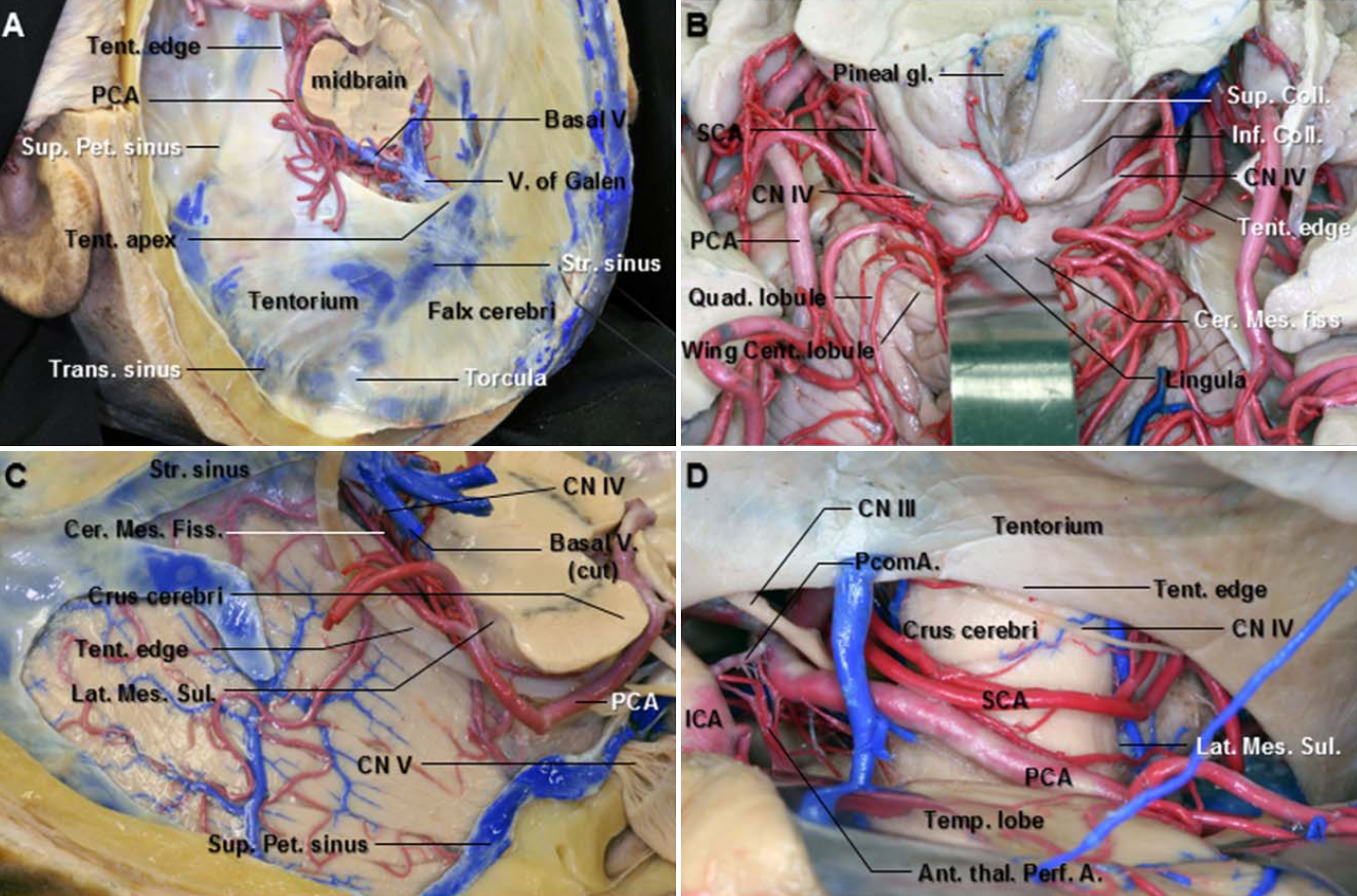
圖1。A.切除大腦後鐮和幕的上外側視圖。腦幕邊緣沿腦梗外側邊緣掃過。幕頂位於蓋倫靜脈和直竇的交界處。帳篷的邊緣從頂部向下傾斜。B.小腦後腦裂正位麵。小腦蚓部的中央小葉和喙部向後收縮,露出小腦後腦裂,它向前延伸到小腦和中腦之間。裂隙的前壁在中線由枕板和舌形成,外側由小腦上梗形成。滑車神經位於下丘下方。滑車神經池段的起始部分向外側延伸至小腦後裂。 C. Superolateral view of the right tentorial surface of the cerebellum. The tentorial edge sweeps along the lateral margin of the cerebral peduncle. The tentorium has been removed while the tentorial venous sinuses and tentorial edge have been preserved. The posterior cerebral artery runs posteriorly along the lateral margin of the midbrain above the tentorial edge. D. Subtemporal view. The right temporal lobe has been elevated to expose the PCA, SCA, and trochlear nerve in the anterior and middle incisural spaces. The PCA passes above and the SCA below the oculomotor nerve. The trochlear nerve courses around the side of the brainstem. It becomes subtentorial at the anterolateral part of the cerebral peduncle. Ant. Thal. Perf. A., anterior thalamoperforating artery; Cent., central; Cer. Mes. Fiss., cerebellomesencephalic fissure; CN., cranial nerve; Coll., colliculus; ICA., internal carotid artery; Inf., inferior; Lat. Mes. Sul., lateral mesencephalic sulcus; PCA., posterior cerebral artery; PcomA., posterior communicating artery; Pet., petrosal; Quad., quadrangular; SCA., superior cerebellar artery; Str., straight; Sup., superior; Tent., tentorium; Temp., temporal; Trans., transverse; V., vein. (Images courtesy of AL Rhoton, Jr.)

圖2。A.優越的視野。向外側收回鉤帶,露出幕邊緣和滑車神經。滑車神經沿著幕邊緣向前延伸穿過硬腦膜,位置在動眼三角後外側邊緣後的幕前外側遊離邊緣處。動眼肌三角的兩個邊緣由前、後岩突硬腦膜褶皺形成,分別從前、後岩突延伸至岩尖。B.幕邊緣向外側收縮,露出滑車神經及其在幕上的溝。滑車神經逐漸在遊離幕邊緣下表麵的凹槽中紮根。槽是從遊離池段到固定硬腦膜入口的平穩過渡。C. b的放大視圖,覆蓋動眼神經和滑車神經的硬腦膜被切開並向外側縮回,露出動眼神經和滑車神經池。動眼神經在硬腦膜穿透部位有一個印痕。 D. The right combined presigmoid approach to expose the upper cerebellopontine angle and petrous apex. The presigmoid dura is opened from the anterior jugular bulb toward the superior petrosal sinus with a cut made superior to the transverse sinus toward the superior petrosal sinus, which can then be ligated and cut. The tentorium is cut posterior to the superior petrosal sinus toward the tentorial edge, taking care to avoid the trochlear nerve. Retractors are then placed using the tentorium between the retractor blade and the temporal lobe to protect the underlying temporal lobe cortex. The trochlear nerve courses under the tentorium behind the superior petrosal sinus. A., artery; Ant., anterior; Bas., basilar; CN., cranial nerve; Interped., interpeduncular; Jug., jugular; Oculom., oculomotor; Parahippo., parahippocampal; penet., penetration; Petroclin., petroclinoid; PCA., posterior cerebral artery; PcomA., posterior communicating artery; PCP., posterior clinoid process; Pet., petrosal; SCA., superior cerebellar artery; Semi., semicircular; Sig., sigmoid; Sup., superior; V., vein. (Images courtesy of AL Rhoton, Jr.)
Iaconetta等人根據腦池段的位置、關係以及臨床和外科方麵的考慮,將其分為四周區和周圍區(Iaconetta et al., 2013)。四頭肌段沿前外側略下的軌跡向橋腦後裂的腦梗外側方向彎曲,並在蛛網膜下腔內保持自由,直到周圍的腦池網(圖1B)。滑車神經穿過小腦中央前膜,形成四頭神經池的側壁,進入周圍池(Lu and Zhu, 2007;Iaconetta et al., 2013)。滑車神經周圍段與小腦上動脈、腦後動脈、羅森塔爾基底靜脈和中腦外側有關係(圖1C和1D)。Herlan等人(2013)研究的所有滑車神經都逐漸固定在遊離幕邊緣下表麵的凹槽中。由於槽是從遊離池段到硬腦膜固定入口的平穩過渡,因此無法準確測量其有效長度(圖2B和2C) (Herlan et al., 2013)。我們建議滑車神經沿著幕下溝的部分可能包括在幕段,如下所述。滑車神經池段的遠端界限表現為神經在完全被硬腦膜管包裹之前開始在幕下表麵的槽內運行的點(圖2C)。因此,池段,包括幕下的神經,不在溝內,相對自由,因此更容易在其過程中因病理而拉伸或扭曲(圖2A和2B)。 However, Tubbs and Oakes defined the cisternal segment as extending from the origin of the trochlear nerve on the dorsal brain stem to its dural entrance. Given this definition, the length of the cisternal segment ranged from 24 to 45 mm (mean 35.6 mm) (Tubbs and Oakes, 1998).
從外科角度看,不需要硬腦膜剝離就可以很容易地暴露池段。用不同的方法觀察四邊形和周圍區(Ammirati等人,2002年;Iaconetta et al., 2013)。四頭區可通過不同的後顱窩入路進入,如中線或幕下-小腦上旁正中入路,而周圍區可通過幕下外側入路進入。極外側幕下-小腦上入路清楚地暴露了四頭神經和周圍神經段之間的連接。使用這種方法,滑車神經通常在周圍池中立即可見,但當滑車神經靠近遊離幕邊緣時,就不容易被識別。在這種情況下,小腦上動脈的小腦間腦段是一個很好的標記;它通常位於中腦上方,沿著中腦的後側和外側(圖1C、1D和2D)。乙狀竇前顳下幕前聯合入路適合暴露腦池和幕段之間的交界處(圖2D) (Ammirati et al., 2002)。
滑車神經在動眼三角後外側邊緣後的小腦幕前外側遊離邊緣處穿過硬腦膜(圖2A-2C)。傳統上,當它刺穿帳篷的自由邊緣時,被稱為海綿段(Bisaria, 1988;Ammirati等,2002;Mercier et al., 2009)。幕段從滑車神經入口延伸到幕到岩突前褶神經在這裏進入海綿竇。幕段的長度和寬度分別為9.3161.49和0.960.4 mm (Iaconetta et al., 2013)。
Rhoton等人注意到,滑車神經在刺穿幕狀肌即滑車池之後,在岩突後褶處移動了一小段距離。在池內,滑車神經直到到達前突頂端才與海綿竇的側壁合並(圖2C) (Rhoton, 2000)。Tubbs等人描述了這部分被蛛網膜褶皺包裹的幕段,並將其稱為三角段。其長度從2.2到6毫米,平均為4毫米(Tubbs等人,2014年)。與相對自由的腦池段相比,固定幕段包括沿幕下溝的神經和三角段,如Tubbs等人所命名。因此,我們認為三角段是滑車神經幕段的遠端部分。
從神經外科的角度來看,打開覆蓋在三角段的硬腦膜可以促進滑車神經在海綿竇外的回縮,因為這個節段沒有靜脈或動脈結構。神經池的存在,而不是神經被硬腦膜緊緊束縛,增加了暴露神經在竇頂的安全邊際(圖2C)。然而,Ammirati等人報道了神經和其幕段的幕硬腦膜之間的緊密關係,使得盡管存在蛛網膜套,解剖仍然非常困難(Ammirati等人,2002)。
在外側切口空間定位滑車神經幕段是困難的,特別是從幕上入路。此外,腫瘤會使其偏離正常的解剖過程。一般來說,滑車神經首先在腦梗後緣的幕下或緊靠幕突(圖1D和2D)。這是外科醫生第一次從幕上入路看不見它的地方。它通常位於外耳道(EAM)最外部部分後方1.5-2厘米處。有趣的是,外耳道與內耳道位於同一冠狀平麵上(圖3D和4A)。因此,從外部聲道到內部聲道畫一條假想的線可以用作參考線。如果停留在這條線後1.5厘米處,並在這一距離的後方切開幕,就可以避免滑車神經受傷(Tubbs and Oakes, 1998)。滑車神經幕部在幕部被切開後就可以看到了。

圖3。A.右側海綿竇硬腦膜外層已從海綿竇外側壁和梅克爾氏洞剝落。由此可見動眼神經和滑車神經進入海綿竇頂並向前穿過眶上裂。岩上竇經過梅克爾氏洞口上方,與海綿竇的後部相連。B.覆蓋海綿竇外側壁的剩餘硬腦膜已被切除。滑車神經沿著動眼神經的下側延伸到前床突的基部。C.切除前床突後,露出頸內動脈的床突段。動眼神經進入竇頂的一個短池,直到到達前床突的下緣才並入外側壁。幕動脈沿著滑車神經的下表麵延伸到眶上裂。d .的概述。 The posterior wall of the cavernous sinus extends laterally from the dorsum sellae to the medial edge of the ostium of Meckel’s cave. The floor of the middle fossa has been removed to expose the infratemporal fossa, which contains the branches of the maxillary artery and the mandibular nerve, the pterygoid venous plexus, and the pterygoid muscle. The trochlear nerve courses around the cerebral peduncle just above the pons, between the posterior cerebral and superior cerebellar arteries. It pierces the dura below the free edge of the tentorium cerebelli, just behind the posterior clinoid process, and then passes forward in the lateral wall of the cavernous sinus below the oculomotor nerve and above the ophthalmic division of the trigeminal nerve. The trochlear nerve crosses the oculomotor nerve at the level of the optic strut, entering the orbit by the superior orbital fissure above the annular tendon and medial to the frontal nerve. A., artery; ACA., anterior clinoid process; Clin., clinoid; CN., cranial nerve; For., foramen; GSPN., great superficial petrosal nerve; IAM., internal acoustic meatus; ICA., internal carotid artery; Lat., lateral; M., muscle; PCA., posterior cerebral artery; PCP., posterior clinoid process; Pteryg., pterygoid; SOF., superior orbital fissure; Tent., tentorial. (Images courtesy of AL Rhoton, Jr.)

圖4。A.形成眶上裂頂部的蝶骨小翼已被切除,以露出裂隙的內容物。滑車神經和額神經是三叉神經眼部的分支,通過眶上裂進入眶環肌腱上方。B.右軌道的軌道頂已被拆除,露出近軌。在眶上裂處,覆蓋中窩和海綿竇的硬腦膜與眶尖的眶周和產生直肌的環形肌腱混合在一起。硬腦膜內襯眶上裂和視神經鞘與眶周融合。C.打開眶周並向外側收縮,暴露眶脂,滑車神經、額神經和淚神經在其中運動。D.眼眶脂肪已被移除。眼神經分為淚神經、額神經和鼻睫神經。滑車神經從提肌上方內側穿過到達上斜肌。 The tendon of the superior oblique muscle passes through the trochlea and below the superior rectus muscle to insert on the globe between the attachment of the superior rectus and lateral rectus muscles. The lacrimal nerve passes above the lateral rectus muscle to innervate the lacrimal gland. E. Enlarged view of the superior orbital fissure from above. The dura lining the right superior orbital fissure has been removed to expose the course of the trochlear nerve from the cavernous sinus to the orbit. ACP., anterior clinoid process; CN., cranial nerve; Falc., falciform; Gl, gland; IAM., internal acoustic meatus; Lac., lacrimal; Lat., lateral; Lev., levator; Lig., ligament; M., muscle; N., nerve; Nasocil, nasociliary; Obl., oblique; PCA., posterior cerebral artery; Pit., pituitary; Rec., rectus; SCA., superior cerebellar artery; SOF., superior orbital fissure; Sph., sphenoid; Sup., superior. (Images courtesy of AL Rhoton, Jr.)
海綿段從滑車神經插入海綿竇外側壁的位置延伸到眶上裂。滑車神經進入動眼三角後外側頂端的海綿竇頂部,在動眼神經入口後麵的8.1262.32 mm(範圍4.52-13.1 mm)和後床突後外側的13.8262.39 mm(範圍10.14-20.1 mm)(圖2A和2B) (Yasuda et al., 2005)。在77.5%的病例中(80個海綿竇中的62個),它在穿透硬腦膜的部位出現明顯彎曲和變平(Bisaria, 1988)。滑車神經在三角段後的海綿段,即幕段的遠端部分,穿過位於前、後岩突硬腦膜褶皺交界處的海綿竇頂後,在動眼神經下方的海綿竇外側壁內延伸。海綿段的近端通過上方的動眼神經和下方的眼神經之間(圖3)。動眼神經和滑車神經之間的距離為1mm,而滑車和眼神經之間的距離為2.5 mm (Iaconetta et al., 2013)。在72.5%的病例中(80個海綿竇中的58個),滑車神經在從入口進入海綿竇到通過眶上裂出口的過程中直徑增加,20%的病例甚至在後顱窩也出現厚度增加(Bisaria, 1988)(圖3C和3D)。滑車神經海綿段的長度和寬度分別為20.3861.95和0.960.4 mm (Iaconetta et al., 2013)。在視神經支柱水平,滑車神經從外側到內側,在動眼神經上表麵和位於前床突和視神經支柱下緣的硬腦膜之間交叉,成為海綿竇的最上層結構(圖3D和4A)。
滑車神經也形成了海綿竇外側壁的帕金森三角上緣。這個三角位於滑車神經的下緣和眼神經的上緣之間。第三個邊緣由一條線構成,連接滑車神經進入硬腦膜的入口點和三叉神經進入Meckel 's穴的位置。頸內動脈後彎和腦膜垂體幹從後彎起的起點位於這個三角形內,除非頸動脈拉長彎曲(圖3D)。
滑車神經在海綿竇的近端由上近端動脈(下外側幹的一個分支)持續供應。這條動脈沿著滑車神經的上表麵沿著海綿竇的側壁。這種恒定的關係可用於經顱顯微外科手術,以幫助通過動脈的存在來識別神經(Lang and Kageyama, 1990;Krisht等人,1994年;d 'Avella et al., 2008)。已經證明,神經的遠端部分在大多數情況下由幕動脈通過內窺鏡下經蝶竇通路供應。幕動脈在其下表麵到達神經,並隨神經延伸至眶上裂(圖3C) (d 'Avella et al., 2008)。
滑車神經穿過動眼神經上表麵,從外側向內側,通過眶上裂進入眶內,是進入眶內神經中最上方的神經(圖4A、4D和4E)。眶上裂是連接中顱窩和眶的一個小但在地形上很重要的區域。它分為上外側和下內側部分(圖4A和4E) (Morard et al., 1994;名取和羅頓,1995年;Govsa et al., 1999)。滑車神經是最靠近眶上裂上緣的結構。上外側部分包括滑車神經、淚神經、額神經和眼上靜脈。下內側部分包括動眼神經、鼻纖毛神經和外展神經的上、下分支和睫狀神經節的交感神經根(Govsa et al., 1999;雷蒙德等人,2008)。滑車神經的眶段出現在眶上裂的外側區域,剛好在Zinn環外側邊緣的外側,向超內側延伸至額神經(圖4D和4E)。 The trochlear, frontal, and lacrimal nerves course in the orbital fat just beneath the periorbita (Figs. 4B and 4C).
滑車神經位於視神經外側靠近眶尖的位置,眶尖經過視神經上方到達上斜肌。從外科的角度來看,如果需要,打開環形肌腱的切口應該在上直肌和內直肌的附著物之間。在打開環空之前,應將滑車神經與眶尖上方的鄰近組織分離,以防止在打開視神經鞘時損傷滑車神經(圖4E) (Natori and Rhoton, 1994;Rhoton, 2002;張等人,2010)。經過眶上裂後,神經向內側和對角線穿過提瞼上肌和直肌上肌到達上斜肌。
上斜肌起於覆蓋蝶骨體的眶周,向視神經管的超內側延伸,並向前延伸,止於穿過滑車的肌腱,這是一個連接到額骨滑車窩的圓形肌腱。肌腱在滑車中繞圈後,從上直肌的外側和後方穿過,插入上直肌和外側直肌之間的鞏膜上。神經平均有6個分支(範圍4-10)(圖4D)。滑車神經末梢的分支位於上斜肌後三分之一的病例占76%(100個眼眶中有76個),位於中間三分之一的病例占24%。滑車神經進入肌肉的起點為17.25 mm,延伸7 mm(上斜肌的平均長度為40.6 mm) (Villain et al., 1993;張等人,2010)。滑車神經終止於滑車內側的上斜肌23例(77%),上緣7例(23%),但從不終止於外側;神經眶段的平均長度為25.1毫米(範圍18-34毫米)(Villain et al., 1993)。滑車神經眶段的血管供應由眼動脈和腦膜前動脈的末端分支提供(Iaconetta et al., 2013)。
滑車神經是顱神經中最小的神經,但它的顱內路徑卻是最長的。它是唯一起源於腦幹背側的腦神經,也是唯一纖維交叉的腦神經。由於滑車神經經過小腦上區、切膜區、海綿竇區和眶區,準確了解滑車神經的解剖結構及其周圍結構對於避免手術過程中損傷滑車神經非常重要。
撰稿人:Wonil Joo和Albert L. Rhoton, Jr
內容來自Joo W, Rhoton AL, Jr.滑車神經的顯微外科解剖。中國阿娜特2015; 28:857 - 864。doi.org/10.1002/ca.22602.
神經外科188bet手机app圖譜很榮幸能夠繼承Albert L. Rhoton, Jr . MD的遺產。
請登錄發表評論。
一定要在社交媒體上關注我們,獲取精彩內容並保持更新生活科恩醫生的會議,關於手術技術的問題,以及更多!


您必須登錄才能查看此材料。
的188bet手机app這幾乎完全取決於你的捐款。
如果沒有你們的大量捐贈,我們就無法繼續開展地圖集。
請承諾每年至少捐贈250美元給Atlas。如果沒有這種承諾,Atlas將很快需要付費訂閱,世界各地的許多外科醫生將無法獲得它,他們的病人的護理依賴於它。
請立即捐款!
如果沒有你們的大量捐贈,我們就無法繼續開展地圖集。請承諾每年至少捐贈250美元給Atlas。
如果沒有這個承諾,Atlas將很快需要付費訂閱世界上許多病人的護理都依賴於它的外科醫生將無法使用它。請立即捐款!