

你可以有所作為。
的188bet手机app這幾乎完全取決於你的捐款。
如果沒有你們的大量捐贈,我們就無法繼續開展地圖集。
請承諾每年至少捐贈250美元給Atlas。如果沒有這種承諾,Atlas將很快需要付費訂閱,世界各地的許多外科醫生將無法獲得它,他們的病人的護理依賴於它。
現在請捐!

最後更新:2021年6月22日
本研究的目的是回顧三叉神經的外科解剖。我們還展示了一些涉及三叉神經及其周圍的結締組織和神經血管結構的圖片。在動脈和靜脈灌注彩色乳膠後,用放大倍數從33到403對10具成年屍體頭部進行了研究。三叉神經是顱神經中最大、最複雜的。它是來自麵部的感覺輸入的主要管道,並為咀嚼肌提供運動神經支配。由於其大小和複雜性,在診斷和治療口麵、顳下頜、顳下和翼齶區的病理過程之前,必須對神經有全麵的了解。三叉神經與顱底手術的影像或手術相接觸。因此,全麵了解三叉神經的解剖結構對實施手術而無明顯並發症是至關重要的。

三叉神經是最大的腦神經,它是對麵部、頭皮、牙齒、口腔和鼻腔的感覺供應,也是對咀嚼肌和其他一些肌肉的運動供應。它還包含來自咀嚼肌和眼外肌的本體感覺神經纖維。三叉神經有三個分支:眼神經、上頜神經和下頜神經。我們將三叉神經沿著其路徑分為腦幹、腦池段、梅克爾洞段、三叉神經節和周圍區(眼區、上頜區和下頜骨區)。
在放大率33 ~ 403的顯微鏡下觀察了10個成年屍體頭部動脈和靜脈注射彩色矽膠的情況。這些屍體的頭部由佛羅裏達解剖委員會捐贈給佛羅裏達大學的神經外科。所有的骨骼工作都是用高速鑽頭(Midas Rex Institute, Fort Worth, TX)完成的。
三叉神經的四個中樞核位於腦幹內:中腦核、主感覺核、運動核、三叉脊髓束和核(圖1A和1B)。中腦核負責輸送眼外肌和咀嚼肌的本體感覺纖維,並調節咬合。中腦核的傳入纖維傳遞來自牙齒、牙周組織、硬齶和顳下頜關節囊的壓力和運動感覺。這個核與控製咬合力的機製有關(Carpenter, 1991b)。它位於中腦下部和橋腦上部,沿導水管周圍灰質外側緣,位於第四腦室前外側,感覺核內側(圖1A和1B) (Go et al., 2001)。
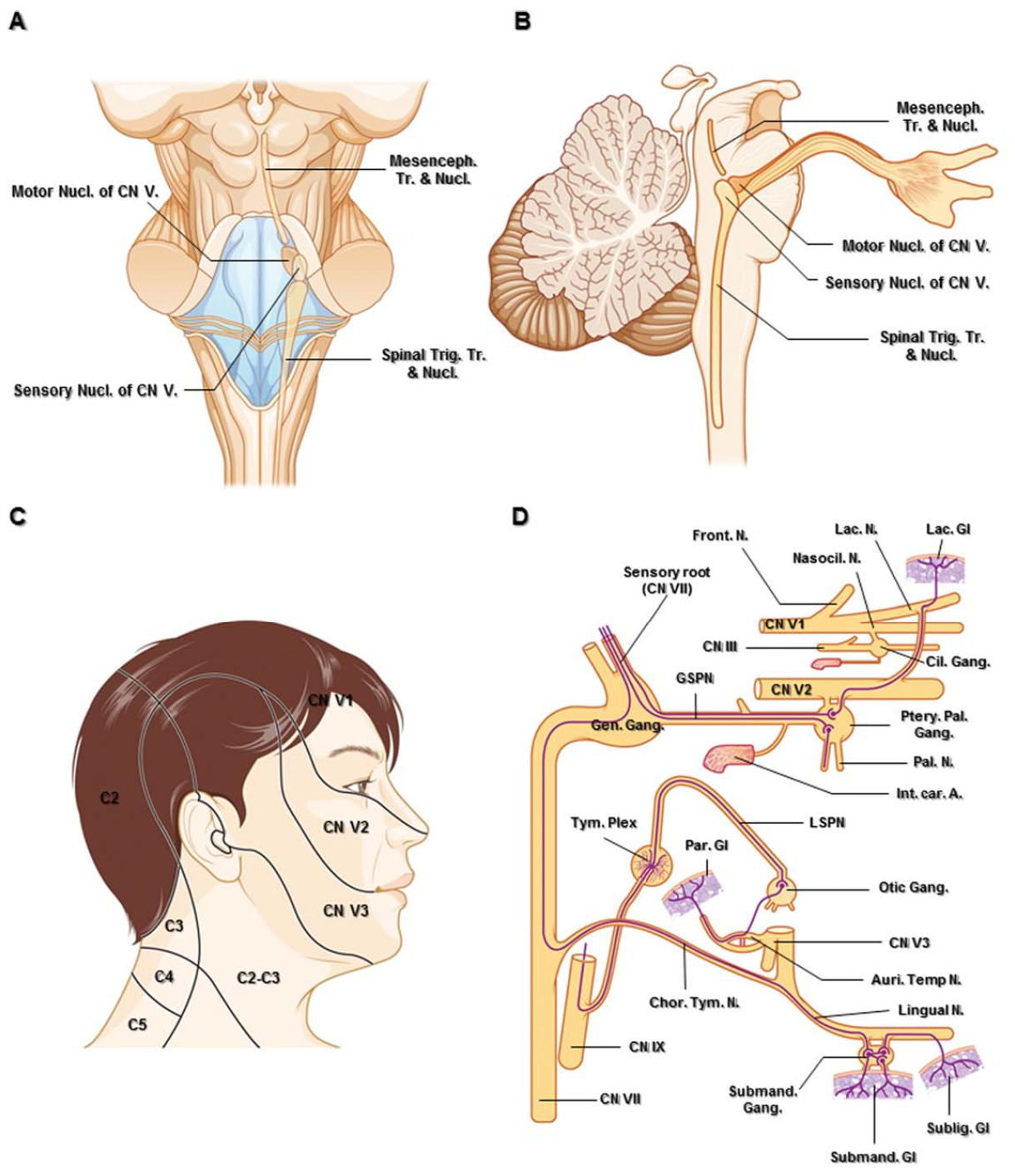
圖1。A.腦幹後側解剖圖和疊加圖,顯示三叉神經核。運動核位於三叉神經感覺核的內側。B.經過右側三叉神經核的矢狀示意圖。C.頭部和上頸部的皮神經支配。三叉神經的三個皮節之間幾乎沒有重疊。D.翼齶、耳部和下頜下神經節的副交感神經連接。副交感神經纖維顯示為紫色線。縮寫:Auri。溫,耳顳神經; C., cervical; Car., carotid; Chor.Tym. N., chorda tympani nerve; Cil., ciliary; CN., cranial nerve; Front., frontal; Gang., ganglion; Gen., geniculate; Gl., gland; GSPN., greater superficial petrosal nerve; Int., internal; Lac., lacrimal; Mesenceph., mesencephalic; N. nerve; Nasocil., nasociliary; LSPN., lesser superficial petrosal nerve; Nucl., nucleus; Par., parotid; Ptery. Pal., pterygopalatine; Subling., sublingual; Submand., submandibular; Tr., tract; Trig., trigeminal; Tym. Plex., tympanic plexus. (Images courtesy of AL Rhoton, Jr.)
主要感覺核傳遞觸覺和壓力的衝動。它呈背前腹側排列。眼部的纖維在腹側終止,上頜部的纖維在中間,下頜骨的纖維大多在背側。這個核位於腦橋上部進入的三叉神經根纖維的外側。來自主要感覺核的三叉神經纖維交叉或不交叉,終止丘腦腹側後內側核(VPM)。起源於核腹側的交叉纖維與對側內側係索一起上升,形成三叉丘腦腹側束。未交叉的纖維起源於核的背內側部分,上升至中腦中央灰質附近,形成三叉丘腦背束(Carpenter, 1991b)。
運動核位於主感覺核的內側。這個核的纖維從腦幹的內側到進入的感覺根,經過三叉神經節的下方,沒有突觸,並並入下頜骨。軸突從中腦核投射到運動核,完成一個反射弧來調節咬傷程度(Go et al., 2001)。
三叉神經束和脊髓核傳遞疼痛和溫度的感覺形態。它從中橋延伸到C2-C4水平的頸髓,位於第四腦室的前外側(Barakos等,1990;木匠,1991 b)。進入三叉脊髓束和神經核的神經根纖維具有明確的地形組織,這是由感覺根進入腦橋時的內側旋轉引起的。眼區纖維多位於腹側,下頜骨區纖維多位於背側,上頜區纖維位於中間,並比其他區纖維向尾端下降得少。三叉神經脊核由三部分組成:(1)口腔部,(2)內插部,(3)尾突部。口腔部主要接收來自鼻和口內部結構的脈衝。內插部主要與皮膚麵部區域有關,而尾部在前額、臉頰和下巴上有較大的感受野。棘三叉神經通路的這種地形解剖可以解釋在棘三叉神經降道中,麵部的洋蔥皮表現(Carpenter, 1991b;昆克和Ceskoslovenská阿卡德米·維哈德,1964)。
三叉神經由一個運動核和三個感覺核組成,它們貫穿腦幹的大部分長度。三叉神經根由大的感覺根和小的運動根組成。感覺根從整個麵部(頸神經叢支配的下頜角除外)、太陽穴、外耳道和遠至頭骨頂點的頭皮前部接受體感感覺(圖1C) (Shankland, 2000)。雖然體感神經元的大部分細胞體位於三叉神經節,但咀嚼肌的本體感覺和拉伸受體的細胞體位於橋背側的中腦核(Dodd and Kelly, 1991)。來自咀嚼肌的本體感覺衝動通過運動根進入中腦核(圖1B) (Gray and Williams, 1989b;讓遊戲,2000)。
三叉神經的大感覺細根從橋中側向小腦梗中間內側出,眼部位於最下方,上頜部位於中間,下頜骨部位於上方(圖2A)。當神經根向前通過前脊池和Meckel 's洞穴到達中窩的三叉神經節時,它們大約旋轉180度橫向旋轉(Jannetta, 1996;讓遊戲,2000)。然而,Rhoton(2000)報道稱,下頜骨的纖維在從神經節到橋腦橋的整個區間內保持在三叉神經根的尾外側位置,眼區位於前內側,而上頜區纖維處於中間位置(Rhoton, 2000)。
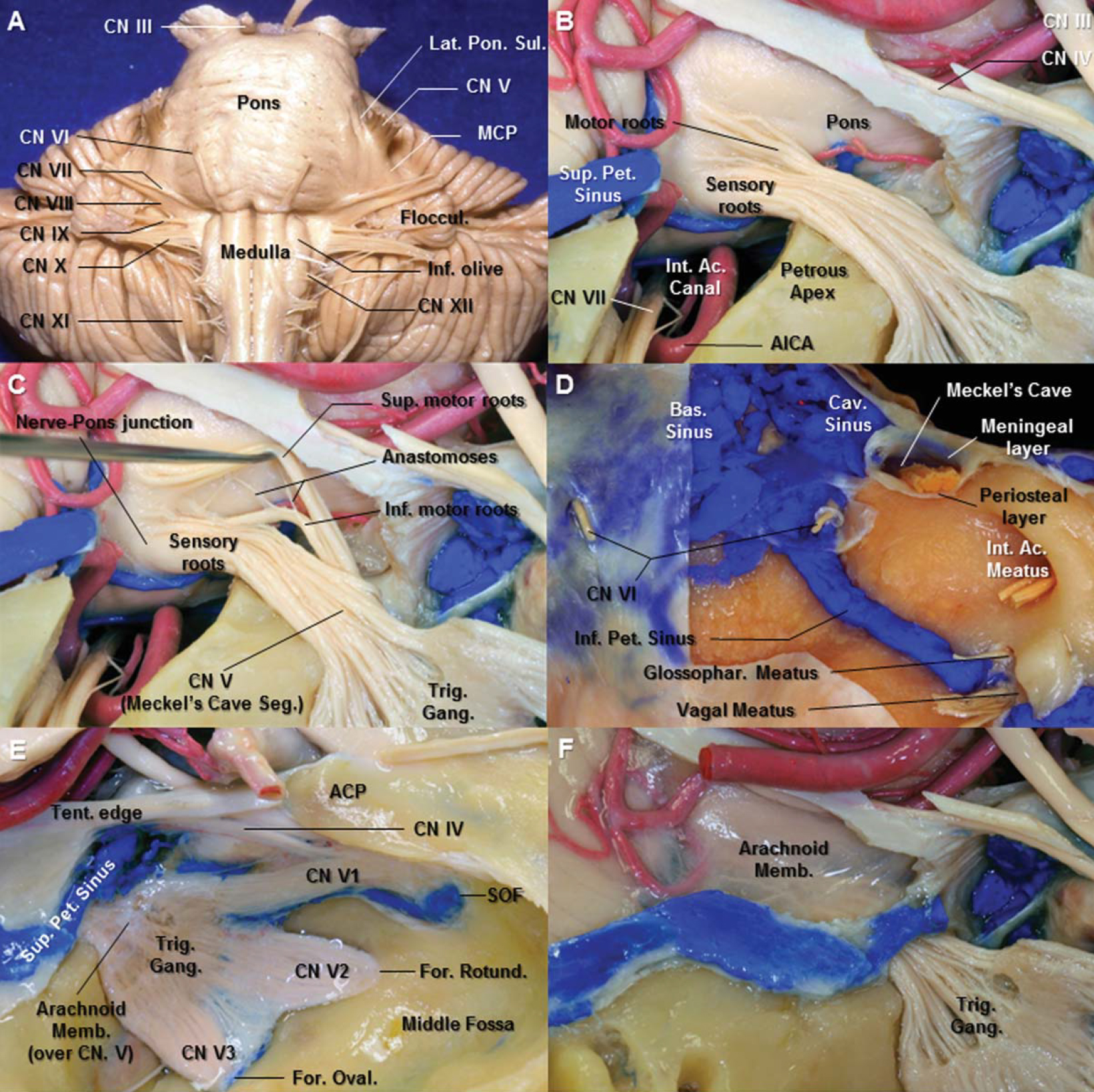
圖2。A.腦幹前視圖。小腦中梗與腦橋之間由腦橋外側溝隔開。就在橋外側溝的外側是三叉神經。從顯微外科的角度來看,三叉神經的明顯起源可以被認為是腦橋和小腦中梗之間的界限。B.右側三叉神經根入區側麵圖。幕和枕葉已經被切除露出三叉神經的根入口區。小的感覺根從橋腦超內側延伸到大的感覺根。內部聲道的屋頂已經被拆除。C.三叉神經的兩個運動小根組,一個主要的上運動小根和一個次要的下運動小根,在感覺根的上運動小根上縮回後可見。 D. The dura of the anterior aspect of the posterior fossa has been removed to expose the basilar plexus, cavernous sinus, and inferior petrosal sinus. Meckel’s cave is a cleft-like dural pocket that originates from the dura propria of the posterior fossa. The cave is situated at the trigeminal impression between the meningeal layer (dura propria) and the periosteal layer of dura. E. The arachnoid membrane from the posterior fossa extends to Meckel’s cave, forming a pocket within the cave, continues along the rootlets of the trigeminal nerve. F. The arachnoid membrane over the trigeminal root has been removed. The superior petrosal sinus extends medially through the upper edge of the porus of Meckel’s cave and above the trigeminal nerve to join the cavernous sinus. Abbreviations: Ac., acoustic; ACP., anterior clinoid process; AICA., anterior inferior cerebellar artery; Bas., basilar; Cav., cavernous; CN., cranial nerve; For., foramen; Gang., ganglion; Glossophar., glossopharyngeal; Inf., inferior; Int., internal; MCP., middle cerebellar peduncle; Memb., membrane; Oval., ovale; Pet., petrosal; Rotund., rotundum; Seg., segment; SOF., superior orbital fissure; Sup., superior; Tent., tentorium; Trig., trigeminal. (Images courtesy of AL Rhoton, Jr.)
小運動根從橋腦前超內側延伸至大感覺根的入口點(Saunders and Sachs, 1970;朗,1981)。有人定義了三叉神經的兩組運動根;一種主要的上細根和一種次要的下細根,它們與感覺根之間有許多連接。上根群位置獨特,與主感覺根相對隔離,代表了運動根的經典起源(圖2B) (Saunders和Sachs, 1970;朗,1981;Yousry等人,2004)。從感覺根收回上運動群後可見下運動根(圖2B和2C)。Pelletier等人(1974)認為下運動根的生理特征與運動纖維的生理特征相同,並服從運動或本體感覺功能,而不是感覺功能(Pelletier等人,1974)。兩個獨立的運動群在離腦幹不遠的地方連接在一起,形成一個單一的神經根,並通過感覺神經根和神經節內側的Meckel’s cave,與下頜骨部相連(圖2C)。
Peker等人報道了100條三叉神經的池部長度為12.3毫米(範圍8-15毫米)。三叉神經痛中提到的三叉神經根出口區(精確的神經-腦橋交界處)和中樞-外周髓鞘過渡區(TZ)是兩個獨立的結構。神經內側的中央髓鞘長度(從腦橋到TZ的距離)(範圍,0.1-2.5 mm;意思是,1.13毫米;中位,1 mm)短於側位(範圍,0.17-6.75 mm;意思是,2.47毫米;中位數,2.12毫米)(Peker等人,2006年)。
位於後窩的三叉神經池段在中硬腦窩的骨膜和腦膜(固有硬腦膜)層之間的幕邊和岩上竇下方向前穿過,進入Meckel’s cave。Meckel’s cave是一種裂狀硬腦膜囊,起源於後窩固有硬腦膜,位於硬腦膜中窩兩層之間,從後窩延伸至中顱窩後內側部分(圖2D-2F)。岩上竇向內側延伸,穿過梅克爾洞孔上緣,在三叉神經上方與海綿竇相連。外展神經穿過硬腦膜後窩下內側至三叉神經孔(圖2D)。
洞穴位於三叉神經印痕處,位於腦膜層(固有硬腦膜)和硬腦膜骨膜層之間,後者似乎與岩尖緊密相連(圖2D)。後窩的蛛網膜延伸到Meckel 's洞穴,在洞穴內形成一個口袋,並沿著三叉神經的小根一直延伸到三叉神經節(圖2E和2F) (Sabanci et al., 2011)。Meckel’s cave的內容是三叉神經的感覺和運動根、三叉神經節和蛛網膜層(圖3C和3D)。三叉神經節的前部緊緊地附著在上麵的蛛網膜和Meckel 's洞穴的固有硬腦膜上,沒有任何潛在的蛛網膜下腔(圖3E)。梅克爾洞穴內的蛛網膜下腔位於三叉神經節後麵,是構成三叉神經池的實際空間(圖2E, 2F, 3C,和3E) (Youssef et al., 2006)。三叉神經節和三叉神經根的上外側有兩層固有硬腦膜。內層的固有硬腦膜構成了梅克爾氏洞的上外側壁。外層是中窩的腦膜層(固有硬腦膜)。解理麵在三叉神經的神經外鞘(內層)和中窩固有硬腦膜(外層)之間橫向形成。這個解理麵是mekel’s cave內容物硬膜間暴露的解剖學基礎(圖3A-3D) (Kawase等人,1996; Yoshida and Kawase, 1999; Al-Mefty et al., 2002; Youssef et al., 2006). However, the limits of the Meckel’s cave decided by its meningeal architecture remain controversial (Taptas, 1982; Kapila et al., 1984; Kawase et al., 1996; Kehrli et al., 1997). According to Sabanci et al. (2011), the meningeal pouch (Meckel’s cave) extended beyond the trigeminal ganglion along the three divisions in some specimens, and in the others, it ended at the level of the ganglion (Sabanci et al., 2011).
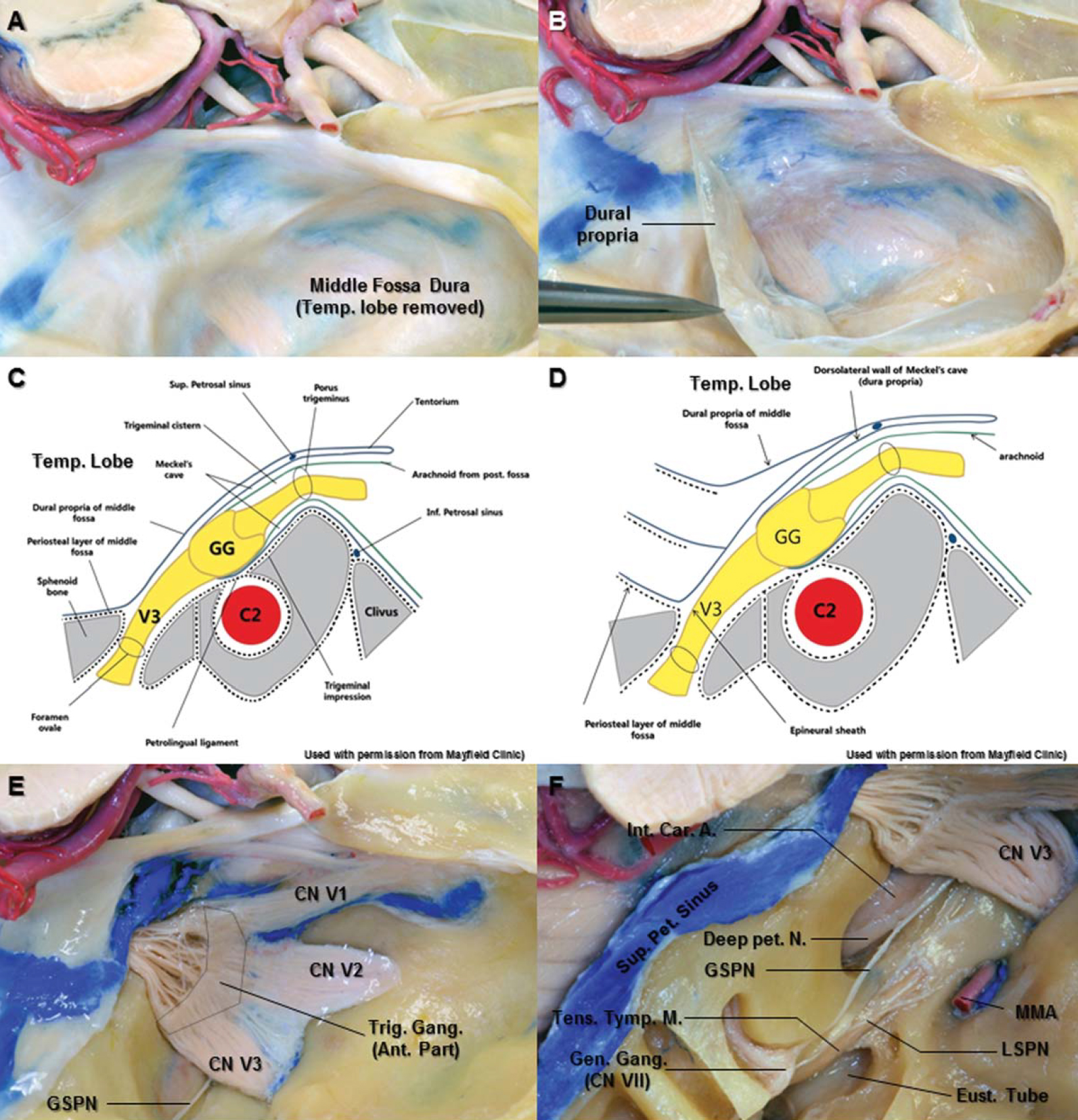
圖3。A.切除右側顳葉,露出中硬腦窩。B.硬腦膜中窩固有硬腦膜已從中窩底向後側剝落。三叉神經已經顯現出來了。C.沿多孔三叉肌和卵圓孔之間的線的斜冠狀切片(經Mayfield診所許可使用)。在三叉神經節的前緣,Meckel 's穴的背外側壁和腹內側壁成為三叉神經每分支的神經外鞘。梅克爾洞的內容是三叉神經的感覺和運動根,三叉神經節和蛛網膜層。D.卵形孔附近的骨內膜切口通向外層(中窩固有硬腦膜、腦膜層)和內層(下頜神經神經外鞘)之間的劈裂平麵(Mayfield診所允許使用)。E.三叉神經節前部與上麵的蛛網膜和Meckel 's洞穴硬腦膜緊密粘附,不存在蛛網膜下腔。由於實際的蛛網膜下腔,蛛網膜層很容易從三叉神經節後部切除。 F. Superior view of the right temporal bone. The bone on the middle fossa floor has been removed laterally to show the petrous segment of the carotid artery, the Eustachian tube, the tensor tympani muscle, and geniculate ganglion of the facial nerve. The greater superficial petrosal nerve is separated from the horizontal segment of the petrous internal carotid artery. It passes under V3 and joins the deep petrosal nerve from the sympathetic carotid plexus to become the vidian nerve in the vidian canal. Abbreviations: A., artery; C2., petrous carotid artery; Car., carotid; CN., cranial nerve; Eust., Eustachian tube; Gang., ganglion; Gen., geniculate; GG., gasserian ganglion; GSPN., greater superficial petrosal nerve; Inf., inferior; Int., internal; LSPN., lesser superficial petrosal nerve; MMA., middle meningeal artery; N., nerve; Pet., petrosal; Sup., superior; Tens. Tymp.M., tensor tympani muscle; Trig., trigeminal; V3., mandibular division. (Images courtesy of AL Rhoton, Jr.)
岩大淺神經(GSPN)位於Meckel’s cave後外側(圖3F)。GSPN起源於膝狀神經節,通過麵部裂孔出,在硬腦膜下向前內側方向延伸至下頜部(V3)。在MC後外側12.8 mm處發現麵神經裂孔(Arslan et al., 2012)。GSPN與岩狀內頸動脈(ICA)的水平段分離。它從V3下經過並從頸交感神經叢與岩深神經相連成為維管中的維神經。維神經的遠端部分會在上頜分裂部分被回顧。
ICA岩的水平段位於MC的下方。水平段開始於後膝的遠端,沿著顳骨岩部的長軸向前內側延伸,大部分位於GSPN下方,位於鼓室張肌和咽鼓管的後方,位於棘孔和卵圓的後方,止於三叉神經和神經節的前膝處。動脈在充滿纖維軟骨的淚孔上方向上轉(圖3E, 12A,和12B) (Osawa等,2008)。
了解三叉神經和自主神經纖維的關係是很重要的。頭部有四個與三叉神經相關的副交感神經節:睫狀神經、翼齶神經、耳部神經和下頜下神經節(圖1D)。
眼神經(V1)是三叉神經的三個分支中最小的。動眼神經、滑車神經、外展神經和V1神經位於海綿竇內。從三叉神經節開始,V1在海綿竇外側壁下部向前穿過,到達眶上裂時呈向上傾斜的狀態(圖4A和4B)。V1在接近SOF時分叉進入淚神經、額神經和鼻纖毛神經。SOF是一個很小但在地形上很重要的區域,它連接中顱窩和眶(圖4A) (Natori and Rhoton, 1995;Govsa et al., 1999)。

圖4。A.右側眶上裂後視圖。SOF上部由蝶骨的小翼包圍,下部由大翼包圍,內側由蝶體包圍。視神經支柱形成裂隙的上內側邊緣。SOF提供了眼眶和中窩之間的通信。B.右側海綿竇側麵圖。海綿竇的外側壁和前床突被切除。動眼肌、滑車、外展肌和眼部(V1)位於海綿竇內。他們通過SOF進入軌道。C.右側額顳區上外側切麵。 After performing the fronto-temporal craniotomy, the dura is elevated. The fronto-temporal dural fold is located on the lateral side of the SOF, between the greater and lesser wings of the sphenoid bone. D. Resecting the dural fold allows further detachment of the dura from the surrounding structures, enabling extradural exposure of the anterior portion of the cavernous sinus and the third, fourth cranial nerves as well as the fifth cranial nerve. E. Superior view of the right orbit and the optic canal. At the SOF, the dura covering the optic nerve, middle fossa, and the cavernous sinus blends into the periorbita of the orbital apex. F. Anteroinferior view of the right orbit. The rectus muscles originate from the annular tendon. The annular tendon surrounds the orbital end of the optic foramen and the adjacent part of the SOF. The fibrous component, which blend together to form the annular tendon, are the periorbita covering the orbital apex, the dura lining the SOF and optic canal, and the optic sheath. G. Superior view of the right SOF. The dura covering the cavernous sinus and the optic sheath blend into the periorbita of the orbital apex and into annular tendon from which the rectus muscles arise. H. Schematic anterior view of the main structures passing through a right SOF. Abbreviations: A., artery; ACP., anterior clinoid process; Cav., cavernous; CN., cranial nerve; Div., division; For., foramen; Front., frontal; Inf., inferior; Lac., lacrimal; Lat., lateral; Lev., levator; M., muscle; Med., medial; N., nerve; Nasocil., nasociliary; Obl., oblique; Oculom., oculomotor; Ophth., ophthalmic; PCP., posterior clinoid process; Rec., rectus; Rotund., rotundum; SOF., superior orbital fissure; Sph., sphenoid; Sup., superior; Temp., temporal; V., vein. (Images courtesy of AL Rhoton, Jr.)
眶上裂.眶上裂(SOF)是眼眶與中顱窩相通的狹窄裂隙。SOF位於大翼和小翼與蝶骨體之間。它有一些三角形的形狀,在蝶體內側有一個寬的基部,在小翼和大翼之間有一個狹窄的頂端(圖4A)。在SOF水平,海綿竇外側壁的外層和內層彼此分離(Kawase et al., 1996)。內層由顱神經周圍神經及周圍結締組織組成,隨神經延伸至後眶。外層由顳硬腦窩形成(圖3A、3B和4B)。在SOF外側緣,硬腦膜的骨膜層與眶周的骨膜層相鄰(Froelich et al., 2007)。硬腦膜橋位於SOF外側邊緣,是額顳部骨膜褶皺,延伸於眶周和顳部硬腦膜褶皺之間。切割額顳骨膜褶皺後,顳硬腦膜和海綿竇外側壁內層之間的解理麵形成(圖4C和4D) (Day et al., 1994; Kawase et al., 1996). Froelich et al. (2007) reported that the lacrimal nerve of the V1 may be at risk during incision of periosteal fold because it is just medial to surgical cleavage plane (Figs. 4F and 4H) (Froelich et al., 2007). The distance between the lacrimal nerve and lateral end of the SOF was 4.21 ± 1.72 mm (Shi et al., 2007).
在裂隙處,覆蓋中窩和海綿竇的硬腦膜與眶尖的眶周和產生直肌的環形肌腱混合(圖4E和4G)。環形肌腱可用於將SOF分為三個區域:中央、外側和下扇區(圖4H) (Natori和Rhoton, 1995;Shi等人,2007)。環狀肌腱包圍視神經管的眶端和SOF的鄰近部分。SOF的中央部分被環狀肌腱包圍,稱為動眼孔,因為它是裂口的一部分,動眼神經通過。外展神經、鼻睫神經和頸動脈交感神經叢的分支也通過動眼孔(圖4H、6A和6C)。鼻纖神經與動眼肌分離,由動眼肌孔內的纖維隔外展神經(圖6D)。外側段被定義為環形肌腱外側,傳遞滑車神經、額神經和淚神經,這些神經都通過環形肌腱外側的裂隙(圖4H和5B)。淚神經位於裂隙最外側,額神經位於內側(圖4F和4H) (Natori and Rhoton, 1995;Shi等人,2007)。 The inferior sector of the SOF is situated below the annular tendon. The inferior rectus muscle arises from the annular tendon at the upper margin of this sector. Orbital fat extends backward below the inferior rectus muscle into this part of the fissure. This sector contains the inferior ophthalmic vein and orbital fat.

圖5。A.移除軌道頂後右側軌道的上方視圖。眶周被切開並縮回,露出額神經、淚神經和滑車神經。B.除去軌道脂肪,露出軌道的主要結構。額神經起於海綿竇外側壁,穿過淚神經和眼上靜脈內側及滑車神經下方的SOF狹窄外側部。它在提肌和眶周之間向前延伸。C.提瞼肌和上直肌向後內側收縮,露出視神經、鼻纖毛神經和外展神經。鼻睫神經在視神經上方向前延伸到達眶內側位於上斜肌和內直肌之間。D, E.右側SOF側位視圖。切除蝶骨的小翼以打開SOF。 At the level of the fissure, the nasociliary nerve ascends laterally to the inferior division of the oculomotor nerve and then crosses medially between the two divisions of the oculomotor nerve. Abbreviations: CN., cranial nerve; For., foramen; Front., frontal; Gl., gland; GSPN., greater superficial petrosal nerve; Lac., lacrimal; Lat., lateral; Lev., levator palpebral; M., muscle; MMA., middle meningeal artery; Nasocil., nasociliary; N., nerve; Obl., oblique; Oph., ophthalmic; Ptery., pterygoid; Rec., rectus; Rotund., rotundum; SOF., superior orbital fissure; Sup., superior; Suporb. N., supraorbital nerve; Sup.-Troch. N., supratrochlear nerve. (Images courtesy of AL Rhoton, Jr.)

圖6。A.右側SOF側麵圖。切除蝶骨小翼和外側壁眼眶,暴露出穿過SOF的結構。額神經已向下外側收縮。外側部分傳遞滑車神經、額神經和淚神經,所有這些神經都通過環肌腱外的裂隙。B.右眶尖上方視圖。視神經已向內側縮回,露出睫狀節。睫狀神經節接收來自下動眼神經的副交感神經運動根,來自鼻睫神經的感覺根,以及來自眼動脈周圍叢的交感神經纖維。C. SOF的放大視圖。(A)中SOF的側緣已被移除。 The oculomotor foramen is the portion of the opening in the annular tendon lateral to the optic foramen through which the superior and inferior divisions of the oculomotor nerve and the nasociliary nerve and abducens nerve pass. The nasociliary nerve is situated above and lateral to the abducens nerve in the anterior part of the cavernous sinus. D. the annular tendon has been divided between the origin of the superior and lateral rectus muscles. The nasociliary nerve separates from the oculomotor and abducens nerves by fibrous septum within the oculomotor foramen. E. The levator and superior rectus muscles have been reflected medially and lateral rectus muscle has been reflected laterally to expose the right optic nerve (removed) and short ciliary nerve arising from the ciliary ganglion. F. Anterior view of the right orbit and extraocular muscles after removing the eye globe and optic nerve. The lacrimal nerve courses along the superior margin of the lateral rectus muscle. The nasociliary nerve gives rise to long ciliary nerves that enter the sclera around the optic nerve with the short ciliary nerves arising from the ciliary ganglion. Abbreviations: A., artery; Cil., ciliary; CN., cranial nerve; Div., division; Front., frontal; Gang., ganglion; Inf., inferior; Lac., lacrimal; Lat., lateral; M., muscle; N., nerve; Nasocil., nasociliary; Obl., oblique; Ophth., ophthalmic; Rec., rectus; SOF., superior orbital fissure; Sup., superior; Sympath., sympathetic. (Images courtesy of AL Rhoton, Jr.)
額葉神經.額神經是V1神經中最大的分支。V1的額支起於海綿竇的外側壁,穿過眶上裂狹窄的外側部分,位於淚神經和眼上靜脈的內側,並在滑車神經下方。它在提瞼肌和眶周肌之間向前延伸。去除眶頂後,眶周可見(圖5A, 5B, 5D)。額神經向環肌腱外側和上外側延伸,並在眼眶內分為滑車上神經和眶上神經(圖5B)。
Supratrochlear神經.滑車上神經與滑車上動脈一起在上斜肌滑車前部的上方運行(圖5B和5C)。神經向鼻纖毛神經的滑車下支發出下降絲。滑車上神經位於滑車和眶上孔之間,位於中線外側16毫米(範圍,12-21毫米)和眶上緣上緣以下7毫米(範圍,6-9毫米)(Jeong et al., 2010)。滑車上神經位於眶上神經內側,位於眶上緣(Cuzalina and Holmes, 2005)。它支配結膜和上眼瞼的皮膚,並在波狀肌和枕額肌的額腹下麵上升,分裂成分支,穿過這些肌肉支配靠近中線的下前額的皮膚(Gray和Williams, 1989b)。
眶上神經.眶上神經位於提瞼肌和眶頂之間。眶上神經從中線外側29mm (25 - 33mm)和眶上緣上緣以下5mm(範圍,4-6 mm)的眶上切跡或孔中出來(Jeong et al., 2010)。神經支配上眼瞼,額骨額竇粘膜,盔狀腱膜和眼輪匝肌。它在前額上升,分為一個更小的內側分支和一個外側分支,它們供應頭皮的皮膚,幾乎與lambdoid縫線一樣遠(Gray and Williams, 1989b;讓遊戲,2001)。它也可能攜帶一些交感神經纖維到眼球和瞳孔擴張器(Rhoton, 2003b)。眶上神經可將自主纖維從頸交感神經節傳遞到葉和瞳孔擴張器(Martins et al., 2011)。
Nasociliary神經.鼻纖神經,大小介於額神經和淚神經之間,位於眼眶較深處。鼻纖毛神經起源於V1內側,位於海綿竇前部外展神經的上方和外側(圖5C-5E)。外展神經和鼻纖毛神經都在V1的內側流過淚神經和額神經。在裂隙水平處,鼻纖毛神經向外側緩慢上升至動眼神經下段,然後在動眼神經兩段之間的內側和視神經上方穿過,到達眼眶內側,在那裏產生篩前和篩後神經和滑車下神經(圖5C和5E)。
鼻纖毛神經與纖毛神經節相連並發出纖毛長神經。睫狀神經節的感覺根起源於鼻睫神經。感覺根在視神經的外側向前延伸並進入睫狀節但不與任何細胞體發生突觸。幾根被稱為短睫神經的細絲離開睫狀神經節,將感覺傳遞到角膜和眼球。長睫狀神經伴短睫狀神經穿過鞏膜靠近視神經附著處。長纖毛神經將交感神經傳遞到眼球和瞳孔擴張器,也可能從眼球和角膜傳遞一些感覺(圖6B, 6E,和6F) (Rhoton, 2003b;讓遊戲,2001)。
淚神經.淚神經是V1的三個分支中最小的。它起源於SOF的水平或後方,從V1的外側緣起,穿過額神經外側裂的外側緣,並在眼上靜脈上方(圖4H和5A-5C)。進入眼眶時,淚神經沿外側直肌上緣移動,作為通往淚腺的副交感神經的橋梁(圖6C和6F)。這些小枝與翼齶神經節(PPG)有關。神經節後副交感神經運動纖維包含淚分泌運動纖維,由上頜部的顴支攜帶,沿眶外側壁延伸,與淚神經相連(圖1D) (Shankland, 2001a;Rhoton, 2003 b;馬丁斯等人,2011)。淚神經也從淚腺前麵的區域傳遞感覺。
纖毛神經節.睫狀神經節位於視神經的下外側和外側直肌的內側。神經節是一個約3mm大小的卵形細胞體集合(圖6B、6E和6F)。神經節與視神經之間的平均距離為2.9 mm(範圍為2.7-3.1 mm),外側直肌與神經節之間的平均距離為10.4 mm(範圍為9.2-11.2 mm) (Izci and Gonul, 2006)。它有三個分支:從下動眼肌分支到下斜肌的副交感神經運動根,來自鼻纖神經的感覺根,以及來自眼動脈周圍叢的交感神經纖維(圖1D, 6B, 6E和6F)。
副交感神經纖維,起源於中腦動眼神經複合體的Edinger-Westphal核,睫狀神經節中的突觸,產生短睫狀神經。這些纖維支配睫狀肌和瞳孔的括約肌。交感神經纖維在頸內動脈表麵上升,通過下下動脈的中央段,與眼動脈相連。交感神經纖維在頸上神經節中轉,在無突觸的情況下穿過睫狀神經節進入睫狀短神經節。來自鼻纖毛神經的感覺根通過短纖毛神經到達眼球,將感覺傳遞到角膜和眼球(圖6B)。
上頜神經(V2)是三叉神經的中間分支,在功能上純粹是感覺的。V2為上頜骨和麵中區域及其周圍的所有結構提供感覺神經支配,包括麵中區域的皮膚、下眼瞼、鼻子一側和上嘴唇;鼻咽、上頜竇、軟齶、齶扁桃體、上頜、上頜牙齦和上頜牙齒的粘膜(圖1C) (Shankland, 2001b)。
對於海綿竇內是否存在V2,文獻中存在相互矛盾的說法。在許多經典教科書中,動眼器、滑車、V1和V2被描述為嵌入海綿竇的側壁(hollinhead, 1982a;Dolenc, 1989;格雷和威廉姆斯,1989a;修複,1995)。然而,一些作者指出,V1是三叉神經中唯一駐留在海綿竇中的部分(Carpenter, 1991a;Rhoton, 2003;塔布斯等人,2008)。V2位於眼(V1)和下頜(V3)之間的三叉神經節中部。V2不像眼神經那樣在海綿竇硬膜外側壁內走行(圖4B和8B)。 It courses beneath the dura of the middle fossa below the level where the medial and lateral walls of the cavernous sinus join at the lower edge of the ophthalmic nerve. As the dura is elevated from the floor of the middle fossa, it can be stripped upward off the lateral aspects of both the V1 and V2 nerves, but only the ophthalmic nerve has the venous space of the cavernous sinus on its medial side (Fig. 4D) (Rhoton, 2003a). The laterotrigeminal venous system, which is the venous system surrounding the foramen ovale and V3, may be extend to the region of the foramen rotundum and lateral aspect of the V2. The venous vascularization of this area may cause a bleeding in surgical intervention of the middle cranial fossa (Figs. 8A and 8B) (Simoes, 1993; Tubbs et al., 2008).
上頜神經通過圓孔進入翼齶窩(PPF),在那裏它變得更加圓柱形和緊湊。後顳下頜骨是位於後翼骨、前內側齶骨垂直板和前外側上頜骨之間的錐體空間。它向外側打開,通過翼上頜骨裂進入顳下窩內側,向上通過眶下裂內側進入眶尖(圖7C和7D)。該窩還通過圓孔後外側與中顱窩相通,後內側通過維丁管與淚孔相通,後內側通過齶陰道管與鼻咽相通,下內側通過齶孔與口腔相通,後內側通過蝶齶孔與鼻腔相通(圖7A, 7B,和8D) (Alfieri et al., 2003)。它包含上頜神經、PPG、上頜動脈及其分支,都嵌入脂肪組織中。前腔室包含上頜內動脈的第三段及其分支(Morton and Khan, 1991)。PPF的後腔室包含PPG及其分支上頜神經(V2)(圖8C-8E和8F) (Alfieri et al., 2003)。
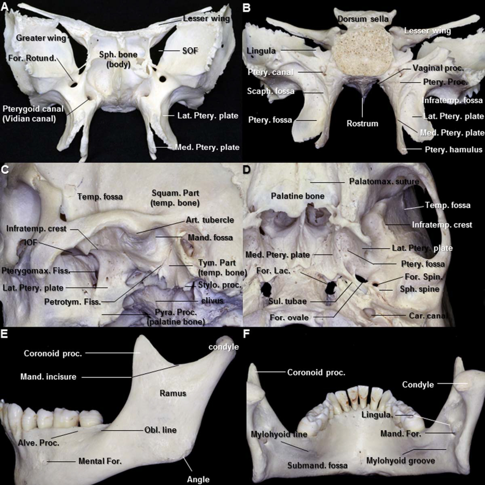
圖7。A.蝶骨前視圖。維管的前開口位於圓孔的內側下方。B.蝶骨後視圖。蝶的翼突從大翅與身體結合的區域垂直下降。每個過程由內側板和外側板組成,其上部在前麵熔接。內側翼板比外側翼板更窄更長;它的下端向外側彎曲,形成一個鉤狀突起,即翼狀hamulus,在翼狀hamulus周圍,齶板張肌的肌腱發生偏轉。C.顳下窩側位視圖。齶骨的錐體突起與翼狀板下端之間的角間距相吻合。 The medial portion of the tympanic part of the temporal bone forms the posterior boundary of the petrotympanic fissure. D. Inferior view of the infratemporal fossa. The sulcus for auditory tube, which is the attachment site of the cartilaginous part of the Eustachian tube to the cranial base, is located on the extracranial surface of the sphenopetrosal fissure, anterolateral to the foramen lacerum and posteromedial to the foramen ovale and spinosum. The foramen ovale, which is irregularly oval, lies close to the upper end of the posterior margin of the lateral pterygoid plate. E. Lateral view of the mandible. The oblique line in the mandible runs upward and backward from the mental tubercle and is continuous with the anterior border of the ramus. The upper border of the ramus is thin and bounds a wide notch, the mandibular incisures, which transmits the masseteric nerve and vessels from the infratemporal fossa. F. Posterior view of the mandible. The mandibular canal runs from the mandibular foramen obliquely downward and forward in the ramus and then horizontally forward in the body. The mylohyoid line is the origin of the mylohyoid muscle. The lingula, the medial border of the mandibular foramen, is the attachment site of the sphenomandibular ligament. Abbreviations: Alve., alveolar; Art., articular; Car., carotid; Condy., condyle; Fiss., fissure; For., foramen; Inf., inferior; Infratemp., infratemporal; IOF., inferior orbital fissure; Lac., Lacerum; Lat., lateral; Mand., mandible; Med., medial; Obl., oblique; Palatomax., palatomaxillary; Ptery., pterygoid; Pterygomax., pterygomaxillary; Petrotym., petrotympanic; Pyra. Proc., pyramidal process; Scaph., scaphoid; SOF., superior orbital fissure; Sph., sphenoid; Spin., spinosum; Squam., squamous; Stylo., styloid; Submand., submandibular; Sul., sulcus; Temp., temporal; Tym., tympanic. (Images courtesy of AL Rhoton, Jr.)
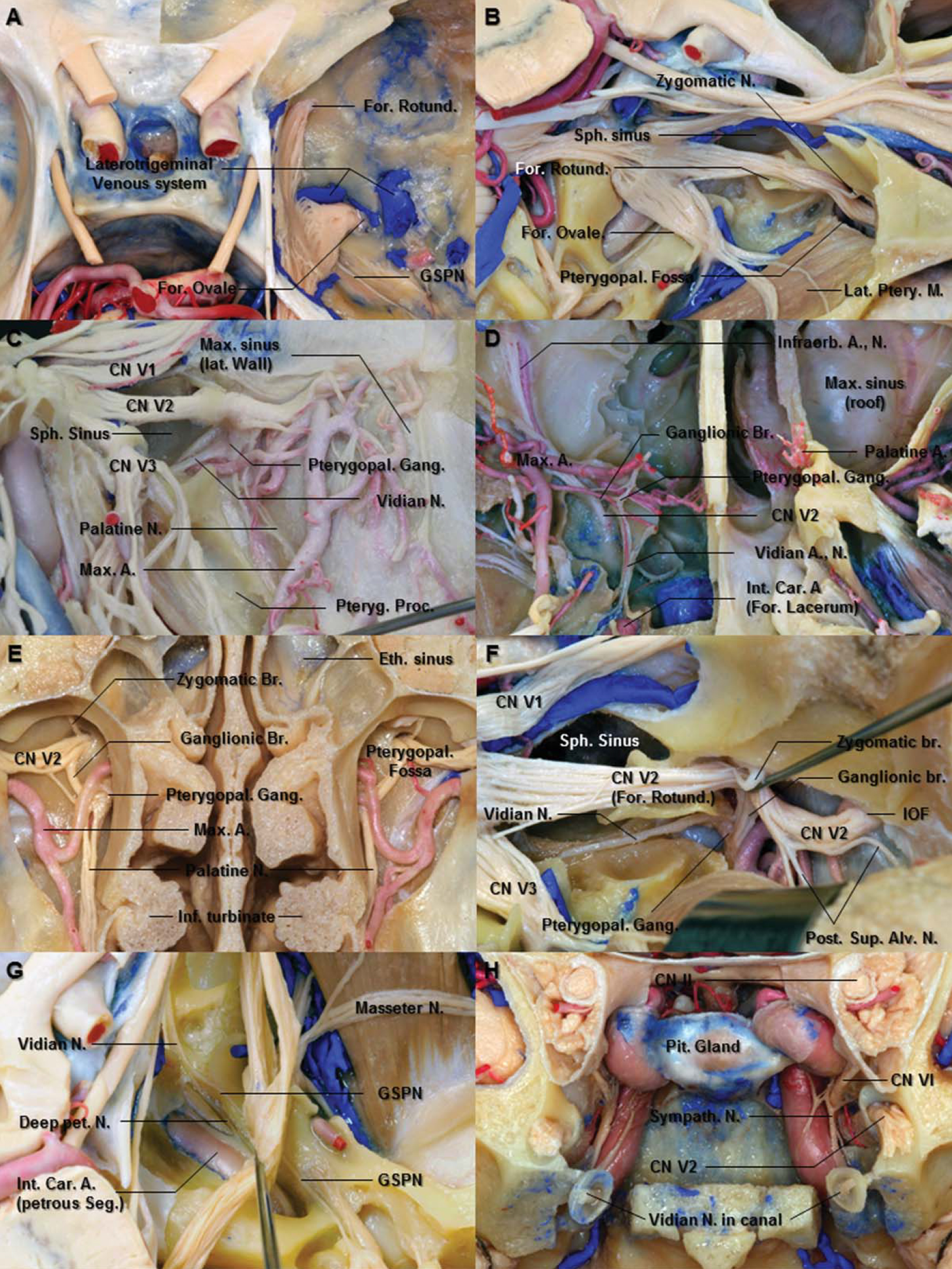
圖8。A.中窩上方視圖。中硬腦窩已被切除,露出三叉神經。三叉側靜脈係統是圍繞卵圓孔和下頜骨的靜脈係統。B.右側三叉神經上外側切麵。圓孔和卵圓孔周圍的骨被切除,分別暴露上頜和下頜骨。移除眼神經和上頜神經之間三角空間的骨頭進入蝶竇。上頜神經通過圓孔進入翼齶窩。C.右側翼齶窩側麵圖。切除顳骨、下頜支和翼狀肌,露出翼齶窩內的上頜神經分支。 The vascular structures are anterior to nervous structures in the pterygopalatine fossa. D. Inferior view of the pterygopalatine fossa. The pterygopalatine ganglion is located inferior and medial to the maxillary nerve. The ganglionic branch in the pterygopalatine fossa connects between the maxillary nerve and pterygopalatine ganglion. After crossing the upper part of the pterygopalatine fossa, the maxillary nerve inclines laterally and then enters the infraorbital groove and canal. E. Cross section through the ethmoidal and maxillary sinuses and the nasal cavity in front of the posterior maxillary wall. The posterior wall of the maxillary sinus has been removed to expose the pterygopalatine fossa and ganglion. The maxillary arteries enter the pterygopalatine fossa from the laterally by passing through the pterygomaxillary fissure and give rise to its terminal branches in the pterygopalatine fossa. F. Lateral view of the right pterygopalatine fossa. The maxillary nerve is situated superior and lateral to the pterygopalatine ganglion. The vidian nerve, which courses the floor of the sphenoid sinus, is inferomedial to the maxillary nerve and joins the pterygopalatine ganglion. The maxillary nerve also gives rise to the zygomatic and the posterior superior alveolar nerves. G. The trigeminal nerve has been reflected laterally to expose the vidian nerve. The vidian nerve is formed by the union of the GSPN and deep petrosal nerve. H. Coronal section through the orbital apex behind the pterygopalatine fossa. The anterior end of vidian canal, which opens into the medial part of the posterior wall of the pterygopalatine fossa, is funnel shaped. Abbreviations: A., artery; Alv., alveolar; Br., Branch; Car., carotid; CN., cranial nerve; Eth., ethmoid; For., foramen; Gang., ganglion; GSPN., greater superficial petrosal nerve; Inf., inferior; Infraorb., infraorbital; Int., internal; IOF., inferior orbital fissure; Lat., lateral; M., muscle; Max., maxillary; N., nerve; Pet., petrosal; Pit., pituitary; Post., posterior; Proc., process; Ptery., pterygoid; pterygopal., pterygopalatine; Rotund., rotundum; Seg., segment; Sph., sphenoid; Sup., superior; Sympath., sympathetic. (Images courtesy of AL Rhoton, Jr.)
V2進入PPF後,向PPG釋放神經節分支。然後它在眶下裂的下方向外側偏移,產生眶周外的顴神經和後上牙槽神經。然後V2在眶下神經的位置向內側轉彎,穿過眶下裂進入眶下溝,在那裏出現前上牙槽神經和中上牙槽神經(圖8B、8D、8E和8F)。
V2的分支根據其起源可分為四組,分別為:頭蓋骨、翼齶窩、眶下管和麵部。
腦膜神經.腦膜神經起源於中顱窩內圓孔附近的V2。它接受來自頸內交感神經叢的分支並伴隨腦膜中動脈的額支為中顱窩的硬腦膜供血。它的前小分支剛好到達前顱窩。這是V2的最小分支(Gray and Williams, 1989b;讓遊戲,2001 b)。
神經節的分支.翼齶窩(PPF)的神經節分支連接V2和PPG。翼齶神經節(PPG)位於上頜神經的下方和內側(V2)(圖8C-8F)。神經節支含有通往淚腺的節後副交感神經纖維,它與V2相連,進入其顴顳神經,經顴顳神經與淚神經相通到達淚腺(圖1D)。它們還包含來自眼眶骨膜和鼻子、上顎和咽的粘膜的感覺纖維(Gray和Williams, 1989b)。
顴骨神經.顴神經起於後腹肌的V2,向前、上、外側通過眶下裂進入眶內。它沿眶外側壁延伸,分為顴顳神經和顴麵神經(圖8B和9C)。

圖9。翼齶窩前斜麵擴大。翼齶神經節接收來自上頜神經的通訊支。蝶齶支通過蝶齶孔進入外側鼻腔。B.由翼齶神經節發出的上頜動脈末梢支和鼻支穿過鼻腔外側壁,沿蝶骨麵前進。C.軌道的前上方視圖。上頜神經產生眶下神經、顴神經和上牙槽神經。翼齶窩前壁由上頜竇後壁構成。D.上頜骨前上麵。上頜神經通過眶下裂進入眶下溝和管,繼續作為眶下神經。 The infraorbital nerve emerges in the face through the infraorbital foramen. E. The posterior wall of the right maxillary sinus has been removed to expose the pterygopalatine fossa. The maxillary artery is located anterior to the pterygopalatine ganglion. The palatine nerves descend through the palatine foramina of the maxilla located at the inferior tip of the pterygopalatine fossa. F. The right buccolabial muscles have been removed to expose the branches of the infraorbital nerve. The branches of the infraorbital nerve communicate with the branches of the facial nerve to form infraorbital plexus in the infraorbital space. G. The branches of the infraorbital nerve have been removed. The infraorbital nerve gives rise to anterior superior alveolar nerve just behind the infraorbital exit through the foramen. Abbreviations: A., artery; Alv., alveolar; Ant., anterior; Br., branch; CN., cranial nerve; For., foramen; Gr., great; Inf., inferior; Infraorb., infraorbital; Max., maxillary; N., nerve; Pit., pituitary; Post., posterior; Pterygopal., pterygopalatine; Rotund., rotundum; SOF., superior orbital fissure; Sph., sphenoid; Sphenopal., sphenopalatine; Sup., superior; Temp., temporal; Zygo., zygomatic. (Images courtesy of AL Rhoton, Jr.)
Zygomaticotemporal神經.顴顳神經沿著眶下外側角,向V1淚神經發出聯絡支,穿過顴骨的骨管,進入顳窩。然後在骨和顳肌之間上升,穿過顴弓上方約2厘米的顳深筋膜,支配顳區皮膚(Totonchi et al., 2005)。有時,當淚神經缺失時,顴顳神經取而代之,支配淚腺(Shankland, 2001b)。
顴麵部神經.顴麵神經也沿著眶下外側角走,通過顴骨上的顴麵孔出現在臉上,支配臉頰突出處的皮膚。它與麵神經的顴支和V2的瞼支形成一個神經叢。
後上牙槽神經.後上牙槽神經起於上頜神經,在下頜骨下腔內穿透眶下管,並向前下方下降,穿過上頜竇的顳下表麵(圖8C和8F)。進入上頜竇後,神經在上頜竇粘膜下向前穿過,為這些膜提供傳入神經支配。起源於顴骨區V2的後上牙槽神經的數量可以從1到3個不等(Moretto et al., 2005)。後上牙槽神經與中上牙槽神經相通,形成分支,形成牙叢(Shankland, 2001b)。它還為上牙齦和臉頰的相鄰部分提供分支(Gray and Williams, 1989b)。
Pterygopalatine神經節.翼齶神經節(PPG)是周圍副交感神經神經節中最大的,位於翼齶神經節深處,靠近蝶齶孔和翼管前方,與V2相連。神經節從上外側的V2(神經節分支)和上內側的維神經接收通信支,並從神經節下表麵產生齶大神經和齶小神經,從內側表麵產生蝶齶神經和咽支,從上表麵產生眶支(圖8C-8F, 9A和9B)。維神經是由岩大神經和岩深神經結合形成的,岩大神經和岩深神經分別傳遞從膝狀神經節水平的麵神經(神經中間)產生的副交感神經纖維,岩深神經從頸動脈叢傳遞交感神經纖維,到達淚腺和鼻黏膜(圖1D、8G和9A)。
齶神經.齶神經分布於上顎、軟齶、扁桃體和鼻粘膜。它們也通過PPG神經和維dian神經將味覺衝動從上顎傳遞到麵神經的中間神經,後者終止於腦幹的孤立束和核(圖1D)。齶大神經向下穿過上頜骨齶大孔,位於下顎下緣的下尖,並在硬齶下表麵的凹槽中向前延伸(圖9A和9E)。它與鼻齶神經的細絲相連,鼻齶神經是蝶齶神經的一個分支。連接PPG的最粗神經總是蝶齶神經和齶大神經(Rusu et al., 2009)。
小齶神經在離開PPG後,向下穿過齶骨的小齶孔,向小舌、扁桃體和軟齶分支。這些神經與舌咽神經的分支吻合,在齶扁桃體周圍形成一個扁桃體叢(圖9E) (Shankland, 2001b)。
神經維迪安.維迪安管連接下腹f和淚孔。它傳遞維神經、動脈和靜脈,並包含脂肪組織。維管的前開口位於後壁圓孔的內側下方(圖8D)。根管是一條從前開口向後外側的直至微彎曲的通路(圖8D和8H) (Kim等人,1996年)。管位於通氣良好的蝶竇底部的下方(圖8D和8F)。後開口位於翼狀內側板後緣上方。它向後打開,進入到前外側邊緣的上部,直至淚孔(圖8D)。維管後開口位於卵圓孔和V3內側邊緣內側6.9 mm (4.8-9.1 mm)處(Rahman等,2009)。
雖然由岩大神經和岩深神經結合而成的維底神經不是V2的分支,但它進入並穿過維底管,終止於位於維底管前開口前方的PPG(圖8G)。岩大神經(GSPN)包含來自齶粘膜的味覺纖維和節前副交感神經纖維,它們起源於腦幹的上唾液腺核,並在神經中間區傳遞。節後副交感神經纖維穿過V2及其顴支,通過眶下裂進入眼眶,並與眼神經淚支相通到達淚腺(圖1D) (Gray and Williams, 1989b;Rhoton, 2003 b;Rahman et al., 2009)。岩深神經包含節後交感神經纖維,起源於頸上神經節向上延伸至頸內動脈的神經叢。因此,維甸神經包含感覺纖維、交感纖維和副交感纖維。然而,副交感神經纖維在PPG中有突觸,感覺和交感神經纖維通過神經節時沒有突觸(Gray and Williams, 1989b;el Shazly, 1991;Rahman et al., 2009)。
維管和神經已成為引導顯微外科手術和內窺鏡入路沿著蝶竇底進入頸動脈岩和海綿竇前內側部分和Meckel’s cave的重要標誌(圖8G和9B) (Rahman等,2009)。
鼻分支.鼻支通過鼻腔側壁的蝶齶孔進入鼻腔。這些分支包括內側和外側上鼻神經和鼻齶神經(圖9B)。鼻後上外側神經。鼻後上外側支通常為6支,支配上鼻甲和中鼻甲後部的粘膜和後篩竇內襯(Gray and Williams, 1989b)。
內側後上鼻神經.內側後上鼻支,數量通常為2 - 3支,穿過蝶竇開口下方的鼻頂,供應鼻頂後部分和鼻中隔的粘膜(Gray and Williams, 1989b)。
Nasopalatine神經.鼻齶神經是PPG的鼻支中最大的神經,它穿過蝶齶孔進入位於開口下方的鼻腔,到達蝶竇,到達鼻中隔。然後,它在骨膜和鼻中隔的粘膜之間向前下延伸,為鼻中隔提供幾根纖維,通過切開孔離開鼻腔,以供應硬齶前部的黏膜而結束,在那裏與齶大神經相連(Gray and Williams, 1989b;讓遊戲,2001 b)。
咽神經.咽神經發源於PPG的後部,與上頜動脈的咽支一起穿過齶陰道管,支配聽管後麵的鼻咽粘膜。齶陰道管是指將齶骨的蝶突與蝶骨的陰道突結合形成的短骨隧道(圖7B和11B) (Wentges, 1975)。齶陰道管位於維甸管的內側和下方。齶陰道管位於鼻咽頂下側下側下壁的後壁上。

圖10。A.右側顳下窩上外側切麵。中窩底已經被切除露出了下頜神經的分支。咬肌和顳深神經經過翼靜脈叢和翼外肌上端。B.下頜支已移至翼狀肌。頰神經向前外側在翼外肌的上下頭之間。C.下頜髁和翼外肌在前方被反射,暴露出上頜動脈和下頜神經的分支。耳顳神經在翼外肌內側向後延伸到下頜骨頸部。腦膜中動脈穿過耳顳神經的兩根之間。D.下頜神經分支的放大視圖。 The anterior division of the mandibular nerve passes in the horizontal plane just below the pericranium of the infratemporal surface of the middle cranial fossa. The chorda tympani nerve enters the infratemporal fossa through the petrotympanic fissure, descends medial to the auriculotemporal and inferior alveolar nerves, and joins the lingual nerve. E. The lateral pterygoid muscle has been removed to pterygoid nerves. The medial pterygoid nerve originates from the medial aspect of the main trunk below the foramen ovale close to the otic ganglion. The tensor veli palatini muscle is located medial to the branches of the mandibular nerve. F. The lesser petrosal nerve emerging through the roof of the tympanic cavity of the temporal bone passes along the tensor tympani muscle anterolaterally to enter the otic ganglion. Abbreviations: A., artery; Acc., accessory; Alv., alveolar; Auri. Temp. N., auriculotemporal nerve; Br., branch; Chor.Tym. N., chorda tympani nerve; CN., cranial nerve; For., foramen; GSPN., greater superficial petrosal nerve; Inf., inferior; Lat., lateral; LSPN., lesser superficial petrosal nerve; M., muscle; Max., maxillary; Med., medial; MMA., middle meningeal artery; N., nerve; Plex., plexus; Ptery., pterygoid; Spin., spinosum; Sph. Mand. Lig., sphenomandibular ligament; Supf., superficial; Temp., temporal; Ten. Tym. M., tensor tympani muscle; Ten. Vel. Pal. M., tensor veli palatini muscle. (Images courtesy of AL Rhoton, Jr.)
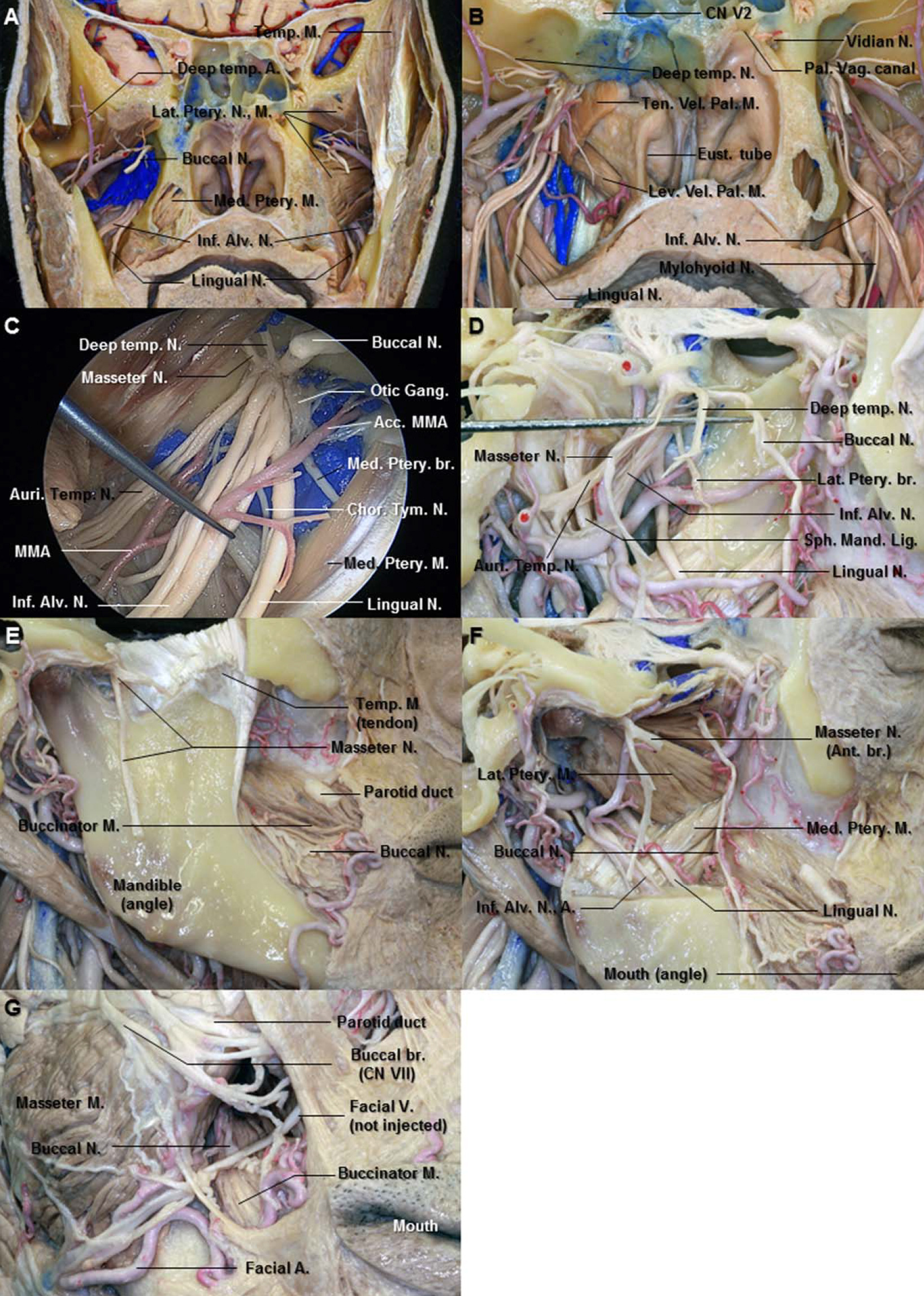
圖11。A.翼齶窩後麵的冠狀麵。右側翼外肌已經從翼靜脈叢和下頜神經後分支中取出。翼外肌的神經起源於頰神經穿過翼外肌的兩個頭之間。B.切除右側翼靜脈叢和翼板,露出下頜神經分支。下頜神經後段位於翼外肌(切除)和齶板張肌(切除)之間。齶陰道管位於維甸管的中間。C.右側下頜神經近端放大視圖。翼肌內側的神經起源於下頜神經靠近耳部神經節的內側。D.右側顳下窩側位麵。 The mandibular ramus and lateral pterygoid muscle have been removed. The buccal, deep temporal, and masseter nerves have been elevated with the dissector. This division passes in the horizontal plane. The auriculotemporal nerve passes backward between the sphenomandibular ligament and the ramus of the mandible (removed). E. The masseter muscle has been removed from the mandible to expose the masseter nerve. The masseter nerve passes laterally above the lateral pterygoid muscle in front of the temporomandibular joint and behind the tendon of the temporal muscle. It runs to the deep surface of the master muscle. F. The mandibular ramus has been removed to expose the lingual and the inferior alveolar nerves. G. Anterior view of the right cheek. The buccal nerve emerges from the undersurface of the ramus of mandible and unites with the buccal branches of the facial nerve. Abbreviations: Acc. MMA., Accessory middle meningeal artery; Ant., anterior; Alv., alveolar; Auri. Temp. N., auriculotemporal nerve; Br., branch; Chor.-Tym. N., chorda tympani nerve; CN., cranial nerve; Eust., Eustachian; Gang., ganglion; Inf., inferior; Lat., lateral; Lev. Vel. Pal. M., levator veli palatini muscle; M., muscle; Med., medial; N., nerve; Pal. Vag., palatovaginal; Sph. Mand. Lig., sphenomandibular ligament; Temp., temporal; Ten. Vel. Pal. M., tensor veli palatini muscle. (Images courtesy of AL Rhoton, Jr.)
在眶下管。上頜神經(V2)在穿過下下緣上部後,向外側傾斜,然後進入眶下溝和管,繼續形成眶下神經(ION)(圖8B和8D)。槽和管的平均長度分別為12毫米(範圍5-22毫米)和14毫米(範圍7-22毫米)(Rahman等人,2009年)。眶下管長軸向下並向內穿過麵部(圖9D)。離子在眶底以下、上頜竇頂部的管道中流動,直到通過眶下緣下方的眶下孔出現在麵部。離子分支進入眶下管內的中、前上牙槽神經(圖9F)。
中上牙槽神經.中上牙槽神經將離子留在眶下溝,即眶下管的後部(圖9C)。它在上頜竇的外側壁向前延伸,以小分支結束,小分支與上牙叢相連,為上前磨牙提供小支。中上牙槽神經與後上牙槽神經吻合(Shankland, 2001b)。
前上牙槽神經.前上牙槽神經比中上牙槽神經大,在通過孔的眶下出口之前離開ION的外側(圖9G)。它穿過上頜竇前壁的管道,分成分支供應門牙和犬牙。它與中牙槽神經相通形成上牙叢,並產生鼻支,鼻支通過下鼻道側壁的一個小管,供給側壁前區和鼻腔底的粘膜(Gray and Williams, 1989b;讓遊戲,2001 b)。
從表麵上.眶下神經(ION)是一種完全感覺神經,是上頜神經(V2)的末端分支。當離子通過眶下孔(通常位於眶緣下緣1厘米內)出現在麵部時,它分為下瞼支、內鼻支、外鼻支和上唇支,它們供應下眼瞼的皮膚和結膜、鼻子的外側部分和上唇的皮膚和黏膜(圖9D和9F) (Gray and Williams, 1989b;摩爾和達利,1999年;Hu等人,2006)。
當離子從眶下孔流出時,除下瞼支外,其餘三個分支向下延伸。眶下空間位於眶下孔下方,在這裏離子向下移動,麵神經橫向移動形成眶下神經叢(圖9F) (Hu等人,2006)。
下眼瞼的分支.下瞼支,通常有兩個或三個,向上深入到眼輪匝肌,刺穿肌肉,供應下眼瞼的皮膚和結膜。這些神經與靠近外側眥的麵神經和顴麵神經相通(圖9F)。
鼻分支.鼻外支支配鼻子外側表麵的皮膚。鼻內支通過眶下孔的內側出現,沿著鼻子向下,並環繞鼻翼(圖9F)。最後,它供給鼻中隔和鼻子前庭。鼻支與篩前神經的外支相連(Gray and Williams, 1989b;Hu等人,2006)。
上唇分支.大而多的上唇支向下延伸至提唇上肌後方,支配臉頰前部、上唇、口腔黏膜和唇腺的皮膚,並與麵神經的顴支相連,形成眶下神經叢(圖9F) (Gray and Williams, 1989b)。
下頜骨分支(V3)是三叉神經的三個分支中最大的。它供應牙齒,下頜骨的牙齦,顳區的皮膚,耳廓的一部分,下唇和臉的下部。然而,與眼區(V1)和上頜區(V2)傳遞純傳入纖維不同,V3區還包含運動或傳出纖維,支配附著於下頜骨的肌肉,包括咀嚼肌、下頜舌骨肌、二腹肌前腹、齶板張肌和鼓室張肌。
V3,三叉神經的是最大的部門,由兩個根源:一個大型的、感官的根,所得側三叉神經節的一部分,出現幾乎立即通過卵圓孔未閉的楔形骨和一個小電機產生的根源在superomedial主要感覺根腦橋的一部分,通過在三叉神經節和統一感覺根就在卵圓孔未閉(無花果。2 b、2 c和5 d)。V3經卵圓孔出中窩,進入顳下窩和翼外肌內側,在那裏主幹分為小的前段和大的後段(圖10A-10C)。
顳下窩是位於顳下脊下方的深部空間,它延伸到顳窩上方包括顳肌和咽旁間隙。顳下窩的骨邊界在前為上頜骨後外側麵,在前為翼突外側板,在外側為下頜支,在後為顳骨的鼓部和莖突(圖7C和7D)。這個窩包含上頜動脈、三叉神經的下頜支(V3)、翼肌和翼靜脈叢(圖10A、10B、11A和11C)。從地形圖上看,下頜部與翼外肌緊密相連,一些下頜神經分支穿過翼外肌(圖10A和10B)。
不可分割的樹幹.小運動根起源於腦橋,經過三叉神經節下方,並與卵圓孔外的大感覺根結合,在卵圓孔外,V3的主幹位於內側齶板張肌和外側翼狀肌之間(圖10C、10E和11B)。在兩個根的連接處,主幹(V3)產生腦膜分支和從內側通向翼肌內側的神經,然後分為一個較小的前幹和一個較大的後幹(圖10E)。
腦膜的分支.腦膜支是一種神經棘,起於竇神經節附近,與腦膜中動脈一起通過棘孔上升支配中顱硬腦窩。它分為前支和後支,伴隨動脈的主支。神經棘包含來自中腦膜叢的交感神經節後纖維(Gray and Williams, 1989b)。
內側翼狀肌神經.翼內神經起於卵圓孔下方靠近耳部神經節的主幹內側,向下支配翼內肌(圖10E和11C)。它包含內側翼肌的感覺纖維、運動纖維和本體感覺纖維。
掌寬神經張量.到齶板張肌的神經像翼內側神經一樣起源於主幹的內側。它通過竇神經節支配位於蝶骨內側翼狀板基部附近的齶狀張肌(Shankland, 2001c)。
張量定音鼓神經.到鼓室張量的神經可能是單獨的,也可能與未分裂的主幹上的齶闊肌張量神經共同生長。它不間斷地穿過耳部神經節。神經穿過聽管軟骨進入鼓室張肌(Kierner et al., 2003;讓遊戲,2001 c)。
耳神經節.小腦神經節是一個小的,橢圓形,扁平,紅灰色的神經節,位於卵圓孔的正下方(圖11C)。它位於下頜骨不可分割幹的內側表麵靠近翼內側神經和齶闊肌張神經的起點。該神經節是一個周圍副交感神經節,在地形上與下頜神經密切相關,但功能上與舌咽神經相連(圖1D)。神經節前副交感神經纖維離開腦幹舌咽神經的唾液腺下核。它進入鼓室支,通過頸靜脈孔和頸動脈管之間頸動脈脊的小管到達鼓室,穿過鼓室叢,從顳骨鼓室頂出,形成岩淺小神經(LSPN)。LSPN向外耳神經節轉移(圖10D和10F)。
在神經節細胞突觸後,神經節後副交感神經分泌運動纖維通過耳顳神經到達腮腺(圖10C, 12C, 12D)。交感神經根起源於腦膜中動脈上的神經叢。它包含來自頸上交感神經節的節後纖維,這些纖維在沒有突觸的情況下穿過耳部神經節。交感神經纖維供應腮腺的血管(Shankland, 2000)。

圖12。A.去除中窩骨後的上視圖。後顳深神經在翼外肌上頭的上表麵。咬肌神經是下頜神經前分支中最靠後的分支。B.顳骨的鱗狀部分已被切除,露出顳骨肌肉的深層表麵。前深顳神經在翼外肌的上、下兩個頭之間。顳深部神經進入顳肌前部的深部表麵。C.右側顳下窩放大視圖。腦膜中動脈穿過耳顳神經的兩根之間。耳顳神經在下頜骨頸後的內側到外側。 D. Lateral view of the right mandibular neck area. The parotid gland has removed to expose the auriculotemporal and facial nerves. The auriculotemporal nerve gives off the parotid branches and ascends in the parotid gland between the temporomandibular joint and external acoustic meatus. It communicates with facial nerve at the posterior border of the mandibular ramus. E. Lateral view of the right preauricular area. The auriculotemporal nerve ascends posterior to the superficial temporal artery over the posterior root of zygoma. The frontal branches of the facial nerve pass over the zygomatic arch. Abbreviations: A., artery; Ac., acoustic; Ant., anterior; Auri. Temp. N., auriculotemporal nerve; Br., branch; Ext., external; Gang., ganglion; Gl., gland; Inf. Alv. N., inferior alveolar nerve; Lat., lateral; M., muscle; Mand. For., mandibular foramen; Max., maxillary; Med., medial; N., nerve; Post., posterior; Post. Auri. A., posterior auricular artery; Ptery., pterygoid; STA., superficial temporal artery; Sup., superior; Temp., temporal; Tr., trunk. (Images courtesy of AL Rhoton, Jr.)
前段.V3在顳下窩分為前小段和後大段。從卵圓孔到V3分叉的未分割主幹的平均距離約為7.7 mm(範圍5-12 mm) (Vrionis et al., 1996)。較小的前分支產生感覺頰神經和運動分支咬肌神經,顳深神經,翼外肌神經。此分隔在正中顱窩顳下表麵的周膜下方的水平麵上進行(圖10A)。
頰神經.感覺頰神經在翼外肌的兩個頭之間向前外側,在顳肌下部下方,深入到下頜骨和咬肌(圖10A, 10B和11A)。它的發展方向是在冠突和上頜骨結節之間。它從下頜骨分支的下表麵和咬肌的前邊緣出現,並與麵神經的頰支結合(圖11E-11G) (Gray and Williams, 1989b)。頰神經將感覺神經支配傳遞到頰肌、頰粘膜和嘴角的皮膚。它通常穿過頰肌的後半部分,廣泛分布於臉頰的頰麵(Tubbs等,2010年)。然而,一些作者報道頰神經是一種混合神經,它為翼外肌和顳肌提供分支(Kim et al., 2003;Piagkou等,2011;戴維斯等人,2012)。
頰神經位於口角外側3cm處(範圍2.8-3.3 cm)。它位於腮腺管進入頰肌入口的尾部7mm處(範圍,4 - 12mm)(圖11E和11G) (Tubbs et al., 2010)。
橫向翼狀肌神經.翼外神經進入肌肉深表麵(圖10E和圖11D)。一般認為翼外神經主要起源於頰神經(圖11A) (Gray and Williams, 1989a;Kim等人,2003;Piagkou等人,2011)。它是整個三叉神經所有分支中最不一致的(Shankland, 2001c)。Foucart等人(1998)報道,翼外肌受一到三個來自下頜神經前幹的分支支配,另外還有來自頰神經、耳顳神經和咬肌神經的分支支配(Foucart等人,1998)。然而,Kim等人觀察到翼外肌不僅受頰神經支配,還受顳深神經和直接來自下頜神經幹的分支支配。起源於耳顳神經或咬肌神經的神經支不支配翼外肌(Kim et al., 2003)。
咬肌神經.咬肌神經在顳下頜關節前麵的翼外肌上方和顳肌肌腱後麵向外側穿過(圖10A)。它穿過下頜切跡後部與咬肌動脈,延伸至咬肌深表麵(圖11D和11E)。咬肌神經在短咬肌中分前支和後支。前支比後支更大更長(圖11E和11F) (Hwang et al., 2005)。咬肌神經是V3前分支中最後的分支(圖10A和11D)。
咬肌運動神經位於耳屏前方3.16±0.30 cm處,顴弓尾緣下方1.08±0.18 cm處。相對於顴弓,神經在遠端進入咬肌時形成了50±7.6°的角度(Borschel et al., 2012)。
顳深神經.神經的前後支通常經過翼外肌上方,進入顳肌深表麵(圖10A) (Gray and Williams, 1989a)。Kwak等人報道,前顳深神經在翼外肌的上、下兩個頭之間穿過,在顳下窩向前移動,然後向上轉,在顱骨外側進入顳窩,進入顳肌前部的深表麵。後顳深神經位於上頭部上表麵,分布於顳肌後部(圖12A、12B)。在所有標本中,前深顳神經起源於頰神經,數量為1或2個(圖12B) (Kwak et al., 2003;讓遊戲,2001 c)。後分支,數量一般為2或3個,與咬肌神經共同出現(Akita et al., 2000;格雷和威廉姆斯,1989a)。
後段.V3的大後節主要是感覺神經,但也有一些來自運動根的纖維。它分為三個主要分支:耳顳神經、下牙槽神經和舌神經,它們沿矢狀麵向下延伸至麵部下部結構(圖10C、10D和11D)。
耳顳神經.耳顳神經起源於V3的後幹,主要有兩個分支,腦膜中動脈通過它們之間(Gray and Williams, 1989b;Joo等,2013;Schmidt et al., 1998)。這些根環繞腦膜中動脈,連接並向後延伸至齶板張肌表麵的翼外肌,在蝶下頜韌帶和下頜骨頸部之間穿過(圖10C、10D、11C和11D)。然而,Gülekon等(2005)報道在16具屍體的32個顳下窩中檢出16例耳顳神經單根(50%),12例雙根(37.5%),3例三根(9.4%),1例四根(3.1%)(Gülekon等,2005)。
耳顳神經在下頜骨頸後的內側向外側延伸,發出腮腺分支,在顳下頜關節和外耳道之間的腮腺內向上改變方向(圖12D)。它從關節後方出現,上升至顳淺血管後方,越過顴骨後根,並分裂為顳淺支(圖12D和12E)。在25具新鮮屍體的解剖中,該神經總是位於太陽穴耳前上區域的顳淺動脈外側(Janis et al., 2010)。
耳顳神經包含CN V3和CN IX的體感和分泌運動纖維(Loughner et al., 1990)。它傳遞分泌運動纖維到腮腺。神經節前副交感神經纖維經舌咽神經的鼓室支離開舌咽神經,在中耳腔海角與鼓室叢彙合,經岩小神經離開耳咽神經叢;這種神經的末端是外耳神經節,神經節前纖維在這裏形成突觸(圖1D)。耳部神經節位於卵圓孔外位於下頜神經的內側並向耳顳神經的根發出纖維耳顳神經通過它從下頜神經發出;這些纖維和感覺纖維一起離開耳顳神經形成該神經的腮腺分支並到達腺體。耳顳神經也與麵神經密切相關。它在下頜支的後緣與麵神經相通,耳顳神經在此穿過髁突頸後方,麵神經出莖突孔後在腮腺內分成兩個主要分支(圖12D)。偶爾,在顳下頜區耳顳神經和下牙槽神經之間可以看到連接的分支(Gülekon et al., 2005;Janis等人,2010)。
文獻回顧顯示,Baumel et al.的研究對耳顳神經的變化提供了最全麵的定量數據(Baumel et al., 1971)。根的長度為15毫米(範圍5-23毫米)。根係之間的距離為6mm(範圍為1 - 13mm)。上根通常位於中腦膜動脈外側,下根位於中間(圖10C和12C) (Baumel et al., 1971)。的average length of the posterior trunk from foramen ovale to origin of the upper root of the auriculotemporal nerve was 3.3 mm (range, 1–7.5 mm) (Joo et al., 2013).
舌神經.舌神經是V3後分支的主要分支它為舌頭的前三分之二和牙齦提供感覺沿著下頜牙齒的舌側變化。在顳下窩內,舌神經起源於V3的後分支。從卵圓孔到舌和下牙槽神經分叉的後幹平均長度為13.5 mm(範圍6-32 mm) (Joo et al., 2013)。Shinohara等報道,卵圓孔到舌下牙槽神經分叉的平均距離為8.7 mm(範圍0-14 mm)。他們發現了高分叉,包括V3分叉和卵圓孔上方舌神經的分叉(Shinohara et al., 2010)。首先,舌神經在齶板張肌和翼外肌之間向下延伸(圖10C, 10D, 11A,和13D)。在翼外肌的下緣,該神經與麵神經的鼓膜索支相連,它包含膝狀神經節和節前副交感神經纖維的感覺纖維,通常還包含下牙槽神經的一個分支(圖10C, 10F和11C)。除此之外,舌神經還包括真正的三叉神經纖維、來自麵神經的味覺纖維和用於下頜下神經節的自主神經纖維(圖1D) (hollinhead, 1982b)。舌神經在下頜支和翼內側肌之間向前向下延伸,位於下牙槽神經的前方略深處,與下牙槽神經相通(圖10B、10C和11B)。 It then passes inferiorly to the attachment of the superior constrictor at the posterior end of the mylohyoid line, where it is covered only by the gingival mucosa (Benninger et al., 2012). According to some reports, the average distance from the lingual alveolar crest to the lingual nerve was 4.45 mm in the retromolar region (Pogrel et al., 1995), and the average distance in the third molar region was 2.28 mm (Kiesselbach and Chamberlain, 1984). It leaves the gingiva and passes on to the side of the tongue, where it crosses the styloglossus and runs on the lateral surface of the hyoglossus and deep to the mylohyoid, above the deep part of the submandibular gland and its duct (Figs. 13A and 13B). The lingual nerve proceeds forward on the side of the tongue, lateral to the hyoglossus and genioglossus, and divides into terminal branches which lie directly under the lingual mucosa (Gray and Williams, 1989b). The nerve distribution in the tongue is actually more complicated. The nerve fibers are complexly intermingled, and communication between the lingual and the hypoglossal nerve have been reported (Shinohara et al., 2010).
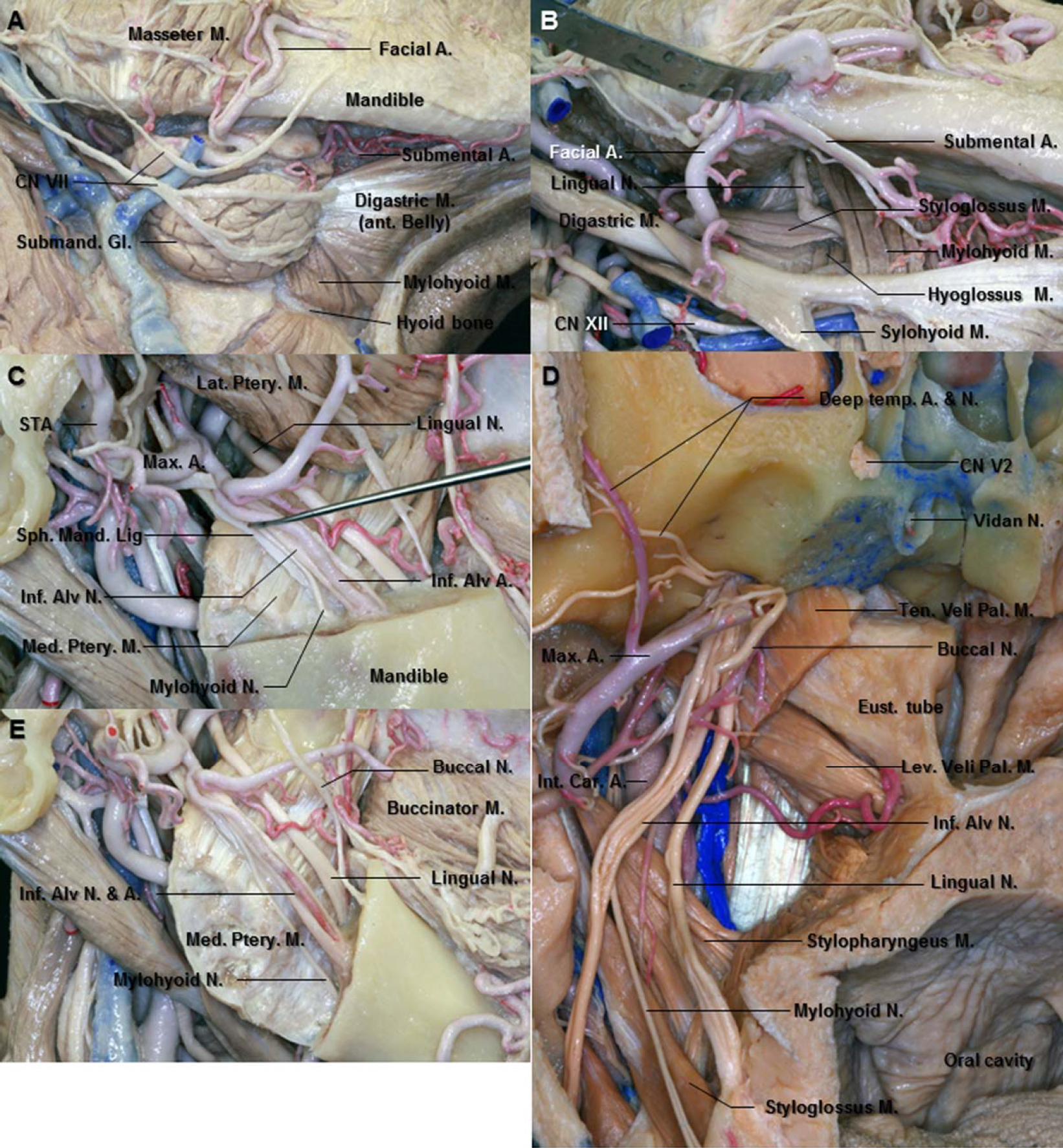
圖13。A.右側下頜下區域的下外側視圖。麵神經在位於二腹肌肌腱上的頜下腺上運行。B.右側下頜下三角的放大視圖。下頜下腺被切除露出下頜下三角的內容物。舌神經經過下頜支和翼突內側肌之間,穿過莖突舌突肌,在下頜下腺深部上方舌突舌肌和下頜舌骨舌肌之間穿行。C.右側顳下窩側位圖。舌神經在下頜支(已移除)和翼內肌之間向前向下延伸,位於下牙槽神經的前方略深處。蝶下頜韌帶是一條扁平的細帶,從蝶骨棘向下延伸,延伸至下頜孔舌部。D.右側顳下窩前視圖。 The lingual nerve is anterior and medial to the inferior alveolar nerve. The inferior alveolar nerve gives rise to the mylohyoid nerve before it enters the mandibular foramen. It then emerges in the submandibular triangle and supplies both mylohyoid and anterior belly of the digastric muscles. E. The angle of mandible has been removed. The mylohyoid nerve runs down and forward between the mandible and the medial pterygoid muscle and lodges in the mylohyoid groove. The buccal nerve emerges from the undersurface of the ramus of the mandible. Abbreviations: A., artery; Alv., alveolar; Ant., anterior; Car., carotid; CN., cranial nerve; Eust., Eustachian; Gl., gland; Inf., inferior; Int., internal; Lat., lateral; Lev. Vel. Pal. M., levator veli palatini muscle; M., muscle; Max., maxillary; Med., medial; N., nerve; Ptery., pterygoid; Sph. Mand. Lig., sphenomandibular ligament; STA., superficial temporal artery; Temp., temporal; Ten. Vel. Pal. M., tensor veli palatini muscle. (Images courtesy of AL Rhoton, Jr.)
大部分副交感神經纖維離開舌神經在頜下腺的位置到達頜下神經節,當它在頜下腺上方運動時從神經上懸空;神經節的纖維分布於下頜下腺和舌下腺,舌神經的感覺纖維分布於舌的前三分之二(圖1D和13A)。
下牙槽神經.下牙槽神經是三叉神經下頜骨分支的最大分支。神經通常有四個分布支和一些交流支,其中三個是感覺支,一個是運動支。感覺分支是敏銳神經,精神神經,以及通向下前磨牙和臼齒的神經。運動支是到下頜舌骨肌的神經(圖13C-13E)。下牙槽神經起於下頜主幹的後外側表麵,向下延伸至翼外肌,然後在翼外肌的下緣,在蝶下頜韌帶和下頜骨支之間穿過,直至下頜孔(圖7E、7F、10C、10F和13C)。下頜切槽到下頜孔的距離為17.4 mm (15-20 mm) (Joo et al., 2013)。在翼外肌下方,伴發於上頜動脈的下牙槽動脈(圖13E)。下牙槽神經進入下頜骨孔在下牙槽動脈的前方和上方。下牙槽神經進入下頜孔前的長度為31.1 mm(範圍13-44 mm) (Joo et al., 2013)。 The nerve passes through the body of the mandible, sometimes enclosed by a thick connective tissue sheath within the bony canal, and gives off branches to the teeth which may form a plexus between the trunk of the nerve and the roots of the teeth (Zoud and Doran, 1993).
下牙槽神經在進入下頜孔之前釋放出下頜舌骨神經(圖13D和13E)。通向下頜舌骨神經的神經穿過蝶下頜韌帶,在下頜舌骨線下向下向前延伸,這是下頜骨分支上的一條溝。然後它出現在二腹肌三角,供應二腹肌的下頜舌骨肌和前腹(Gray and Williams, 1989b;庫馬爾等人,2011;讓遊戲,2001 c)。然而,Kumar et al.(2011)報道稱,他們在50具印度南部屍體中的5具中發現了通向下頜舌骨神經的神經,該神經起源於下頜神經幹,通過後內側到達下牙槽神經(Kumar et al., 2011)。最近,Kim等人(2004)在12.5%的病例中描述了下頜舌骨和舌神經之間的交流,他們首次提到這種交流可以為側支感覺傳遞到舌頭提供另一種途徑(Kim等人,2004)。
下牙槽神經的牙支支配磨牙、前磨牙、門牙和犬齒。在進入牙根之前,它們相互交流並形成一個下牙叢,它位於下頜管和下頜牙根之間。精神神經,純粹的感覺神經,是下牙槽神經的一個大分支,它離開下頜骨的內部支配著下巴和下唇的皮膚、嘴唇的粘膜和鄰近的牙齦。頦孔通常位於從眶上切跡向下畫出的垂直線上,位於前磨牙水平以下(圖7E) (hollinhead, 1982b)。精神神經從它的孔中出來,在口凹肌下麵分成三個分支:一個分支向下延伸到下巴的皮膚,另兩個分支向上延伸到下唇的皮膚和粘膜。這些分支與麵神經的下頜分支相連。
頜下神經節.周圍副交感神經節是一個小的梭狀體,位於舌舌肌的上部。它位於頜下腺深部上方,舌神經下方,由前絲和後絲從舌神經懸垂(圖13A和13B)。雖然神經節與舌神經有關,但它在功能上與麵神經和鼓膜脊索相連。神經節前副交感神經纖維離開腦幹的麵神經上涎核。它進入鼓膜索它離開了莖突孔上方的麵神經在它的小管中向上和向前循環穿過後壁進入鼓膜腔。它穿過鼓膜上部在錘骨和砧骨之間,並通過鼓膜岩裂中的一個通道離開腔體。然後它以銳角連接舌神經的後側(圖10F和11C)。鼓膜脊索中含有節前副交感神經分泌運動纖維,這些纖維進入下頜下神經節,節後纖維從那裏傳遞到下頜下和舌下腺體(圖1D)。
交感神經根起源於麵部動脈上的神經叢。它包含來自頸上神經節的節後纖維,它穿過下頜下神經節,但沒有突觸。它們是頜下腺和舌下腺血管的血管舒縮器。
三叉神經是最大的腦神經,包含感覺和運動兩部分。它是在顱底手術的成像或手術中遇到的。隨著外科手術的進展,許多涉及三叉神經的病變可以通過手術治療。因此,全麵了解三叉神經的解剖結構對實施手術而無明顯並發症是至關重要的。
投稿人:Wonil Joo, Yoshioka Fumitaka, Funaki Takeshi, Mizokami Koji, Albert L. Rhoton, Jr
內容來自Joo W, Yoshioka F, Funaki T, Mizokami K, Rhoton AL, Jr.三叉神經的顯微外科解剖。中國阿娜特2014; 27:61 - 88。doi.org/10.1002/ca.22330.
神經外科188bet手机app圖譜很榮幸能夠繼承Albert L. Rhoton, Jr . MD的遺產。
請登錄發表評論。
一定要在社交媒體上關注我們,獲取精彩內容並保持更新生活科恩醫生的會議,關於手術技術的問題,以及更多!


您必須登錄才能查看此材料。
的188bet手机app這幾乎完全取決於你的捐款。
如果沒有你們的大量捐贈,我們就無法繼續開展地圖集。
請承諾每年至少捐贈250美元給Atlas。如果沒有這種承諾,Atlas將很快需要付費訂閱,世界各地的許多外科醫生將無法獲得它,他們的病人的護理依賴於它。
現在請捐!
如果沒有你們的大量捐贈,我們就無法繼續開展地圖集。請承諾每年至少捐贈250美元給Atlas。
如果沒有這個承諾,Atlas將很快需要付費訂閱世界上許多病人的護理都依賴於它的外科醫生將無法使用它。現在請捐!