


你可以做出改變。
的188bet手机app幾乎完全取決於你的捐款。
我們不能繼續阿特拉斯沒有重大捐贈。
請提交至少每年250美元捐贈到阿特拉斯。沒有這一承諾,阿特拉斯將很快需要付費訂閱,將成為訪問世界各地許多外科醫生的病人的護理依賴於它。
現在請捐!

最後更新:2021年4月7日
堅硬的,顳骨分為鱗狀骨乳突,鼓膜的,莖突部分(無花果。8.1和8.2)。鱗狀骨部分幫助大腦封閉。乳突的部分是小梁和pneumatized變量學位和包含乳突腔。堅硬的部分是緊湊,包含耳蝸,門廳,半圓形,麵部、頸動脈運河(圖8.3)。牆的鼓膜的一部分一部分的鼓室和外耳道。莖突項目向下和作為附件的幾個網站的肌肉。這部分更詳細地檢查這些零件和定義的解剖基礎方法直接通過顳骨顱後窩和岩斜區。方法研究了中間窩,translabyrinthine, transcochlear,超越和infratentorial presigmoid, subtemporal前經岩,subtemporal preauricular顳顬骨下的,和postauricular transtemporal方法。
方法直接通過顳骨的表麵形成中間窩層包括1)非常有限的中間窩接觸內部聲學道;2)前petrosectomy方法指示內側內部聲學道穿過岩石的頂端訪問上麵的前顱後窩和斜坡的一部分;3)擴展中央窩的方法,這可能不僅包括切除的屋頂內部聲學道和岩石的頂端,但橫向延伸到內部聲學道包括切除,根據需要,半規管的技工,屋頂的乳突腔和鼓室和後顳骨的臉;和4)subtemporal preauricular顳顬骨下的窩中間窩接觸的方法結合暴露顳顬骨下的窩,如果需要,堅硬的頸動脈,堅硬的頂端,pterygopalatine窩,和軌道。
方法直接通過乳突乙狀竇前不同的顳骨切除。包括1)最小乳突切開術變體中,隻夠presigmoid硬腦膜暴露打開前麵的硬腦膜乙狀結腸沒有公開迷宮;2)retrolabyrinthine方法暴露了迷宮的骨膠囊;3)部分迷路切除術,包括刪除一個或多個半圓形凹槽;4)translabyrinthine方法,其中包括切除的半規管和技工;和5)transcochlear修改,其中包括刪除所有的迷宮中,可能包括耳蝸和岩石的頂端。這些變異transmastoid方法都可以結合起來,在需要時,超越和infratentorial presigmoid中間,顱後窩的方法。
最後審查的方法是postauricular transtemporal方法,它允許病變涉及乳突、鼓室,堅硬的頂端,和頸靜脈孔是落後地區暴露retrosigmoid和far-lateral顳顬骨下的方法和轉發,pterygopalatine中間窩,橫向上頜骨和軌道。選擇一種方法直接通過顳骨需要了解其複雜的解剖及其關係岩斜區,顳顬骨下的窩,腫塊空間。保護和保留麵神經,堅硬的頸動脈,內耳的感覺器官,是重要的元素包含在顳骨手術方法直接通過顱側方麵的基礎。

當頭骨和顳骨從橫向的角度來看,一些地標有用在執行方法指導,通過顳骨可以被識別(圖8.2)。結束後顳線繼續劣等supramastoid脊和顴弓的上邊緣融合在一起。supramastoid波峰位於中間的地板窩的水平。的結supramastoid波峰與鱗狀縫合位於側麵的堅硬的山脊。的會議點parietomastoid和鱗狀縫合線位於幾毫米低於岩石的側麵的山脊。的前邊緣結乙狀結腸和橫向鼻竇鱗狀位於結和parietomastoid縫合。
乳突腔,pneumatized空間打開進入鼓室,位於約1.5厘米深suprameatal三角形,蕭條的乳突表麵位於外耳道後上的邊緣之間,supramastoid脊,縱切後道的邊緣。亨利suprameatal脊柱位於外結束後上的外部邊緣運河沿線的前邊緣suprameatal三角形對應側的水平半規管和麵神經的鼓膜的部分的深度約1.5厘米。幾個地標也有助於確定橫向連接的位置和乙狀竇後部的乳突。星點位於人字形的結,occipitomastoid, parietomastoid縫合線通常位於交界處的下部橫向和乙狀竇。一個15放在這個網站通常會暴露這個結的下緣。結的治療15位於supramastoid脊和鱗狀骨縫合將位於中央窩的後部地板上方的上邊緣和前結的橫向和乙狀竇。

點擊這裏查看這張圖片的交互模塊和相關內容。
圖8.1。顳骨。A和B,劣質的觀點。顳骨的鱗狀骨部分,形成的一些地板和側壁中顱窩。也是這個網站的下頜窩在下頜髁。鼓膜的部分形成前低,後壁的一部分外部運河,鼓室的牆壁的一部分,骨性部分咽鼓管,下頜窩的後壁。乳突部分包含空氣乳突細胞和乳突腔。堅硬的部分是聽力和前庭的迷宮,頸動脈管,內部聲學道,麵神經管。堅硬的部分也形成的前牆和穹頂頸靜脈窩。莖突向下項目一部分,作為附件三的肌肉。 B, inferior view of the temporal and surrounding bones. The squamosal and petrous parts articulate anteriorly with the greater wing of the sphenoid. The petrous apex faces the foramen lacerum and is separated from the clival part of the occipital bone by the petroclival fissure. The occipital bone joins with the petrous part of the temporal bone to form the jugular foramen. The mandibular fossa is located between the anterior and posterior roots of the zygomatic process. C and D, superior views. C, the medial part of the upper surface is the site of the trigeminal impression in which Meckel’s cave sits. Farther laterally is the prominence of the arcuate eminence overlying the superior semicircular canal. Anterolateral to the arcuate eminences is the tegmen, a thin plate of bone overlying the mastoid antrum and epitympanic area. The temporal bone articulates anteriorly with the sphenoid bone, above with the parietal bone, and posteriorly with the occipital bone. The zygomatic process of the squamosal part has an anterior and a posterior root between which, on the lower surface, is located the mandibular canal. D, temporal and surrounding bones. The squamosal part of the temporal bone joins anteriorly with the sphenoid bone to form the floor of the middle cranial fossa. Posteriorly, it articulates with the occipital bone to form a portion of the anterior wall of the posterior fossa. Medially, it articulates with the clival portion of the occipital bone at the petroclival fissure. The sigmoid sulcus descends along the posterior surface of the mastoid portion and turns forward to enter the jugular foramen. The foramen lacerum is located at the junction of the temporal, sphenoid, and occipital bones. The porus of the internal acoustic meatus is located in the central part of the posterior surface. Ac., acoustic; Ant., anterior; Arc., arcuate; Car., carotid; Cond., condyle; Digast., digastric; Emin., eminence; For., foramen; Gr., greater; Impress., impression; Int., internal; Jug., jugular; Mandib., mandibular; N., nerve; Occip., occipital; Pet., petrosal; Post., posterior; Proc., process; Sig., sigmoid; Stylomast., stylomastoid; Trig., trigeminal; Tymp., tympanic.

點擊這裏查看這張圖片的交互模塊和相關內容。
圖8.2。模擬。顳骨。後右顳骨的看法。地板的一部分鱗狀骨部分和中間窩側壁。乙狀溝是沿著乳突的後表麵部分。內部聲學道進入中央部分的堅硬的骨骼的一部分。三叉神經的印象和弓狀隆起位於上表麵堅硬的部分。前庭導水管連接門廳內淋巴的囊在堅硬的部分,位於後岩石的表麵下側的內部聲音道。B,放大圖。 The transverse crest separates the meatal fundus into a superior part where the facial canal and superior vestibular areas are situated, and an inferior part where the cochlear and inferior vestibular areas are located. The vertical crest separates the facial and superior vestibular areas. C, enlarged view of another internal acoustic meatus. The transverse crest divides the meatal fundus into superior and inferior parts. The anterior part above the transverse crest is the site of the facial canal and the posterior part is the site of the superior vestibular area. Below the transverse crest, the cochlear area is anterior and the inferior vestibular area is posterior. D, another internal acoustic meatus. The view is directed to expose the singular foramen, for the singular branch of the inferior vestibular nerve that innervates the posterior ampullae. The inferior vestibular nerve also has a saccular and, occasionally, a utricular branch. Ac., acoustic; Arc., arcuate; CN, cranial nerve; Coch., cochlear; Emin., eminence; Ext., external; For., foramen; Impress., impression; Inf., inferior; Int., internal; Mandib., mandibular; Occipitomast., occipitomastoid; Parietomast., parietomastoid; Proc., process; Sig., sigmoid; Sp., spine; Sup., superior; Supramast., supramastoid; Trans., transverse; Trig., trigeminal; Vert., vertebral; Vest., vestibular.

點擊這裏查看這張圖片的交互模塊和相關內容。
圖8.2。情況。E、顳骨的側麵圖。鱗狀骨部分中間窩側壁的一部分,顴弓的後一部分,下頜窩的上方。鼓膜的部分形式下頜窩的後壁和幾乎所有的牆外管。基部莖突是放入鞘中下行鼓膜的部分和項目,作為附件的幾個肌肉。乳突部位於後方,包含空氣乳突細胞凝聚、乳突腔。F,放大視圖的外耳道。亨利的脊柱,一個優秀的具有裏程碑意義的定位深側向運河和麵神經鼓室的段,沿後上的邊緣位於外部的運河。乳突竇位於深到貧困地區,稱為suprameatal三角形,坐落在亨利的脊柱。 The view into the canal exposes the tympanic cavity, which has the promontory overlying the basal turn of the cochlea and the oval and round windows in its medial wall. G, lateral surface of the temporal bone in the intact skull. The tympanic part forms the anterior and lower and part of the posterior wall of the external canal. The mandibular fossa is formed above and anteriorly by the squamosal part and behind by the tympanic part. The mastoid antrum is located posterosuperior to the spine of Henley, between the spine of Henley and the anterior part of the supramastoid crest. The asterion, the junction of the lambdoid, parietomastoid, and occipital mastoid sutures, is usually located over the lower half of the junction of the sigmoid and transverse sinuses. The midpoint of the parietal mastoid suture is usually located at the anterior margin of the junction of the transverse and sigmoid sinuses, and the lateral edge of the petrous ridge is located at the junction of the squamosal suture and the supramastoid crest. H, the supra- and infratentorial areas have been exposed while preserving the bone at the site of the sutures. The asterion, located at the junction of the lambdoid, occipitomastoid, and parietomastoid sutures, overlies the lower half of the junction of the transverse and sigmoid sinuses. The junction of the supramastoid crest and the squamosal suture is located at the posterior edge of the middle fossa and slightly anterior and above the junction of the transverse and sigmoid sinuses.

點擊這裏查看這張圖片的交互模塊和相關內容。
圖8.3。模擬。顳骨的後表麵。,內部道位於頸靜脈孔附近的中心和較低的後表麵的邊緣。乙狀竇下降沿的後表麵乳突 向前,在乙狀結腸枕骨通過頸靜脈孔的一部分。下堅硬的竇下降沿岩斜裂縫和穿過堅硬的頸靜脈孔的一部分。subarcuate窩位於上外側和前庭導水管側麵的孔內部聲學道。三叉神經的印象是一個淺槽的上表麵背後的顳骨卵圓孔未閉。弓狀隆起覆蓋上半規管。B,顳骨神經保留。 The abducens nerve ascends to enter Dorello’s canal. The trigeminal nerve passes above the petrous apex to enter the porus of Meckel’s cave. The facial and vestibulocochlear nerves enter the internal acoustic meatus, and the glossopharyngeal, vagus, and accessory nerves enter the jugular foramen. The posterior and superior semicircular canals have been exposed. C, enlarged view. The upper end of the posterior canal and the posterior end of the superior canal share the common crus. The endolymphatic duct extends downward from the vestibule and opens into the endolymphatic sac located beneath the dura inferolateral to the meatus. The endolymphatic ridge, the bridge of bone forming the posterior lip of the vestibular aqueduct, has been preserved. The jugular bulb can be seen through the thin bone below the internal meatus. D, enlarged view of the fundus of the meatus after removal of the posterior wall. The upper edge of the porus has been preserved. The subarcuate artery enters the subarcuate fossa. The inferior vestibular nerve gives rise to the singular branch to the posterior ampullae, plus utricular and saccular branches. The superior vestibular nerve innervates the ampullae of the superior and lateral semicircular canals and commonly gives rise to a utricular branch. A., artery; Ac., acoustic; Arc., arcuate; Car., carotid; CN, cranial nerve; Coch., cochlear; Emin., eminence; Endolymph., endolymphatic; Fiss., fissure; For., foramen; Hypogl., hypoglossal; Impress., impression; Inf., inferior; Int., internal; Intermed., intermedius; Jug., jugular; Lat., lateral; N., nerve; Nerv., nervus; Pet., petrosal, petrous; Petrocliv., petroclival; Post., posterior; Semicirc., semicircular; Sig., sigmoid; Subarc., subarcuate; Sup., superior; Trig., trigeminal; Vest., vestibular.
鼓膜的顳骨的一部分是一個弧形板前乳突(無花果。8.1、8.2和8.4)。其前壁凹後表麵形式,地板,部分外耳道後壁。屋頂和上後壁鱗狀骨形成的部分。其表麵包含附件的一部分鼓膜的溝的鼓膜,關閉的內側端外部運河。的前表麵凹,形成下頜窩的後壁。它的橫向邊界形態外耳道的邊緣。內側,它加入了堅硬的部分在鼓索通過岩鼓裂。頸動脈管和頸靜脈孔位於鼓室的內側部分。
莖突,細長的小穗狀花序下邊界的鼓骨放入鞘中,項目進入顳顬骨下的窩,莖突舌肌的依戀,突咽,莖突舌骨的肌肉(圖8.5)。它位於立即前的出現麵神經從莖突乳突的孔和腮腺橫向覆蓋。莖突乳突的孔,外部麵神經管,打開莖突與乳突之間的過程。麵部神經穿過莖突的側麵,和頸外動脈穿過小費。成就莖突和反映連接肌肉向下暴露頸內靜脈退出頸靜脈孔和頸動脈,進入頸動脈管內側鼓骨。

點擊這裏查看這張圖片的交互模塊和相關內容。
圖8.3。情況。顳骨的後表麵。E,堅硬的頂端內側內部聲學道被揭露了堅硬的頸動脈。岩石的頸動脈的外側膝,位於連接的垂直和水平段岩石的頸動脈,位於耳蝸下方內側。頸靜脈球向上延伸向前庭、半規管毗鄰後道的牆。下堅硬的竇課程沿著岩斜裂縫和進入堅硬的頸靜脈孔的一部分,和乙狀竇下乙狀溝和進入乙狀結腸孔的一部分。舌咽神經、迷走神經和輔助神經穿過孔的中心或intrajugular部分乙狀結腸和堅硬的部分。F,骨頭被移除的前邊緣道的底部打開耳蝸,以及沿後緣暴露門廳。頸靜脈球向上延伸的半規管和技工。 G, enlarged view. The cochlear nerve penetrates the modiolus of the cochlea where its fibers are distributed to the turns of the cochlear duct. The basal turn of the cochlea communicates below the modiolus with the vestibule. H, enlarged view of the vestibule and cochlea. The stapes has been removed from the oval window. The promontory in the medial wall of the tympanic cavity is located lateral to the basal turn of the cochlea. A silver fiber has been introduced into the superior canal, a red fiber into the lateral canal, and a blue fiber into the posterior canal. The ampullated ends are located at the bulbous ends of the three fibers. The common crus of the superior and posterior canals is located at the site where the tips of the blue and silver fibers overlap. The superior vestibular nerve passes to the ampullae of the superior and lateral canals. The singular branch of the inferior vestibular nerve innervates the posterior ampullae. A small black fiber has been introduced into the opening of the endolymphatic duct into the vestibule.
的外部凸表麵鱗狀骨給附件部分顳肌(無花果。8.1、8.2和8.5)。supramastoid脊向後延伸在它的後部分,對顳肌肌肉和筋膜。suprameatal三角形,一個貧困地區,位於下麵的前部分波峰的後上的邊緣外耳道的背後,是深深的乳突腔的位置。的大腦表麵鱗狀骨部分是凹,容納顳葉和加入更大的翼的蝶在前麵。鱗狀骨的顴骨突起部分項目向前和顴弓顴骨完成。附件的顴骨突起鱗片寬給它前部和後部邊緣,稱為前部和後部的根源。顳肌筋膜高度的優越的邊境拱和咬肌高度較低的邊界。的後根顴骨突起suprameatal脊後方融合。前根位於顳下頜關節的前邊緣與聯合形成一個圓形窩的下緣顴骨突起之間的前部和後部的根源。顴骨突起的上緣之間的兩個根給附件後顳肌的一部分。 The mandibular fossa, located on the lower margin of the process between the two roots, is delimited in front by the articular tubercle and posteriorly by the postglenoid tubercle adjacent to its junction with the tympanic bone. The squamotympanic fissure is located between the medial part of the squamosal part of the mandibular fossa and the medial part of the tympanic bone. The petrotympanic fissure is situated between the tympanic plate and the petrosal part and leads into the tympanic cavity; it contains the anterior ligament of the malleus and the anterior tympanic branch of the maxillary artery. The anterior canaliculus for the chorda tympani exits the tympanic cavity in the petrotympanic fissure. The rootlets of the temporal branch of the facial nerve cross the lateral aspect of the zygomatic arch and course through the subcutaneous tissues on the superficial layer of the temporal fascia. During resection of the zygomatic arch, the superficial temporalis fascia should be carefully dissected from the underlying deep fascia, starting as close as possible to the tragal cartilage, and carried forward, reflecting the superficial fascia anteriorly to avoid damage to the filaments of the temporal branch to the frontalis muscle.

點擊這裏查看這張圖片的交互模塊和相關內容。
圖8.4。鼓室乳突腔。前鼓骨形成,低,外耳道後壁的一部分。麵部神經退出頭骨通過莖突乳突的孔,位於內側tympanomastoid縫合。亨利的脊柱接近深的鼓膜的麵部段和外側運河。乳突竇位於後上的牆之間的外部管和中間窩地板深大蕭條背後亨利的脊柱。B、乳突切開術已經完成暴露後的膠囊和橫向運河和鼓膜的乳突麵部段。C、後和外耳道和鼓膜的上壁已被移除,同時保留錘骨和鼓索。麵神經乳突段的下降通過麵神經管產生鼓索,通過向上和向前在鼓膜和錘骨的脖子。D,放大圖。 The head of the incus articulates with the head of the malleus, the short process of the incus points backward toward the facial nerve, and the long process attaches to the stapes, which sits in the oval window. The stapedial muscle passes forward below the tympanic segment of the facial nerve and attaches to the neck of the stapes. E, the incus has been removed to expose the stapes sitting in the oval window. The chorda tympani crosses the neck of the malleus. The promontory is located superficial to the basal turn of the cochlea. The labyrinth and fundus of the internal meatus are located medial to the tympanic cavity. A line directed medially through the skull along the long axis of the external meatus will also approximate the site of the long axis of the internal meatus on the medial side of the promontory and acousticovestibular labyrinth. F, the stapes has been removed from the oval window. The handle of the malleus attaches to the tympanic membrane, the neck is crossed by the chorda tympani, and the head articulates with the incus, which has been removed. The tendon of the tensor tympani attaches to the upper part of the handle of the malleus. The stapedial muscle is housed within the pyramidal eminence and its tendon inserts on the stapedial neck. Chor., chorda; CN, cranial nerve; Emin., eminence; Endolymph., endolymphatic; Epitymp., epitympanic; Eust., eustachian; Jug., jugular; Lat., lateral; Long., longus; M., muscle; Mast., mastoid; Memb., membrane; N., nerve; Post., posterior; Proc., process; Seg., segment; Sig., sigmoid; Sp., spine; Squamomast., squamomastoid; Temp., temporal; Tymp., tympani, tympanic; Tympanomast., tympanomastoid.
顳骨乳突後部分的(無花果。8.1、8.2和8.4)。It項目向下形成過程的依戀,從淺到深,胸鎖乳突肌,夾肌和longissimus夾肌的肌肉,以及二腹肌後腹的肌肉(圖8.5)。較低的內側表麵乳突由乳突槽切口二腹肌後腹的高度。內側切口,枕槽給枕動脈通道。前筋膜覆蓋邊緣二腹肌後腹的是連續的在前麵與周圍的結締組織的出現的麵神經乳突段莖突乳突的孔,可以用作識別最初的一項具有裏程碑意義的顱外段的神經。退出莖突乳突的孔後,神經分裂的物質腮腺顳,顴骨,頰,邊際下頜,頸分支(圖8.5)。時間和顴骨分支穿過顴弓和顳淺筋膜肌肉。保持周圍的結締組織神經動員期間莖突乳突的孔完好無損的麵部神經將減少麵部神經損傷的風險。乳突的後緣是由一個或多個穿孔小孔通過導靜脈的乙狀竇,從枕動脈通過硬腦膜的分支。
乳突的內側方麵是槽由乙狀竇(無花果。8.1 - -8.4)。竇代表後乳突腔的限製。竇滿足腔的屋頂在堅硬的山脊。堅硬的優越,乙狀竇之間的角度和中產窩硬腦膜劃入硬腦膜的空間稱為sinodural角。sinodural角是一個重要的裏程碑,當暴露乳突的內容。下級,乙狀竇曲線內側和前進,穿過枕骨頸靜脈孔。上級的頸靜脈孔對應的頂點頸靜脈球和乳突腔構成了下限。
內側乳突腔的限製是由固體塊骨頭,包含骨迷路的耳軟骨囊,(無花果。8.4和8.6)。顱後窩硬腦膜的麵積,可以通過乙狀結腸和上級之間的乳突腔堅硬的鼻竇,耳軟骨囊和頸靜脈球被稱為特羅特曼的三角形。這個硬腦膜的三角形的大小是重要的手術中硬腦膜分隔的三角形必須打開乙狀竇內側。的前邊緣的距離乙狀竇的耳軟骨囊後半規管平均水平的8毫米(範圍,6 - 9毫米)在右邊,和7毫米(範圍,第4 - 9毫米)左邊(44)。
頸靜脈球的頂點之間的距離和上堅硬的竇性也是一個重要的決定性風險的大小,可以通過打開特羅特曼的三角形。如果有這個距離是減少高頸靜脈球。頸靜脈球通常是不如後半規管的壺腹,但它可以項目優的水平側半規管(27)。平均距離頸靜脈球上堅硬的竇是14毫米(範圍,10 - 19毫米)在右邊,和16毫米(範圍、乳毫米)左邊(44)。
乳突室內小梁骨組成,聯合起來形成一個空腔,乳突腔,通過開放通信,入口,讓前進的epitympanic部分在鼓室(無花果。8.4和8.6)。半規管內側到外側epitympanic休會。內側壁腔後半規管。屋頂是由外殼中顱窩的地板上。麵神經管課程的乳突段相鄰的下邊緣腔。的乳突腔的側壁,通常是接近手術,是由postmeatal鱗狀顳骨的一部分。腔的側壁位於深suprameatal三角形,這是劃分優suprameatal脊,位於中央窩的地板的水平;下後上的保證金的聲學道,這表明大約下降的位置或乳突麵神經管的一部分;和後方的後垂直切後緣外耳道。背後的空氣乳突細胞可能延長乙狀竇和鱗狀骨顳骨的一部分,後根的顴骨突起,骨性外耳道屋頂,地板上鼓室的頸靜脈球附近和頸動脈管周圍的岩石的頂端,咽鼓管和迷宮。
鼓室是狹窄的充氣鼓膜外側之間的空間和包含聽覺和前庭迷路的海角內側(無花果。8.4、8.6和8.7)。它溝通後方乳突腔,通過咽鼓管與鼻咽在前麵。它包含錘骨、砧骨和鐙骨。鼓室打開向上epitympanic休會,其中包含錘骨和砧骨的頭。鼓室的屋頂是由薄鋼板,外殼的定音鼓,分離中間窩和鼓膜的蛀牙,同時屋頂乳突腔和張量定音鼓。鼓室的薄層分離從頸靜脈球腔。內側地板穿孔的一部分開放舌咽神經的鼓膜的分支。側牆是由鼓膜和骨性環膜的高度。上麵的環缺陷附近的空缺的前部和後部淚小管鼓索(無花果。8.4和8.6)。後小管的鼓索起源於麵神經管幾毫米以上乳突孔和提升在麵神經管前開到鼓室的上部層麵的錘骨的處理。 The chorda tympani passes in close relation to the tympanic membrane and the medial aspect of the neck of the malleus and forward to enter its anterior canaliculus at the medial aspect of the petrotympanic fissure, and descends vertically medial to the sphenoid spine and lateral pterygoid muscle to join the lingual nerve.
鼓室的內側牆,形成橫向邊界的內耳和堅硬的顳骨的一部分,是網站的海角,橢圓形和圓形的窗戶,和突出的麵部神經(無花果。8.2和8.4)。鼓膜的神經叢凹槽的海角上覆外側隆起基底的耳蝸。耳蝸的頂端靠近內側壁腔前到海角。橢圓形窗口後上的海角和連接鼓室技工,並占據了鐙骨的踏板。圓形窗口posteroinferior卵圓窗和打開的突出優勢下海角。突出的麵神經管上方的橢圓形窗口。鼓室的後壁主要是網站的入口,鼓室的開幕式,乳突腔。內側的入口有一個圓形突出覆蓋側半規管。的金字塔的隆起,這房子鐙骨的肌肉,位於後方卵圓窗和前乳突麵神經管的一部分。卓越的stapedius向前延伸附加鐙骨的脖子。 The fossa incudis is a small depression low and posterior in the epitympanic recess; it contains the short process of the incus, which is fixed to the fossa by ligamentous fibers.
鼓室前壁的縮小和通向咽鼓管,後者將鼻咽和鼓室(無花果。8.4、8.7和8.8)。骨和軟骨部分。骨部分開始前鼓室的一部分,是直接在前麵和內側。它加入了軟骨交界處一部分鱗狀和顳骨的堅硬的部分。的軟骨部分管相連的下邊界sphenopetrosal槽,它坐落在堅硬的骨頭和大蝶骨翼,和它的基礎是直接的粘膜下nasaopharynx的側壁。岩石的頸動脈和咽鼓管直接入,咽鼓管是位於頸動脈管的前邊緣(無花果。8.7和8.8)。張定音鼓肌肉和骨semicanal位於咽鼓管上方,水平段平行的堅硬的頸動脈。張量的運河定音鼓優和咽鼓管劣等的骨性部分開放的前壁的上部鼓室。這些運河是斜向下的,在以前,內側;他們開放的夾角鱗狀和顳骨的堅硬的部分,由一個薄,骨隔。 The canal for the tensor tympani extends posterolaterally on the medial wall of the tympanic cavity, to end above the oval window where the posterior end of the canal curves laterally to form a pulley, the trochleariform process, around which the tensor tympani tendon turns laterally to attach to the handle of the malleus.

點擊這裏查看這張圖片的交互模塊和相關內容。
圖8.5。f。肌肉和骨的關係。已被移除,皮膚和皮下組織公開腮腺和麵神經分支課程深腮腺的麵部肌肉。咬肌有兩個頭:更膚淺的前負責人,側麵的角度向下傳遞的下巴,和更深的後頭部,起源於顴弓的內側表麵,通過下頜體。胸鎖乳突肌連接項線的橫向部分和乳突,降臨在前方向,由耳大神經交叉。顳肌筋膜的上表麵顴弓的高度重視。斜方肌連接項線的內側部分。後三角的脖子,位於胸鎖乳突肌和斜方肌,半棘肌,夾肌和肩胛提肌在地板上。枕動脈的分支和枕大神經到達皮下組織通過附件之間的斜方肌和胸鎖乳突肌肌肉項線。 B, enlarged view. The facial nerve branches are exposed along the anterior edge of the parotid gland. C, the parotid gland has been removed to expose the facial nerve and its branches distal to the stylomastoid foramen. The nerve passes lateral to the styloid process, the external carotid artery, and mandibular neck. The superficial and deep heads of the masseter muscle are exposed. This lower end of the sternocleidomastoid muscle has been reflected posteriorly by dividing its attachment to the clavicle and sternum. The superficial temporal artery ascends in front of the ear. D, the upper part of the mandibular ramus and the lower part of the temporalis muscle and its attachment to the coronoid process have been removed while preserving the inferior alveolar nerve. The infratemporal fossa is located medial to the mandible and on the deep side of the temporalis muscle. The upper and lower heads of the lateral pterygoid, which insert along the temporomandibular joint, and the superficial head of the medial pterygoid, which extends from the lateral pterygoid plate to the angle of the jaw, have been exposed. The structures in the infratemporal fossa include the pterygoid muscles, branches of the mandibular nerve, the maxillary artery, and the pterygoid venous plexus. The sternocleidomastoid muscle has been reflected out of the exposure to expose the splenius capitis muscle. E, posterolateral view. The splenius capitis has been reflected downward to expose the longissimus capitis, superior oblique, and semispinalis capitis. The occipital artery passes along the occipital groove on the medial side of the digastric groove. F, the longissimus capitis has been reflected downward to expose the rectus capitis posterior minor and major, which descend from the occipital bone to attach to the spinous process of C1 and C2, respectively; the superior oblique, which passes from the occipital bone to the transverse process of C1; and the inferior oblique, which extends from the spinous process of C2 to the transverse process of C1. The vertebral artery, in its ascent from C2 to C1, is exposed medial to the attachment of the levator scapulae to the C1 transverse process. The C1 transverse process is situated immediately behind the internal jugular vein and a short distance below and behind the jugular foramen. A., artery; Alv., alveolar; Ant., anterior; Aur., auricular; Brs., branches; Cap., capitis; Car., carotid; CN, cranial nerve; Cond., condyle; Constr., constrictor; Eust., eustachian; Ext., external; Gl., gland; Gr., greater; Inf., inferior; Int., internal; Jug., jugular; Lat., lateral; Lev., levator; Long., longus; Longiss., longissimus; M., muscle; Maj., major; Mandib., mandibular; Max., maxillary; Med., medial; Memb., membrane; Min., minor; N., nerve; Obl., oblique; Occip., occipital; Pal., palatini; Parapharyng., parapharyngeal; Pet., petrosal; Post., posterior; Proc., process; Pteryg., pterygoid; Pterygopal., pterygopalatine; Rec., rectus; Scap., scapula; Semispin., semispinalis; Splen., splenius; Sternocleidomast., sternocleidomastoid; Suboccip., suboccipital; Sup., superior; Superf., superficial; Temp., temporal, temporalis; Tens., tensor; TM., temporomandibular; Trans., transverse; Tymp., tympanic; V., vein; Veli./Vel., veli; Vert., vertebral.

點擊這裏查看這張圖片的交互模塊和相關內容。
圖8.5。G-L。肌肉和骨的關係。G,下頜髁和分支被公開莖突和附著的肌肉。翼狀肌肌肉和一些下頜神經的分支被揭露耳顳部的神經,分裂成兩根圍繞腦膜中動脈。提肛肌使,咽鼓管的高度較低的保證金,在內側接觸的一部分。長肌capitis暴露內側頸內動脈在咽後的地區。H,附著在莖突的肌肉一直處於分裂狀態的起源。麵部神經穿過莖突的側麵。附件的張量拉齶顱底延伸卵圓孔未閉與咽鼓管。 I, the external auditory canal has been removed, but the tympanic membrane and cavity have been preserved. The levator veli palatine and part of the tensor veli palatine have been removed and the membranous part of the eustachian tube opened. The eustachian tube crosses anterior to and is separated from the petrous carotid by a thin shell of bone. The jugular bulb and lateral bend of the petrous carotid are located below the osseous labyrinth. The pterygopalatine fossa is exposed anteriorly. J, the eustachian tube has been resected and the mandibular nerve divided at the foramen ovale to expose the petrous carotid. This exposes the longus capitis and rectus capitis anterior, both of which are located behind the posterior pharyngeal wall. K, the petrous carotid has been reflected forward out of the carotid canal to expose the petrous apex medial to the carotid canal. L, the petrous apex and upper clivus have been drilled and the dura opened to expose the anterolateral aspect of the pons below the trigeminal nerve. The sigmoid sinus and the jugular bulb have been removed to expose the nerves exiting the jugular foramen.

點擊這裏查看這張圖片的交互模塊和相關內容。
圖8.6。模擬。Translabyrinthine曝光。插入顯示該網站的,導演通過乳突。亨利的脊柱的後上的邊緣外部道是一個膚淺的地標,接近深側半規管和麵神經鼓室的段。乳突切開術已經完成。堅硬的優越,乙狀竇、頸靜脈球和麵部神經通常場大病方法,留下一層薄薄的骨。的半規管,位於內側皮質骨鬆質乳突和乳突腔,已經暴露了。乙狀結腸和上級之間的硬腦膜堅硬的鼻竇,頸靜脈球,和迷宮,臉小腦橋腦角,被稱為特羅特曼的三角形。B,乳突腔打開通過入口進入epitympanic鼓室的一部分,其中包含的上部錘骨和砧骨。 The tympanic segment of the facial nerve passes between the lateral canal and the stapes in the oval window and then turns downward as the mastoid segment. The chorda tympani arises from the mastoid segment of the facial nerve and passes upward and forward along the deep surface of the tympanic membrane crossing the neck of the malleus. The incus, the head of which is located in the epitympanic area, has a long process that attaches to the stapes. C, the semicircular canals and vestibule have been removed and the dura lining the internal acoustic meatus has been opened to expose the vestibulocochlear nerve. D, the dura has been opened to expose the petrosal cerebellar surface and the structures in the cerebellopontine angle. Anatomic variants that limit the exposure include an anterior position of the sigmoid sinus, a high jugular bulb, or a low middle fossa plate. The jugular bulb may extend upward into the posterior wall of the internal acoustic meatus and be encountered as the posterior meatal wall is being removed by either the translabyrinthine or retrosigmoid approaches. Ac., acoustic; A.I.C.A., anteroinferior cerebellar artery; Chor., chorda; CN, cranial nerve; Coch., cochlear; Inf., inferior; Int., internal; Intermed., intermedius; Jug., jugular; Laby., labyrinthine; Lat., lateral; Mast., mastoid; N., nerve; Nerv., nervus; Pet., petrosal; P.I.C.A., posteroinferior cerebellar artery; Post., posterior; Seg., segment; Sig., sigmoid; Sup., superior; Tymp., tympani, tympanic; V., vein; Vest., vestibular.

點擊這裏查看這張圖片的交互模塊和相關內容。
圖8.6。情況。Translabyrinthine曝光。E、小腦橋腦角接觸的放大圖。在這種情況下,舌咽神經和迷走神經,雖然在translabyrinthine暴露,頸靜脈球經常阻礙神經進入頸靜脈孔的觀點。F,蝸神經已經升高暴露麵神經。鼓膜的G,錯綜複雜的,和麵神經乳突段已經暴露在準備換位的神經transcochlear方法。H,麵部神經已經向後轉置和骨前道的眼底已經被暴露的耳蝸transcochlear耳蝸的方法被進入的斜坡和腦幹的前麵。耳蝸神經分裂。耳蝸神經纖維支配耳蝸管通過耳蝸軸。

點擊這裏查看這張圖片的交互模塊和相關內容。
圖8.7。模擬。中間窩顳骨的接觸。上外側視圖。除了邊緣,小腦幕被移除。硬腦膜已被刪除從中間窩地板和海綿竇壁暴露更堅硬的神經,腦膜中動脈,竇壁的神經。B,中間窩地板已打開暴露耳蝸,半圓的運河,堅硬的頸動脈,和麵部,耳蝸,優越的前庭神經道。上級管向上隆起成下麵的中間窩弓狀隆起。耳蝸神經通過麵部神經進入耳蝸,以下是上方岩石的頸動脈的外側膝pregeniculate麵部之間的角度和更大的堅硬的神經。C,另一個顳骨鑽孔揭露內部聲學道,耳蝸、前庭、半規管,鼓室,外耳道。 The vestibule is located posterolateral and the cochlea is anteromedial to the fundus of the internal meatus. The vestibule communicates below the meatal fundus with the cochlea. The tensor tympani muscle and eustachian tube are layered along, but are separated from, the anterior surface of the petrous carotid by a thin layer of bone. The tegmen has been opened to expose the head of the incus and malleus in the epitympanic area. The internal acoustic meatus lies directly medial to, but is separated from, the external meatus by the tympanic cavity and the labyrinth. D, the nerves in the meatus have been separated to expose the superior and inferior vestibular, facial, and cochlear nerves. A., artery; Ac., acoustic; A.I.C.A., anteroinferior cerebellar artery; Car., carotid; CN, cranial nerve; Coch., cochlear; Eust., eustachian; Ext., external; Gang., ganglion; Genic., geniculate; Gr., greater; Inf., inferior; Lat., lateral; M., muscle; Men., meningeal; Mid., middle; N., nerve; Pet., petrosal, petrous; Post., posterior; S.C.A., superior cerebellar artery; Sup., superior; Tens., tensor; Tent., tentorial; Tymp., tympani, tympanic; Vert., vertebral; Vest., vestibular.

點擊這裏查看這張圖片的交互模塊和相關內容。
圖8.7。情況。中間窩顳骨的接觸。E,放大視圖。前庭、半規管的開放、溝通與耳蝸在道的眼底。縱脊,通常被稱為比爾的酒吧,把上級前庭和麵部神經道的眼底。張量的肌腱定音鼓使直角轉trochleariform進程的內側邊緣鼓室錘骨插入。F,放大視圖。上級運河項目中間的地板上窩向上。外側運河位於鼓室的上麵部分的麵神經後中的epitympanic區域的一部分,和後運河位於外側的後壁內部聲學道。 G, bone has been removed below the greater petrosal nerve to expose the petrous carotid. The tensor tympani muscle above and the eustachian tube below are layered along the anterior surface of the petrous carotid. H, enlarged view. Suture has been placed in the three semicircular canals. The anterior end of the superior and lateral canals and the lower end of the posterior canal are the site of the ampullae. The posterior end of the superior canal and the upper end of the posterior canal join to form a common crus. The facial and superior vestibular nerves have been removed to expose the cochlear and inferior vestibular nerves. The singular branch of the inferior vestibular nerve innervates the posterior ampullae. The superior vestibular nerve innervates the superior and lateral ampullae.

點擊這裏查看這張圖片的交互模塊和相關內容。
圖8.8中,優越的顳骨顳顬骨下的窩和軌道。中央窩的地板已被刪除暴露顳肌顳窩和翼狀肌肌肉和第三三叉神經分支部門顳顬骨下的窩。的後一部分中間窩形成顳下頜關節的上表麵被暴露了下頜髁。內部聲學道延伸顳骨的後表麵的外側。背後的乳突位於外部運河和橫向的半規管和技工。B,放大圖。三叉神經已經反映向前咽鼓管和骨已被刪除,張量定音鼓肌肉,堅硬的頸動脈,和內部聲學道。硬腦膜被移除的側壁海綿竇公開滑車,三叉神經,眼球運動的神經在竇壁和外展神經傳遞以下petrosphenoid韌帶和通過Dorello的運河。更大的堅硬的神經是加入了深層岩石般的頸交感神經叢的分支形成神經,維迪安通過維迪安的運河,是露天的。較小的岩石般的神經起源於舌咽神經的鼓膜的分支,通過在海角的鼓膜的神經叢和重整旗鼓越過中間的地板窩,退出頭骨提供通過耳神經節交感神經支配,腮腺。 The tensor tympani muscle and eustachian are layered along, but are separated from, the anterior surface of the petrous carotid by a thin layer of bone. A., artery; Car., carotid; Cav., cavernous; Chor., chorda; CN, cranial nerve; Cond., condyle; Eust., eustachian; Gang., ganglion; Gen., geniculate; Gr., greater; Lat., lateral; Less., lesser; Lig., ligament; M., muscle; Mandib., mandibular; Max., maxillary; N., nerve; Ophth., ophthalmic; Pet., petrosal, petrous; Pteryg., pterygoid; Semicirc., semicircular; Sphen., sphenoid; Temp., temporal; Tens., tensor; Tymp., tympani, tympanic.

點擊這裏查看這張圖片的交互模塊和相關內容。
圖8.9。劣質的觀點一個頭骨的軸向部分基地。一、顳顬骨下的窩周圍是上頜竇在前麵,下頜骨橫向,入蝶翼狀的過程,對咽旁間隙原發性和後中的,和包含下頜神經和上頜動脈及其分支,內側和外側翼狀肌肌肉,翼狀的靜脈叢。B,翼狀肌外側肌肉被移除的一部分暴露了三叉神經的分支追逐在下麵的顳顬骨下的窩蝶翼就越大。後之間的pterygopalatine窩位於上頜骨牆在前麵,蝶翼狀的流程後方,鼻腔內側,顳顬骨下的窩外側。Rosenmuller咽休會(窩)橫向項目的後外側的角落裏側的鼻咽頂麵對頸內動脈外側和上麵的破裂孔。鼻咽後壁分開下斜坡和上部頸椎長肌,鼻咽頂靠在上斜坡和蝶竇的後部分地板上。C,蝶翼狀的過程中被移除暴露上頜神經穿過孔rotundum進入pterygopalatine窩產生眼眶下的神經,在上頜竇的屋頂課程。pterygopalatine窩內的上頜神經發出溝通rami pterygopalatine神經節。的聯盟形成的神經,維迪安深岩石般的從頸交感神經叢和堅硬的大神經,課程通過運河維迪安加入pterygopalatine神經節。 The terminal part of the petrous carotid is exposed above the foramen lacerum. D, enlarged view with highlighting of the pre- (red) and poststyloid (yellow) compartments of the parapharyngeal space. The styloid diaphragm, formed by the anterior part of the carotid sheath, separates the parapharyngeal space into pre- and poststyloid parts. The prestyloid compartment, a narrow fat-containing space between the medial pterygoid and tensor veli palatini, separates the infratemporal fossa from the medially located lateral nasopharyngeal region containing the tensor and levator veli palatini and the eustachian tube. The poststyloid compartment, located behind the prestyloid part, contains the internal carotid artery, internal jugular vein, and the cranial nerves IX through XII. A., artery; Cap., capitis; Car., carotid; CN, cranial nerve; Cond., condyle; Eust., eustachian; For., foramen; Gl., gland; Gr., greater; Infraorb., infraorbital; Infratemp., infratemporal; Int., internal; Jug., jugular; Lat., lateral, lateralis; Lev., levator; Long., longus; M., muscle; Mandib., mandibular; Max., maxillary; N., nerve; Nasolac., nasolacrimal; Occip., occipital; Pal., palatini; Parapharyng., parapharyngeal; Proc., process; Pteryg., pterygoid; Pterygopal., pterygopalatine; Rec., rectus; Tens., tensor; V., vein; Vel., veli.

點擊這裏查看這張圖片的交互模塊和相關內容。
圖8.10。模擬。Preauricular subtemporal-infratemporal窩的方法。頭皮皮瓣向前一直反映。皮瓣是頸部解剖定位,以及額顳葉顱骨切開術可以完成。頭皮皮瓣向前一直反映同時保護麵部神經及其分支。頸部解剖腮腺下麵已經完成。腮腺的麵神經分支傳遞深度一直保存了下來。B,周圍的解剖進行腮腺暴露麵神經的分支。頸內靜脈和內部下麵暴露頸動脈腺。 C, the parotid gland has been removed to expose the branches of the facial nerve distal to the stylomastoid foramen. D, a segment of the mandibular ramus has been removed, leaving the mandibular condyle in the mandibular fossa, to expose the maxillary artery and pterygoid muscles in the infratemporal fossa. Branches of the third trigeminal division pass between the lateral and medial pterygoid muscles. The inferior alveolar nerve descends to enter the inferior alveolar foramen and canal.

點擊這裏查看這張圖片的交互模塊和相關內容。
圖8.10。E-F。E,額顳葉穿顱術已經完成和海綿竇的硬腦膜的側壁已經升高。此外,軌道外側牆被移除暴露全球,眼外肌肉,淚腺。F,海綿竇區域的放大圖。PCA和SCA已經暴露追逐上方和下方動眼神經的滑車神經,分別。視神經暴露頸內動脈之上。開放已經進入蝶竇的側壁之間的第一和第二部門。上頜神經傳遞期待加入上頜動脈的分支pterygopalatine窩。上頜神經繼續向前在地麵上的軌道眼眶下的神經。 The superior ophthalmic vein descends across the origin of the lateral rectus muscle and enters the anterior portion of the cavernous sinus. A., artery; A.I.C.A., anteroinferior cerebellar artery; Alv., alveolar; Bas., basilar; Brs., branches; Cap., capitis; Car., carotid; Cav., cavernous; CN, cranial nerve; Ext., external; Front., frontal; Gl., gland; Inf., inferior; Infraorb., infraorbital; Int., internal; Jug., jugular; Lac., lacrimal; Lat., lateral; Long., longus; M., muscle; Max., maxillary; Med., medial; N., nerve; Ophth., ophthalmic; P.C.A., posterior cerebral artery; Pet., petrosal, petrous; Pteryg., pterygoid; Pterygopal., pterygopalatine; Rec., rectus; S.C.A., superior cerebellar artery; Sphen., sphenoid; Submandib., submandibular; Sup., superior; Temp., temporal; Tens., tensor; TM., temporomandibular; Tymp., tympani; V., vein; Vert., vertebral.
岩石的顳骨的一部分是夾在蝶與骨頭枕(無花果。8.1和8.3)。它包含聲和前庭迷路的頸靜脈窩和麵部和頸動脈運河(無花果。8.3、8.4和8.7)。它有一個基地,先端3表麵和利潤率。頂點位於之間的角度更大的翼的蝶和枕骨頸運河的內側。它形成的後外側的極限破裂孔。前表麵麵臨中顱窩的地板,其表麵由三叉槽三叉神經節的印象;前外側的,它的屋頂形式頸動脈管(無花果。8.1和8.7)。側三叉神經的印象是一個很淺的抑鬱症,這部分屋頂內部聲學道和有限橫向弓狀隆起,覆蓋上半規管。弓狀隆起的後坡覆蓋後,側半規管。遠外側,屋頂覆蓋門廳和麵神經管的一部分。 The tegmen extends laterally from here and roofs the mastoid antrum and tympanic cavities and the canal for the tensor tympani. Opening the tegmen from above exposes the heads of the malleus, incus, the tympanic segment of the facial nerve, and the superior and lateral semicircular canals (Fig. 8.7). The tympanic segment of the facial nerve begins at the geniculate ganglion and ends at the level of the stapes, where the nerve turns downward below the lateral semicircular canal. The tegmen anteriorly is grooved by the greater petrosal nerve extending anterior and medial from the area in front of the arcuate imminence and crossing the floor of the middle fossa toward the foramen lacerum (Figs. 8.7 and 8.8). The greater petrosal nerve can be identified medial to the arcuate eminence as it leaves the geniculate ganglion by passing through the facial hiatus to reach the middle fossa floor. It runs beneath the dura of the middle fossa in the sphenopetrosal groove formed by the junction of the petrous and sphenoid bones, immediately superior and anterolateral to the horizontal segment of the petrous carotid. In a previous study, we found that bone of the middle cranial fossa was absent over the geniculate ganglion in 16% of the specimens, thus exposing the facial nerve and geniculate ganglion to the danger of injury during elevation of the dura from the floor of the middle fossa (31). Facial nerve injury can also result from damaging the branch of the middle meningeal artery, which passes through the facial hiatus to supply the nerve, or from traction applied to the ganglion when manipulating the greater petrosal nerve (30).
小從鼓膜的堅硬的神經叢通過鼓膜的小管,位於前入的麵部中斷和課程方向平行於更大的堅硬的神經(圖8.8)。中間的地板下麵的耳蝸謊言窩角之間錯綜複雜的部分的麵部神經和更大的堅硬的神經,隻是內側膝狀神經節,眼底的內部聲學道前,和後上的岩石的頸動脈的外側膝。耳蝸是分開的堅硬的頸動脈,厚度2.1毫米(範圍0.6 - -10.0毫米)期間受傷的骨頭,可以暴露的岩石的頸動脈。腦膜中動脈,一個重要的裏程碑,當接近中央窩的結構,進入顱腔的棘孔楔形骨。棘孔平均4.5毫米(範圍,3 - 6毫米)前外側的頸動脈管和14.0毫米(範圍11.0 - -17.0毫米)的前外側膝狀神經節(44)。
後麵對顱後窩的堅硬的部分表麵和小腦橋腦角與乳突表麵連續(無花果。8.1 - -8.3)。內部的開放耳道位於之間的中途基地和後表麵上的頂點。側麵的道分為高級和低級半橫脊。橫脊以上的麵積進一步除以縱脊,也叫比爾的酒吧,把前方位於麵神經管從後方位置優越前庭區(29)。耳蝸和前庭神經穿過側麵的低劣道橫脊以下,與耳蝸神經坐落在前麵。道的後壁,橫向porus的網站是一個小骨,subarcuate窩,使通道subarcuate動脈,下小腦動脈的分支(AICA),通常結束該地區盲目上半規管。下側的的porus道是前庭導水管,傳輸下麵打開的內淋巴的導管入內淋巴的囊位於硬鋁層之間的。耳蝸導水管的開放,也叫做耳蝸小管並占領了perilymphatic管,位於porus不如內在道的雙側頸靜脈孔的邊緣,隻是上級和橫向的舌咽神經進入intrajugular頸靜脈孔的一部分。
下表麵非常不規則。斜坡的頂端連接內側由纖維軟骨和給附件的提肛肌使和咽鼓管軟骨部分(無花果。8.1和8.9)。後麵這是頸動脈管的開放,即頸背後包含頸靜脈球窩。小的孔的鼓膜的分支舌咽神經位於頸動脈管之間的山脊和頸靜脈孔(圖9.2)。側壁的頸靜脈球的乳突小管是耳迷走神經的分支。上邊界,位於沿岩石的山脊,由堅硬的竇優越,槽作為附件的小腦幕,除了內側後三叉神經根交叉的地方。較低的後緣,位於岩斜裂縫,是槽中駐留的網站下堅硬的竇連接海綿竇和頸靜脈球的內側壁。這背後,頸靜脈窩的顳骨連接頸切口在枕骨頸過程形成的頸靜脈孔的邊緣。
頸靜脈孔位於petro-occipital低端的裂縫,分為一個更大的橫向開口,乙狀結腸,收到乙狀竇的排水係統,和一個小內側部分,堅硬的部分,傳輸下堅硬的竇(圖9.1)。intrajugular部分位於乙狀結腸和堅硬的部分,傳輸舌咽神經、迷走神經和輔助神經。前加入橫向邊界是在petrosquamosal縫合和內側顳鱗一端蝶的大翅膀。
骨迷路包含三個部分:前庭、半規管,耳蝸。門廳,位於中央骨迷路的一部分,是一個小腔的融合的半規管的壺狀腺和nonampullated結束。它位於道的眼底外側,內側鼓室,後耳蝸,優於頸靜脈球的頂點(無花果。8.3、8.4和8.7)。
門廳的地板是分開的頸靜脈球頂的骨厚度平均6毫米(範圍,4 - 8毫米)和右邊8毫米(範圍、4到10毫米)左邊(44)。這個距離是特別重要的在translabyrinthine方法由於頸靜脈球的高度是一個主要決定因素的暴露的小腦橋腦角的大小可以通過這種方法來實現。高層頸靜脈球可能是麻煩的出血和空氣栓子的來源,如果是開在迷宮的接觸或內部聲音道。
半規管是位於後上的門廳(無花果。8.3、8.4和8.7)。側半規管的前部位於麵神經鼓室的上麵部分,可以用作指導定位段的神經。後半規管是平行和附近的後表麵堅硬的骨骼在該地區僅次於和橫向側麵的內部聲音道。的地板上半規管項目向中間窩,通常在弓狀隆起關係密切。每個管有一個壺狀腺和nonampullated端打開到門廳。前外側和優越的運河和下結束後壺腹的運河,由前庭神經支配。後的優越和後運河、壺腹,對麵的兩端連接,形成一個共同的小腿,打開到門廳。優越的前庭神經中樞的壺腹優越和橫向運河,和奇異的分支下前庭神經中樞後壺腹。前庭神經也有分支的小囊,小囊位於前庭。內聽道可以發現內側弓狀隆起約60度角,內側的長軸上半規管。 The superior canal is the most susceptible to damage in completing the middle fossa approach to the internal acoustic meatus. The posterior canal may be damaged in removing the posterior wall to expose the meatal contents by the retrosigmoid approach (Fig. 8.3).
在手術方法的小腦橋腦角後道的唇,應該小心避免打開前庭導水管,前廳、後半規管,或共同的小腿(無花果。8.2和8.3)。在我們的研究中,我們發現有一組常數的後道的嘴唇周圍的結構之間的關係。的共同小腿後部和上級的半規管位於外側的入口subarcuate動脈subarcuate窩。前庭導水管的斜方向。它離開了前庭,運行方向後打開硬腦膜下水平相應的後半規管。後半規管之間的平均距離,在結的常見的小腿,和外側邊緣porus 7毫米(範圍,5 - 9毫米)(44)。
頸動脈,進入頸動脈管,周圍是一個強大的結締組織層,此時很難調動動脈(無花果。8.9和8.10)(38、39)。向上的垂直段動脈通過運河向膝,曲線入形成水平段。咽鼓管和張量定音鼓肌肉位於平行和水平段的前邊緣,在那裏他們是分開一層薄薄的骨的動脈。
三叉神經節和相鄰的一部分後根及其周圍的硬鋁和arachnoidal洞穴,叫美克耳氏洞穴,坐在一個印象的上表麵堅硬的頂端上方內側部分岩石的頸動脈(無花果。8.1、8.7和8.8)。水平段的長度可以通過刪除暴露的岩石的頸骨外側三叉神經節平均8.1毫米(範圍4.0 - -11.0毫米)(44)。長度可以接觸可以增加如果三叉神經下頜支的收回或分裂,之後的平均長度,可以暴露增加到20.1毫米(範圍17.5 - -28.0毫米)(無花果。8.7和8.8)(10、17)。獲得這種風險可以特別有用在外科手術,通過岩石的頂端直接完成血管吻合,使閉塞動脈出血的控製,允許動員的垂直和水平段動脈(40)。可變大小的靜脈叢,一個擴展的海綿竇內的遠端骨膜覆蓋運河的一部分,圍繞著動脈。
麵神經顳骨,經常屏蔽顳骨內病變和深度,分為三個部分(無花果。8.4、8.5和8.7)。首先,或複雜的部分,位於堅硬的部分,從道的眼底延伸到膝狀神經節和坐落在耳蝸入和半規管後外側的。錯綜複雜的部分結束的網站更大的表麵堅硬的神經起源於麵神經膝狀神經節的水平。從那裏,神經外側和後方的內側表麵鼓室,因此給名字鼓膜的段神經的一部分。鼓膜的段運行之間的側半規管上麵和下麵的橢圓形窗口。如下神經傳遞的中點側半規管,結果垂直向下,課程通過堅硬的部分毗鄰乳突顳骨的一部分;因此第三段,結束在莖突乳突的孔,稱為乳突或垂直段。
這些transtemporal手術方法通常是針對岩斜區位於的後表麵堅硬的顳骨滿足clival枕骨沿著岩斜裂縫的一部分。兩根骨頭的交界處形成一條直線,從頸靜脈孔到堅硬的頂端(圖8.1)。從手術的角度來看,岩斜區硬膜內的隔間是沿著這岩斜行分為1)的劣質空間與髓質和結構枕骨大孔的周邊地區;2)中間空間與腦橋和prepontine中的結構和橋小腦角;和3)優越的空間相關的內容interpeduncular水箱,sellar和parasellar區域。
下岩斜空間對應的前表麵髓質和相鄰的斜坡和枕骨大孔前緣(4)。這個地區的神經與血管的結構中包含那些premedullary水箱。的上限是結腦橋和髓質。的下限是吻側邊緣第一頸神經根結的脊髓和髓質。下岩斜空間包括低四個顱神經、小腦的下部,椎動脈及其分支,和枕髁周圍的結構。

點擊這裏查看這張圖片的交互模塊和相關內容。
圖8.10。G-J。Preauricular subtemporal——顳顬骨下的窩的方法。G,中間的地板窩被切除的水平張定音鼓肌肉和咽鼓管和堅硬的頸動脈。退出頸靜脈孔神經和舌下運河之間傳遞橫向頸內動脈和頸內靜脈到達他們的器官。H,咽鼓管和張量定音鼓切除和卵圓孔未閉的骨側移除。這讓整個岩石的頸動脈的長度。我,向前反映了岩石的頸動脈頸動脈管暴露的岩石的頂端內側頸靜脈孔,側壁的斜坡。J,堅硬的頂端和相鄰的一部分斜坡內側頸靜脈孔和耳蝸已被移除和硬腦膜的結打開暴露椎動脈和基底動脈和AICA的起源。
中間岩斜空間對應於腦橋、小腦前外側。它的上限是pontomesencephalic溝和下限pontomedullary溝。橫向限製的後表麵形成堅硬的骨頭和小腦橋腦角的內容包括三叉神經,外展神經,麵部,蝸神經、基底動脈,AICA和優越的堅硬的靜脈。
優越的岩斜空間位於前中腦和對應的前一部分幕的切跡。它在前麵和橫向延伸至sellar和parasellar區域。它的屋頂是由間腦的結構形成第三腦室的地板。後限製是由大腦總花梗和後多孔物質。上麵的下限位於三叉神經的起源pontomesencephalic溝。它包括硬膜內的段動眼神經的和滑車神經,基底動脈及其分支進入大腦後動脈(PCA)和小腦上動脈(SCA)和海綿頸動脈及其intracavernous分支的硬腦膜上斜坡。的內側邊緣小腦幕上岩斜空間分為下文,幕上的隔間。
訪問顳骨的結構重要的從後方和橫向已經審核。本節回顧了結構位於顳骨前到達病變涉及重要的骨頭或涉及骨和地區前。包括一些肌肉,顳肌和嚼肌,顳顬骨下的窩,腫塊空間。
顳肌和顳深血管,通過之間的差距形成的顴弓的地板和顳窩(圖8.5)。肌肉高度下頜骨的喙突。淺和深顳肌筋膜連接,分別的外側和內側方麵顴弓上緣。下級,腮腺筋膜投資腮腺咬肌和顴弓的高度較低的邊界。咬肌兩層疊加。的表麵層的顴骨突起高度的上頜骨前部的下邊界顴弓和深層連接整個顴弓內側的方麵。下級它插入到下頜骨的角度和分支。
腮腺、腮腺導管和麵部神經的分支機構都位於表麵的咬肌(無花果。8.5、8.9和8.10)。在外科手術中,下頜髁切除或流離失所的下級,腮腺,隨著麵部神經的分支機構,可以從底層咬肌解剖避免過度牽引麵部神經,減少麵部麻痹的風險(33)。
肌肉的手術方法對該地區常見顳骨包括二腹肌後腹的肌肉和肌肉附著在莖突。二腹肌後腹起源於二腹肌溝,外側枕枕動脈課程槽,並插入到舌骨上。肌肉附著在莖突,莖突舌肌、莖突和突咽肌、舌骨延長,舌頭,和咽壁,分別。
顳顬骨下的窩,一個路由通過一些顳骨病變可以達到,是一個不常見的網站參與的病變,還包括顳骨(11)。顳顬骨下的窩是骨性邊界的後外側的上頜骨表麵在前麵,翼狀肌外側板入,下頜支外側,鼓膜的部分顳骨莖突後方。窩是顳顬骨下的圓頂在前麵的大的蝶翼,表麵的小孔那棘,後方的鱗狀顳骨的一部分(無花果。8.8 - -8.10)。下,後中的上外側方麵沒有骨牆是開放的。
結構位於顳顬骨下的窩是翼狀肌肌肉和靜脈叢和上頜動脈的分支和下頜神經。翼狀肌外側肌肉十字架的上部顳顬骨下的窩,來自上下頭;表麵上頭來自顳顬骨下的更大的蝶翼,和底蓋源於翼狀肌外側板(無花果。8.8 - -8.10)。頭後外側的傳遞和插入的脖子上下頜髁的過程和顳下頜關節關節盤的。內側翼狀肌肌肉十字架的下部顳顬骨下的窩和出現淺和深頭;淺頭來自齶側方麵的錐體過程和上頜結節並將表麵傳遞到翼狀肌外側的底蓋;和深源於內側翼狀肌外側板和表麵之間的翼狀肌窩兩個翼狀的板塊,通過深低的翼狀肌外側頭。頭下落後和橫向附著的內側表麵下頜支下頜孔的下麵。sphenomandibular韌帶位於內側下頜髁的過程,源於蝶脊柱附著的海豆芽下頜孔。之間的結構位置或通過sphenomandibular韌帶和下頜骨翼狀肌外側和耳顳神經優,和下牙槽神經、腮腺、上頜動脈及其下級劣質肺泡分支。
上頜動脈分為三個部分:下頜,翼狀肌,pterygopalatine(無花果。8.8 - -8.10)。下頜部分來自附近的頸外動脈的後緣髁的過程,通過過程和sphenomandibular之間的韌帶,劣質邊境的翼狀肌外側,並產生了深深的耳,前鼓膜的中產和配件腦膜,下牙槽動脈。中間腦膜提升內側翼狀肌外側進入棘孔,配件腦膜來自上頜或中間腦膜進入卵圓孔未閉,和下肺泡下降進入下頜孔。通常翼狀的部分課程外側,但偶爾內側,外側的底蓋翼狀肌引起顳深,翼狀肌,masseteric,頰動脈。pterygopalatine段課程兩個頭之間的橫向翼狀肌和進入pterygopalatine窩通過pterygomaxillary裂縫。它的分支將被描述pterygopalatine窩。
翼靜脈叢位於顳顬骨下的窩,有兩部分:一個膚淺的部分位於顳肌和橫向翼狀肌之間;和深部分位於外側,內側翼狀肌前部之間,和對咽旁間隙原發性翼狀肌外側和後方。深是更加突出,與海綿竇的使者靜脈穿過小孔那棘,偶爾通過楔形的使者孔(孔Vesalius)。的主要水係翼狀的上頜靜脈叢是通過頸內靜脈。
下頜神經進入顳顬骨下的窩,經過對咽旁間隙原發性外側的卵圓孔未閉,引發了許多小樹枝,然後分成小前軀幹和一個更大的後樹幹(無花果。8.8 - -8.10)。前軀幹產生深時間和masseteric神經,供應顳肌和嚼肌,分別翼狀肌外側的神經。傳達的頰神經感覺纖維,經過前外側的兩個頭之間的橫向翼狀肌,和下側的底蓋頰肌和頰粘膜。後樹幹發出舌,劣質的肺泡,和耳顳部的神經,這下內側翼狀的外側。舌和下牙槽神經,前者追逐前後者,通過外側和內側翼狀肌之間。耳顳神經通常分割包圍腦膜中動脈,通過後外側的下頜支和sphenomandibular韌帶。鼓索神經,其中包含的味道纖維前三分之二的舌頭和副交感神經纖維secretomotor頜下和舌下唾液腺,通過岩鼓裂進入顳顬骨下的窩,是耳顳部的內側和下牙槽神經、舌神經連線。下麵的立即耳神經節位於卵圓孔未閉的內側下頜神經。神經節接收到較小的堅硬的神經,它穿過中間的地板窩前外側的大岩石般的神經退出通過卵圓孔未閉或越後方位於小管innominatus和傳達副交感secretomotor腮腺通過耳顳部的神經纖維。內側翼狀肌下頜神經的神經起源於內側方麵靠近耳神經節和下降供應內側翼狀肌和張量肌使。 The nervus spinosus, a meningeal branch, also arises near the otic ganglion and ascends through the foramen spinosum to innervate the middle fossa dura.
腫塊空間位於咽壁外側,形狀像一個倒金字塔,它的基地在顱底優和頂點在舌骨相接。腫塊空間分為prestyloid poststyloid隔間的莖突的隔膜,纖維表也構成了頸動脈鞘的前一部分(無花果。8.5和8.9)。prestyloid部分,坐落在前麵之間的筋膜覆蓋的反對表麵內側翼狀肌肌使和張量,是一個薄充滿了艙分離顳顬骨下的窩從咽鼓管的結構張量和提肛肌使鼻咽壁外側的肌肉。上部prestyloid部分位於兩個筋膜之間的床單,麵向在矢狀麵。外側板來自內側表麵內側翼狀肌,向上傳遞,落後,和內側下頜神經和腦膜中動脈,將sphenomandibular韌帶後方,到達retromandibular腮腺深葉。內側板是由筋膜肌使上覆的側表麵張量與筋膜是連續的下級在上級咽縮和後方粗莖突的隔膜,信封突咽,莖突舌肌、莖突和混合到頸動脈鞘。優越的邊界位於兩筋膜表融合在一起並插入顱底延伸向後從翼狀的過程橫向張量肌使的起源,內側的小孔那和棘蝶脊椎和關節窩的後緣。尖尖的下邊界位於結二腹肌後腹和舌骨大角。poststyloid部分,其中包含頸內動脈,頸內靜脈,和最初的顱外段顱神經通過第十二第九,分開後外側的部分的顳顬骨下的窩prestyloid部分。舌咽神經退出通過頭骨intrajugular頸靜脈孔的一部分,前迷走和輔助神經,並傳遞,內側的莖突關係密切的側麵頸動脈的動脈進入頸動脈管(圖8.9)。 Care is required to avoid injury to the glossopharyngeal nerve if the artery is to be mobilized at the carotid canal. The vagus nerve leaves the skull through the anteromedial edge of the intrajugular part of the foramen and courses deep within the carotid sheath, between the internal carotid artery and the jugular vein. The accessory nerve exits the intrajugular part and runs backward, lateral to the jugular vein and medial to the styloid process and the posterior belly of the digastric muscle, to innervate the sternocleidomastoid muscle.
舌下神經出口通過舌下運河,深頸靜脈和神經從頸靜脈孔,並向下運行,頸動脈和頸靜脈(無花果。8.9和8.10)。變得膚淺的下巴的角度與內部和外部的頸動脈,頸總動脈分叉的水平,舌頭。
pterygopalatine窩,打開外側向內側顳顬骨下的窩的一部分,是有界的後方的蝶翼狀的過程,由齶垂直板內側,橋梁之間的時間間隔上頜骨和翼狀的過程,並打開優通過內側眶下裂到軌道頂端的一部分(無花果。8.5,8.9,和8.10)(11)。窩包含上頜神經,pterygopalatine神經節,上頜動脈及其分支,所有嵌入在脂肪組織中。橫向邊界,pterygomaxillary裂縫,打開進入顳顬骨下的窩,並允許通過的上頜動脈顳顬骨下的pterygopalatine窩,動脈產生它的分支。窩的下部漏鬥形,劣質頂端打開更大的和較小的齶運河,傳輸更大的和較小的齶神經和血管,與口腔和交流。蝶齶骨的孔,位於上部的窩內側牆,傳達了蝶齶骨的神經和血管,並打開到上級鼻道略高於中間鼻外耳的根源。孔rotundum打開下麵眶上裂通過優越的窩的後壁的一部分。翼狀的運河打開孔通過蝶翼狀的過程inferomedial rotundum和傳達了pterygopalatine神經節神經攜帶自主纖維維迪安。上頜神經,進入窩後,發出pterygopalatine神經節神經節的分支。然後它背離外側眶下裂之下形成,在秩序,顴骨和後上的牙槽神經periorbita之外的。然後把內側的眼眶下的神經,通過眶下裂進入眼眶下的溝,優越的牙槽神經的前部和中部出現的地方。 Finally, it exits the infraorbital foramen to terminate on the cheek. The pterygopalatine ganglion, located in front of the pterygoid canal and inferomedial to the maxillary nerve, receives communicating rami from the maxillary nerve and gives rise to the greater and lesser palatine nerves from the lower surface of the ganglion, the sphenopalatine nerve and pharyngeal branch from the medial surface, and the orbital branch from the superior surface. The vidian nerve is formed by the union of the greater petrosal nerve, which conveys parasympathetic fibers arising from the facial nerve at the level of the geniculate ganglion, and the deep petrosal nerve, which conveys sympathetic fibers from the carotid plexus, to reach the lacrimal gland and nasal mucosa. The parasympathetic fibers synapse in the pterygopalatine ganglion, whereas the sympathetic fibers do not. The sympathetic fibers synapse in the superior cervical sympathetic ganglion.
第三或pterygopalatine段上頜動脈進入pterygopalatine窩通過pterygomaxillary裂縫。這部分課程在前、內側和優越的方向和產生眼眶下的動脈,通過眼眶下的神經的眶下裂和課程;後上的肺泡動脈,這是皮爾斯上頜骨後外側的壁;複發性腦膜分支,通過孔rotundum;更大的和較小的齶動脈,通過越來越小齶運河下降;動脈維迪安翼狀的運河;咽分支palatovaginal運河;最後蝶齶骨的動脈,通過蝶齶骨的孔到達鼻腔和被認為是上頜動脈的分支,由於其大直徑。動脈結構pterygopalatine窩位於前神經結構。
可能參與動脈病理異常涉及顳骨包括頸上部和頸內動脈的堅硬的部分,後方指揮頸外動脈的分支,和上部椎動脈。
頸總動脈分叉形成的內部和外部的頸動脈的甲狀軟骨上緣水平。頸內動脈最初提升相對膚淺的頸動脈三角的脖子,但假定一個更深的位置經過二腹肌後腹的內側(無花果。8.9和8.10)。下麵二腹肌,越過舌下神經和頸袢,舌和麵部靜脈。內側二腹肌,越過莖突舌骨肌和枕和耳後動脈。優於二腹肌,頸內動脈與頸外動脈分離莖突和肌肉。頸動脈管入口處,動脈由致密結締組織鞘和涉及分開頸內靜脈舌下神經和頸靜脈孔神經退出。
頸內動脈,幾乎直向上,後入頸外動脈和頸內靜脈,頸動脈管。在顱底,頸內靜脈課程後頸內動脈,頸脊分開了。舌咽神經位於外側和迷走神經,配件和舌下神經內側。
頸內動脈進入後頸動脈管頸交感神經和周圍的靜脈叢,它提升短距離(垂直段),達到下麵的區域,身後的耳蝸,結果入在一個直角(橫向彎曲的網站)和橫向(水平段)課程向堅硬的頂端(無花果。8.8 - -8.10)。破裂孔的內側邊緣,它大幅上行的內側彎曲進入海綿竇的後部分。
前頸外動脈提升頸內動脈的後中的邊緣腮腺和內側二腹肌和莖突的肌肉。近端終端分岔到上頜骨和顳淺動脈,它給上升到六個枝子,可以分為前部和後部組根據他們的方向。後者與顳骨的地區。
咽升動脈,第一個分支後,通常供應提供了最突出的頸靜脈孔周圍的腦膜(18)。它出現在分岔或從最低的外部或內部頸動脈的一部分。很少,它起源於枕動脈的起源。它向上課程之間的內部和外部的頸動脈,引起眾多分支鄰近肌肉,神經和淋巴結。腦膜分支通過破裂孔分布到硬腦膜襯砌中間窩和通過頸靜脈孔或供應的舌下神經管周圍的顱後窩硬腦膜。咽升動脈也產生下鼓室的動脈,在鼓室通過鼓膜的小管舌咽神經的鼓膜的分支。
枕動脈,第二個最大的分支後,來自頸外動脈的後表麵和課程斜向上二腹肌後腹之間的肌肉和頸內靜脈,然後內側乳突和淺或深longissimus肌肌肉(圖8.5)。課程深度後者肌肉如果課程枕乳突骨槽,二腹肌溝位於內側。後通過longissimus capitis肌肉,枕動脈課程深頭夾肌肌肉,最後達到一個皮下穿刺位置的筋膜之間的依戀胸鎖乳突肌和斜方肌的肌肉項線。枕動脈產生一些肌肉和腦膜的分支,頸外動脈的吻合與其他分支包括咽升和椎動脈的分支。腦膜的分支,通過頸靜脈孔或進入後窩髁的運河,腫瘤可能做出重大貢獻的頸靜脈孔。
耳後動脈,最後一個分支後,出現以上二腹肌後腹的肌肉和腮腺和莖突之間傳播。在乳突的前邊緣,它分為耳枕分支,這是分布式postauricular和枕葉區域,分別。莖突乳突的分支,它出現在莖突乳突的孔,進入莖突乳突的孔提供麵部神經。其損失會導致麵部癱瘓,即使它吻合與堅硬的腦膜中動脈的分支。耳後分支可能共享一個公共與枕動脈主幹,或有時是缺席,在這種情況下,枕動脈引起莖突乳突的動脈。前組的成員,其起源可能可視化在暴露在該地區的病變,包括甲狀腺上、舌、動脈和麵部。
顳淺動脈的頸外動脈起源於物質背後的腮腺脖子的下頜骨,跨越了時間和顴麵部神經的分支(圖8.5)。提升在顴骨的後根,分為前部和後部分支運行與顳淺靜脈和耳顳神經淺顳肌筋膜。
後脊髓,椎動脈及其腦膜和posteroinferior小腦分支,這可能是暴露於通過顳骨方法指導,詳細在章枕骨大孔(4、20、24)。
顱底的引流靜脈的結構是通過頸內靜脈,在硬腦膜鼻竇,一係列的使者靜脈溝通內部和顱外的隔間(25)。上堅硬的竇位於岩石的山脊和連接海綿和橫向鼻竇。它接收支流從顳葉的下表麵和堅硬的靜脈漏小腦和腦幹。下堅硬的竇課程沿著petro-occipital裂縫和下水道clival區域。它由一個或多個通道,在其下端,當然吻側或尾神經通過頸靜脈孔之間。進入內側壁的頸靜脈球隻是前顱神經下顯示出,在雙側頸靜脈球牆(18)。它連接的海綿竇上緣。橫竇開始在內部的上,通過橫向和期待的後外側的部分顳骨,它連接上堅硬的乙狀竇竇並繼續。它接收來自小腦幕的表麵排水通過幕的鼻竇和顳葉拉貝風的靜脈。底靜脈叢由多個層之間的連接通道位於斜坡上的硬腦膜。 It forms the largest communication between the paired cavernous sinus and communicates through the inferior petrosal sinuses with the sinuses in the region of the foramen magnum (10).
枕下retrosigmoid方法,傳統的神經外科路線硬膜內的病態中出現的區域小腦橋腦角,較低的斜坡,枕骨大孔,在小腦橋腦角上的章了。這裏的方法回顧了那些通過顳骨執導。

點擊這裏查看這張圖片的交互模塊和相關內容。
圖8.11。中間窩內部聲學道的方法。垂直線顯示該網站的,頭皮切口和點畫區域輪廓骨皮瓣邊緣中間窩地板上。B,硬腦膜已經提升到公開腦膜中動脈,堅硬的大神經、弓狀隆起。C,骨頭被移除暴露的結堅硬的大神經和膝狀神經節。部分的上牆內部道被移除。弓狀隆起的上表麵鑽孔揭露上半規管。中間窩的方法,聽神經瘤,耳蝸和半規管不打開,就像這解剖說明一些重要的結構,要避免開口道。D,放大圖。耳蝸,位於中間窩樓以下的麵部之間的角度和更大的堅硬的神經,在該地區已打開入道的眼底。 The roof of the meatus has been opened to expose the superior vestibular nerve, which innervates the ampullae of the superior and lateral canals and the meatal segment of the facial nerve. E, the vestibule and semicircular canals are located posterolateral and the cochlea is located anteromedial to the meatal fundus. The tensor tympani is layered along the anterior edge and the greater petrosal nerve above the petrous carotid. F, enlarged view. The vertical crest (Bill’s bar) separates the facial and superior vestibular nerves at the meatal fundus. The superior and inferior vestibular nerves are located posteriorly and the facial and cochlear nerves anteriorly in the meatus, with the cochlear nerve passing below the facial nerve to enter the modiolus. The labyrinthine segment of the facial nerve courses superolateral to the cochlea. A., artery; Ac., acoustic; Arc., arcuate; Car., carotid; CN, cranial nerve; Coch., cochlear; Emin., eminence; Gang., ganglion; Genic., geniculate; Gr., greater; Inf., inferior; Int., internal; Laby., labyrinthine; M., muscle; Meat., meatal; Men., meningeal; Mid., middle; N., nerve; Pet., petrosal, petrous; Post., posterior; Seg., segment; Sup., superior; Tens., tensor; Tymp., tympani; Vert., vertebral; Vest., vestibular.

點擊這裏查看這張圖片的交互模塊和相關內容。
圖8.12。模擬。前petrosectomy和擴展 中央窩的方法。骨瓣的,這個網站是 一樣如圖8.11所示。硬腦膜的高架地板的 中間窩。骨被移除 暴露膝狀神經節,硬腦膜襯裏 內部聲學道,張定音鼓,一些 堅硬的頸動脈,和上半規管。B, 骨之間的岩石的頂端三叉神經和 內部聲學道被移除暴露 的斜坡。C,三叉神經延伸到下的暴露下堅硬的竇的邊緣。D,顱後窩硬腦膜已經打開暴露prepontine水箱,基底動脈和外展神經。一個。, artery; Ac., acoustic; A.I.C.A., anteroinferior cerebellar artery; Bas., basilar; Car., carotid; Cav., cavernous; Chor., chorda; CN, cranial nerve; Ext., external; Gang., ganglion; Gen., geniculate; Genic., geniculate; Inf., inferior; Int., internal; Laby., labyrinthine; Lat., lateral; M., muscle; Mast., mastoid; Men., meningeal; Mid., middle; N., nerve; P.C.A., posterior cerebral artery; Pet., petrosal, petrous; P.I.C.A., posteroinferior cerebellar artery; Post., posterior; S.C.A., superior cerebellar artery; Seg., segment; Sup., superior; Tens., tensor; Tymp., tympani; Tent., tentorial; Trig., trigeminal; Tymp., tympani, tympanic.

點擊這裏查看這張圖片的交互模塊和相關內容。
圖8.12。情況。前petrosectomy中產窩和擴展的方法。E,額外的骨頭被移除在內部聲學道和硬腦膜打開暴露麵部,蝸神經。F,曝光已經擴展橫向內部聲音道。外殼被打開暴露的砧骨epitympanic地區。迷宮的骨性膠囊打開暴露的半規管。背後的presigmoid硬腦膜迷宮已經暴露,打開了。G, translabyrinthine方法直接通過中間窩已經完成刪除的半規管和技工。硬腦膜已經開放給曝光中間窩與presigmoid方法類似。 The labyrinthine, tympanic, and mastoid segments of the facial nerve have been exposed. H, this extended middle fossa exposure extends from the lateral wall of the cavernous sinus, across the trigeminal nerve to the area lateral to the internal acoustic meatus, and provides wide access to the anterior part of the posterior fossa.
本節的重點是中間窩內部聲學方法道而不是更廣泛的方法直接通過岩石的頂端岩斜區或更擴展方法直接通過顳骨橫向內部聲音道。中間窩內部聲學方法道通常是選擇小腫瘤內部聲中主要位於道有機會保護聽力。使用這種方法,從上麵走近道,通過開顱顳上方的耳朵和顴骨(無花果。8.7和8.11)(2),硬腦膜下顳葉是高架地板的中間顱窩直到弓狀隆起和更大的堅硬的神經識別。內部表的距離頭骨的麵部中斷,堅硬的大神經走過的範圍從1.3到2.3厘米(平均1.7厘米)(42)。當分離的硬腦膜的地板中間窩,每個人都應該記住,骨頭可能逾膝狀神經節的全部或部分。100年我們之前研究顳骨頭,全部或部分的膝膝狀神經節和麵部神經被發現受到中間的地板上窩在15個骨頭(15%)(31)。在其他15個標本,膝狀神經節完全覆蓋,但沒有骨擴展更大的堅硬的神經。最大的長度更堅硬的神經被骨頭是6.0毫米。超過50%的標本不到2.5毫米更大的堅硬的神經了。它也是重要的是要記住,頸動脈的堅硬的部分可能沒有覆蓋暴露的骨頭中間的地板上窩深大岩石般的神經(17)在先前的研究中,我們發現7毫米長度的堅硬的頸動脈可能暴露沒有骨覆蓋在該地區低於下麵的堅硬的大神經通過三叉神經節的側緣維迪安到運河的前緣的前邊緣破裂孔(30、31)。 The foramen spinosum and middle meningeal artery and the foramen ovale and third trigeminal division are situated at the anterior margin of the extradural exposure. The extradural exposure can usually be completed without obliterating the middle meningeal artery at the foramen spinosum.
兩種不同的方法被用於暴露內部的聲音道。一是去除骨頭堅硬的大神經和跟隨它膝狀神經節和麵神經膝。從這裏,錯綜複雜的部分麵神經後側麵的內部聽覺運河,之後,運河是露天的。另一種方法是開始前通過鑽井岩石的脊區內側弓狀隆起。長軸之間的角度上半規管或堅硬的大神經和長軸的內部聲學道有助於選擇鑽探的網站。中央部分的長軸的內部聲學道位於平均61度的長軸後麵更大的堅硬的神經和平均37度的長軸內側弓狀隆起和優越的半規管。鑽探是指示前外側的縱脊的道的眼底。
道的附近的側骨切除的一部分由上半規管眼底是有限的後方,這是位於幾毫米,麵向平行於錯綜複雜的部分麵神經(無花果。8.7和8.11)。雙側耳蝸邊緣接觸有限,坐落隻有幾毫米的網站前骨切除,在錯綜複雜的部分麵部神經之間的角度和更大的堅硬的神經。耳蝸和半規管應該避免在這種方法如果聽力保留。縱脊,確定道的眼底的上部邊緣,提供了一個寶貴的具有裏程碑意義的識別麵部神經。在最後階段的骨切除,上牆的內部聽覺運河被暴露的硬腦膜襯裏整個優越的表麵內部聽覺運河porus垂直脊。打開硬腦膜暴露病理。
擴展的中央窩的方法用於大型聽神經瘤的切除包括更廣泛的開放的後部分岩石的金字塔(42歲,21日,28日43)。這種方法結合了不同程度的切除骨迷路的subtemporal transtentorial路線(圖8.12)。擴大切除的堅硬的骨後方乳突和骨迷路使整個intrapetrous麵部神經,並提供訪問的小腦橋腦角subtemporal、translabyrinthine,和presigmoid路線,直接通過後的地板中間窩的一部分。

點擊這裏查看這張圖片的交互模塊和相關內容。
圖8.13。f。Subtemporal曝光正確的中間,顳顬骨下的,後窩。,頭皮切口插入顯示的一麵。額顳葉穿顱術已經完成和硬腦膜已經從中間窩高架地板和海綿竇的側壁。B,放大圖。骨性膝狀神經節和內部道屋頂已經被移除和硬腦膜襯裏道打開暴露麵部和優越的前庭神經。C,額外的中間窩地板被暴露了堅硬的頸動脈,耳蝸的夾角越大堅硬的神經和pregeniculate麵部神經的一部分,半規管和鼓室。定音鼓張肌和咽鼓管暴露在堅硬的頸動脈。D,骨頭上和後運河之間被暴露的前庭的兩端的半規管溝通。 The vestibule contains the utricle and saccule and communicates below the fundus of the meatus with the cochlea. The meatal segment of the facial nerve courses in the internal acoustic meatus, the labyrinthine segment between the semicircular canals and the cochlea, the tympanic segment between the anterior margin of the lateral canal and the oval window on the medial side of the tympanic cavity, and the mastoid segment descends to exit the stylomastoid foramen. E, the petrous apex, medial to the cochlea and extending under the trigeminal nerve, has been removed to expose the lateral edge of the clivus and the posterior fossa dura. F, the medial tentorial edge has been divided behind the petrous ridge to expose the oculomotor, trochlear, and trigeminal nerves and the basilar artery. A., artery; A.I.C.A., anteroinferior cerebellar artery; Alv., alveolar; Ant., anterior; Bas., basilar; Car., carotid; Chor., chorda, choroidal; CN, cranial nerve; Comm., communicating; Eust., eustachian; Gang., ganglion; Gen., geniculate; Genic., geniculate; Gr., greater; Inf., inferior; Int., internal; Jug., jugular; Laby., labyrinthine; Lat., lateral; M., muscle; Mandib., mandibular; Mast., mastoid; Max., maxillary; Meat., meatal; Men., meningeal; Mid., middle; N., nerve; P.C.A., posterior cerebral artery; Pet., petrosal, petrous; Post., posterior; S.C.A., superior cerebellar artery; Seg., segment; Sup., superior; Temp., temporal; Tens., tensor; Trig., trigeminal; Tymp., tympani, tympanic; V., vein; Vert., vertebral; Vest., vestibular.
圖8.13。G-L。Subtemporal曝光正確的中間,顳顬骨下的,後窩。G,硬腦膜的開放已經擴展向下暴露的外側邊緣斜坡和下堅硬的竇沿著岩斜裂縫。外展神經和AICA暴露的下緣。H、顴弓的截骨術、下頜窩周圍的中央窩的地板已經完成幫助揭露顳顬骨下的窩。我的下頜窩和地板中間窩,內側延伸到卵圓孔未閉的水平,已被移除。下頜神經的分支和上頜動脈暴露顳顬骨下的窩。更堅硬的神經連接深堅硬的維迪安從頸交感神經叢形成了神經,傳遞維迪安在運河到達pterygopalatine窩。J,上部的頸動脈暴露頸靜脈孔的內側。 The petrous carotid crosses behind the eustachian tube and tensor tympani. K, the eustachian tube and tensor tympani have been resected, the petrous carotid reflected forward out of the carotid canal, the petrous apex removed, and the posterior fossa dura opened to expose the vertebral artery and the AICA. L, enlarged view. The right vertebral artery has been displaced forward to expose the left vertebral artery. The AICA passes toward the nerves entering the internal acoustic meatus.

點擊這裏查看這張圖片的交互模塊和相關內容。
圖8.14。模擬。Presigmoid 方法。,插入顯示 temporo-occipital顱骨切開術, 乳突曝光。 的乳突切開術 完成和致密皮層迷宮周圍的骨骼 已經暴露出來。的鼓膜的麵神經和 段外側運河位於深 亨利的脊柱。特羅特曼的三角形, 硬腦膜的補丁乙狀竇前,麵對小腦橋腦角 。 B, 打開presigmoid硬腦膜和優越的堅硬的 竇和小腦幕分裂, 的照顧保護靜脈拉貝風加入 橫竇,滑車神經, 進入前邊緣小腦幕。外展和麵部神經暴露內側 蝸神經。 的 posteroinferior cerebellar artery courses in the lower margin of the exposure with the glossopharyngeal and vagus nerves. The SCA passes below the oculomotor and trochlear nerves and above the trigeminal nerve. C, the semicircular canals have been opened. The superior canal is located under the middle fossa’s arcuate eminence and the posterior canal is located immediately lateral to the posterior wall of the internal acoustic meatus. D, labyrinthine exposure in another specimen. The tympanic segment of the facial nerve courses below the lateral canal and turns downward as the mastoid segment where it gives origin to the chorda tympani, seen ascending along the inner surface of the tympanic membrane and neck of the malleus. The head of the malleus and incus are located in the epitympanic area above the level of the tympanic membrane. The mastoid antrum communicates through the aditus with the epitympanic area and tympanic cavity. A., artery; Ac., acoustic; A.I.C.A., anteroinferior cerebellar artery; Bas., basilar; Br., branch; Chor., chorda; Cist., cisternal; CN, cranial nerve; Coch., cochlear; Gang., ganglion; Genic., geniculate; Inf., inferior; Int., internal; Jug., jugular; Laby., labyrinthine; Lat., lateral; Marg., margin; Mast., mastoid; Meat., meatal; Memb., membrane; N., nerve; Pet., petrosal; P.I.C.A., posteroinferior cerebellar artery; Post., posterior; S.C.A., superior cerebellar artery; Seg., segment; Sp., spine; Sup., superior; Tymp., tympani, tympanic; V., vein; Vert., vertebral; Vest., vestibular.

點擊這裏查看這張圖片的交互模塊和相關內容。
圖8.14。情況。Presigmoid方法。E,迷路切除術已經完成暴露內部的聲音道。F、硬腦膜襯裏道已經開了,麵部神經轉置後方。麵部段池狀的部分位於水箱內側的道的porus,道的段橫向擴展從porus道的眼底,錯綜複雜的部分,位於基底和膝狀神經節,鼓膜的部分出現在神經節和急轉彎,膝,和通過側半規管和橢圓形窗口,和乳突段下降退出莖突乳突的孔。之間的錯綜複雜的部分課程的半規管和技工的後外側的側耳蝸入保證金。上級偽劣前庭神經器官失去了最後的半規管的鑽探和技工。耳蝸神經傳遞橫向進入耳蝸,仍保留了骨入眼底的道。G,耳蝸神經分裂,反映和骨頭暴露耳蝸中刪除。 H, the transcochlear exposure, completed by removing the cochlea and surrounding petrous apex, provides access to the front of the brainstem and vertebrobasilar junction, but at the cost of loss of hearing due to the labyrinthectomy and almost certain temporary or permanent facial weakness associated with the posterior transposition of the facial nerve.
這種方法是通過顳部顱骨切開術,延伸到中間的地板窩(無花果。8.12和8.13)(19)。硬腦膜是仔細高架地板的中央窩暴露腦膜中動脈,這可能是淹沒在棘孔和分裂。進一步提升硬腦膜對岩石的山脊將揭露弓狀隆起和更大的堅硬的神經後方。耳蝸這被保留下來,前壁的內部聽覺運河構成的橫向極限接觸通過岩石的頂端。骨層內部聽覺運河的上壁,平均5毫米厚度(範圍,3 - 7毫米),可以刪除與鑽頭提高曝光(44)。岩石的頸動脈的前極限形式曝光。上述限製內側骨切除術是三叉神經的一部分氏洞穴。鑽井是在堅硬的頸動脈,通過岩石的頂端內側耳蝸和三叉神經。岩石的頂端被移除和骨切除是擴展到斜坡的外側,暴露下堅硬的竇外側邊緣的斜坡。需要護理,以防損壞的外展神經,因為它經過Dorello運河位於岩斜裂縫的上邊緣。 The width of the bone resection from the trigeminal impression to the posterior wall of the internal auditory canal averages 13 mm (range, 9–14 mm) (44). The depth of the exposure, from the trigeminal ganglion to the petroclival fissure, averages 13 mm (range, 9–17 mm). The cochlea lies below the floor of the middle fossa near the apex of the angle formed by the greater petrosal nerve anteriorly and the internal acoustic meatus posteriorly. The cochlea is to be avoided if hearing is to be preserved.
骨刪除完成後,上級堅硬的竇閉塞和劃分的區域是側三叉神經,硬腦膜的切口是在小腦幕擴展。小腦幕的硬腦膜的傳單都收回了縫合線和硬腦膜的切口進行向下的下邊緣開堅硬的頂端。之後,方法是將上麵的三叉神經,下緣之間的和內部聲學道下級和外側(20)。
暴露很小,如上所述,可能需要重要的顳葉收縮,尤其是如果目標是到達腦幹的較低的方麵。達到腦橋前的方麵,上述觀點必須直接從外側向內側內部聽覺運河。的角度來看,通過區域petrousectomy可以增加上級如果頭蓋骨走近進行開顱手術結合顴弓額顳葉切除術。

點擊這裏查看這張圖片的交互模塊和相關內容。
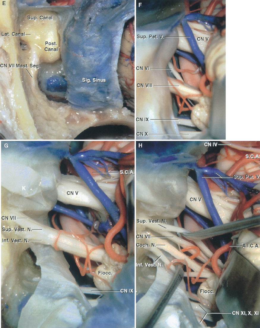
圖8.15。模擬。retrosigmoid方法的比較和最小的乳突切開術, retrolabyrinthine, translabyrinthine, transcochlear 的方法修改presigmoid方法。一個,retrosigmoid方法。左側小腦 已經發展到了 暴露通過XI顱神經V 小腦橋腦角。每一步的插圖是 與視圖方法的其他修改 。B,麵部 蝸神經和小葉的側 收回暴露底動脈。C, 最小乳突切開術隻夠骨切除 乙狀竇前 打開presigmoid硬腦膜 劃分上堅硬的竇和小腦幕。D, 打開presigmoid硬腦膜和乙狀竇 已經收回了後方。觀點大約是一樣的,看到 retrosigmoid曝光。 The retrosigmoid approach provides a better view of the nerves entering the jugular foramen. A., artery; A.I.C.A., anteroinferior cerebellar artery; Bas., basilar; Cist., cisternal; CN, cranial nerve; Coch., cochlear; Flocc., flocculus; Inf., inferior; Laby., labyrinthine; Lat., lateral; Mast., mastoid; Meat., meatal; N., nerve; Pet., petrosal; P.I.C.A., posteroinferior cerebellar artery; Post., posterior; Presig., presigmoid; S.C.A., superior cerebellar artery; Seg., segment; Sig., sigmoid; Suboccip., suboccipital; Sup., superior; Tymp., tympanic; V., vein; Vest., vestibular.
點擊這裏查看這張圖片的交互模塊和相關內容。
圖8.15。情況。retrosigmoid方法的比較和最小的乳突切開術,retrolabyrinthine, translabyrinthine, transcochlear presigmoid方法的修改方法。E,周圍的骨膠囊的半規管和麵部神經受到retrolabyrinthine presigmoid方法的變體。F,曝光retrolabyrinthine版本並沒有顯著的不同,實現最小的乳突切開術。G,半規管和技工已被移除,硬腦膜襯砌內部聲學道已打開完成translabyrinthine曝光。這一收益率的暴露內部聲學道但隻提供最低限度的改善結構內側的曝光條件的道.H,神經分離開始橫向的眼底道和擴展的解理麵內側向腦幹。麵部神經和背後的優越的前庭神經下前庭耳蝸神經背後的神經。

點擊這裏查看這張圖片的交互模塊和相關內容。
圖8.15。我和j . retrosigmoid方法的比較和最小的乳突切開術,retrolabyrinthine, translabyrinthine, transcochlear presigmoid方法的修改方法。我,錯綜複雜的、鼓膜的麵神經乳突段的接觸,準備所需的神經後換位完成transcochlear曝光。J,麵部神經已經轉置和耳蝸堅硬的頂點刪除完成transcochlear暴露前的腦幹和基底動脈。
translabyrinthine方法,內部聲學道和小腦橋腦角接近通過乳突切開術和迷路切除術(圖8.6)(38)16日,29日有兩個目標骨切除的方法。首先是揭露特羅特曼的三角形的硬腦膜的後表麵上顳骨麵對小腦橋腦角。第二是消除足夠的骨骼能夠識別神經外側的腫瘤通過內部聽覺運河和橫向和垂直的波峰。的方法也可以結合retrosigmoid或超越和infratentorial presigmoid方法。
retroauricular切口開始在耳廓和乳突尖(3)擴展了劣等。骨膜皮瓣和軟組織覆蓋乳突和retromastoid地勢較高的地區。在乳突皮質骨鑽,空氣乳突細胞移除,揭露乳突腔,迷宮周圍的皮質骨,二腹肌脊主要在前麵的麵神經乳突段退出莖突乳突的孔和sinodural角度。鑽探是繼續公開的半規管,記下概要乙狀竇,中間窩硬腦膜,麵神經乳突段,以及頸靜脈球的上表麵,隻留下骨頭在這些結構的薄殼。側半規管是最外側突出運河和是第一個遇到的這種方法。它提供了一個寶貴的具有裏程碑意義的識別麵部神經的鼓膜的部分和其他的運河。下麵發現神經外側運河。retrofacial空氣細胞移除和頸靜脈球的穹頂是劣等。在去除骨背後的內部聲音道,重要的是要記住,背後的頸靜脈球可能向上隆起後半規管或內部耳道。前庭導水管和內淋巴的囊可能打開和刪除在道之間的骨切除,頸靜脈球。耳蝸小管會深到前庭導水管視為骨之間的區域移除道和頸靜脈球。 The lower end of the cochlear canaliculus is situated just above the area where the glossopharyngeal nerve enters the medial half of the jugular foramen. The labyrinthectomy portion of the procedure involves removing the semicircular canals and the vestibule to expose the dura lining the internal auditory canal. The lateral and posterior semicircular canals are drilled away. As the bone removal proceeds medially, the ampullae of the lateral and superior semicircular canals are exposed. At this point some bleeding can occur as the subarcuate artery is encountered in the bone near the center of the superior semicircular canal. The vestibule is an oval-shaped cavity located immediately lateral to the internal acoustic meatus, which forms the communication between the semicircular canals and the cochlea. Bone is removed medial and posterior to the vestibule, completely exposing it anterior and inferior to the facial nerve. Care is required to avoid injury to the facial nerve as it courses below the lateral canal and the ampullae of the posterior canal and around the superolateral margin of the vestibule.
前內部聽覺運河位於內側和麵神經鼓室的的部分。硬腦膜襯砌內部管暴露通過鑽井的半規管、技工和優越,周圍的骨頭後,低利潤的內部運河。進一步年底外側骨切除道暴露了橫向和縱向波峰(圖8.2)。的intrameatal部分麵神經分開月底上級前庭神經外側縱脊的運河,也叫比爾的酒吧,可以用來積極識別麵部神經(13、16)。最初的錯綜複雜的麵部神經的一部分,位於前垂直脊,暴露在道的眼底。識別麵部神經,硬腦膜後打開道。硬腦膜的切口特羅特曼的三角形的頂點V型切口的“V”擴展沿道的硬腦膜。下麵的“V”擴展一個肢體上堅硬的竇和其他肢體延伸頸靜脈球以上。硬腦膜的皮瓣然後反映後方暴露道和小腦橋腦角的結構。subarcuate動脈,或AICA,可能遇到的硬腦膜特羅特曼的三角形。 Usually, the subarcuate artery arises from the AICA and passes through the dura on the upper posterior wall of the meatus as a fine stem. Occasionally, however, the subarcuate artery, along with its origin from the AICA, may be incorporated into the dura on the posterior face of the temporal bone. The approach may include transection of the external canal and obliteration of the middle ear with packing of the eustachian tube at closure.

點擊這裏查看這張圖片的交互模塊和相關內容。
圖8.16。f。比較的retrosigmoid和各種修改presigmoid曝光。presigmoid方法的修改包括最小乳突切開術,retrolabyrinthine,部分迷宮,translabyrinthine,修改transcochlear, transcochlear與麵神經換位的方法。,頭皮切口(插入)定位上,通過開顱temporo-occipital infratentorial曝光。開顱temporo-occipital已經完成和硬腦膜打開暴露顳葉和retrosigmoid區域。橫向和乙狀竇一直保存了下來。小腦已經收回了暴露小腦橋腦角的神經。B的放大視圖retrosigmoid接觸與獲得的曝光的各種修改presigmoid方法。C, retrosigmoid曝光蝸神經已經升高和舌咽神經抑鬱的基底動脈暴露在AICA的起源。 D, subtemporal exposure. The temporal lobe has been elevated to expose the optic tract and oculomotor nerve and the PCA, internal carotid, and anterior choroidal arteries. E, the tentorium has been opened while preserving the trochlear nerve. The SCA courses below and the PCA above the oculomotor and trochlear nerves. F, minimal mastoidectomy modification of the presigmoid approach. The minimal mastoidectomy approach is completed by removing only enough bone in the front of the sigmoid sinus so that the presigmoid dura can be opened to expose the posterior cranial fossa. The bony capsule of the labyrinth is not exposed in the minimal mastoidectomy as it is in the retrolabyrinthine approach. The exposure shown with the minimal mastoidectomy in this figure is to be compared with the retrosigmoid exposure shown in B. A., artery; Ac., acoustic; A.I.C.A., anteroinferior cerebellar artery; Ant., anterior; Bas., basilar; Car., carotid; Chor., choroidal; CN, cranial nerve; Comm., communicating; Inf., inferior; Int., internal; Lat., lateral; Mast., mastoid; P.C.A., posterior cerebral artery; Ped., peduncle; Pet., petrosal; P.I.C.A., posteroinferior cerebellar artery; Post., posterior; S.C.A., superior cerebellar artery; Seg., segment; Sig., sigmoid; Sup., superior; Temp., temporal; Tent., tentorial; Tr., trunk; Trans., transverse; V., vein; Vert., vertebral.
入transcochlear方法主要是一個擴展的translabyrinthine方法(圖8.6)(3、15、16)。它通常包括部門和關閉外部運河,至少後切除骨性外耳道的一部分,和鼓膜和鼓膜,咽鼓管的消亡。暴露硬腦膜後襯砌內部聽覺運河,作為translabyrinthine描述方法,刪除砧骨和麵部神經暴露從膝狀神經節莖突乳突的孔。表麵堅硬的大神經切斷,麵部神經轉置後方。在最後階段,完成骨切除麵神經管,神經換位後,耳蝸和相鄰的部分岩石的頂端是鑽(圖8.6)。
骨切除,向上延伸到斜坡的邊緣,暴露下從頸靜脈球下麵堅硬的竇上堅硬的竇。的提升部分岩石的頸動脈暴露的前極限解剖。骨切除,現在延伸到外側邊緣的斜坡,可以很容易地進行內側到斜坡。延長硬鋁在這個區域允許可視化的外展神經內側內部聲學道,三叉神經的下緣,進入頸靜脈孔神經,的基底動脈段,AICA的起源和初始段。
替代置換麵神經是完整的一個廣泛的骨切除hypotympanic頸動脈管和retrofacial地區向前延伸,從而使成骸骨麵神經乳突段和離開它懸浮在骨殼,所述由Gantz和費(7)。在這種方法中,外耳道關閉作為盲囊和鼓膜,砧骨,和身體錘骨的刪除(7)執行乳突切開術,包括切除retrofacial retrolabyrinthine, supralabyrinthine隔間。麵神經鼓室的段和標識在莖突乳突的孔。劣質部分鼓骨暴露infralabyrinthine艙中移除,頸靜脈球,intrapetrous頸動脈。retrofacial解剖進行內側和優,刪除的半規管和技工。顱後窩硬腦膜的解剖進行下級在內部聽覺運河和麵神經管下。耳蝸是鑽了劣質和麵神經管前工作。然後左橋在麵神經管手術領域之間的硬腦膜暴露頸動脈和頸靜脈球。

點擊這裏查看這張圖片的交互模塊和相關內容。
圖8.16。(。比較的retrosigmoid和各種修改presigmoid曝光。G,深接觸最小乳突切開術和收縮的蝸舌咽肌神經,與c所示的retrosigmoid方法相比獲得的曝光類似於retrosigmoid方法。H, retrolabyrinthine方法更廣泛的鑽探的乳突已完成公開骨性半規管的膠囊。我,硬腦膜一直向前折疊後完成retrolabyrinthine曝光。用最小的接觸不同的小乳突切開術暴露所示F和g J,運河部分迷路切除術後的接觸是類似於實現最小的乳突切開術。K,部分迷路切除術已被移除擴展上級運河除了切除後運河。L, infratentorial暴露並沒有顯著的不同,實現最小的乳突切開術,見F和g的優越運河減少所需的顳葉收縮和艾滋病暴露在中間窩地板和岩石的頂端。米,translabyrinthine暴露的半規管和門廳已被移除。 This adds the internal auditory canal to the exposure, but does not improve the exposure of the structures medial to the meatus, as compared with the minimal mastoidectomy or even the retrosigmoid approach. N, the facial nerve has been transposed posteriorly out of the field and the cochlea has been removed to complete the transcochlear approach. This approach greatly improves access to the front of the brainstem, clivus, and basilar artery, but is done at the cost of a temporary or permanent facial paralysis and loss of hearing.
presigmoid方法結合了超越和infratentorial顱骨切開術集中在乳突和不同程度的乳突和迷宮般的切除(圖8.14)。最小程度的乳突切除術,我們稱之為最小乳突切開術,暴露了presigmoid隻有足夠的硬腦膜打開硬腦膜在乙狀竇前暴露的小腦橋腦角(無花果。8.15和8.16)。下一個更廣泛的程度的乳突切除術,retrolabyrinthine修改,是一個更完整的乳突切開術暴露的骨膠囊的半規管和至少部分麵部神經變得極瘦。在部分迷路切除術,一個或兩個的半規管,一般上級和/或後運河,與外側運河保護切除。移除這些運河可能,但並非總是如此,與聽力的損失(37)。後運河可以增加訪問顱後窩,和刪除上級運河僅給出了一個更直接的接觸到堅硬的頂端中間窩。下一個更廣泛的修改是translabyrinthine方法,半規管和前庭切除均勻,導致聽力的喪失。translabyrinthine方法提供了很好的訪問內部聽覺運河。下一個更廣泛的修改是transcochlear方法,在耳蝸入位於底部的道是移除,因此提供的內側部分岩石的頂端和斜坡的一邊。另一個修改,我們稱之為擴展translabyrinthine方法,和類似於transcochlear方法,涉及鑽井骨前部和後部的麵部神經,留下一個列的麵部神經場大病骨和工作前和後的麵部神經去除耳蝸和訪問的斜坡。 Gaining access for drilling the cochlea anterior to the facial nerve commonly requires that at least part of the posterior part of the external canal be removed, that the tympanic cavity be obliterated, and that the internal carotid artery be exposed below the promontory.
在評估這些方法在我們的實驗室中,我們發現,最小乳突切開術給大約相同的曝光retrolabyrinthine方法,但在風險降低後半規管和麵部神經不是場大病(無花果。8.14和8.15)。刪除後管增加訪問顱後窩,但訪問隻是稍微增加retrolabyrinthine實現的方法。刪除上級管增加中間窩和岩石的頂端,減少了需要收縮的顳葉。translabyrinthine方法並不顯著增加訪問區域內的porus內部聲學道,實現最小的乳突切開術或retrolabyrinthine方法,但提供的內部聽覺運河。transcochlear修改,骨頭切除斜坡的邊緣,並顯著增加訪問前的腦幹和斜坡,實現較小程度的骨切除。retrosigmoid, presigmoid最小乳突切開術,retrolabyrinthine方法相比,取得了幾乎相同的暴露小腦橋腦角,但retrosigmoid方法沒有提供額外的暴露的窩和岩石的頂端,可以實現超越和infratentorial presigmoid方法。
皮膚切口在顴骨上方的顳區開始,向下延伸超過耳朵和枕骨下的區域的內側乳突(無花果。8.14、8.15和8.17)。皮瓣是反映的外耳道。顳肌升高和反映在前麵,肌肉在乳突和枕骨下的區域是劣等。執行一個temporooccipital顱骨切開術和橫竇暴露。骨瓣後升高,乳突切開術進行沒有進入迷宮。乙狀竇是場大病sinodural角到頸靜脈球。骨被優暴露的地板中間窩和優越的堅硬的竇。特羅特曼的三角形是暴露在耳軟骨囊外側的區域。
然後切開硬腦膜顳顱骨切開術的基礎,同時保留的靜脈交界處拉貝風橫竇。打開後窩硬腦膜的乙狀竇前特羅特曼的三角形。硬腦膜的切口延長在上級堅硬的竇加入顳硬腦膜的硬腦膜的切口。上堅硬的竇分工後,小腦幕下切平行,僅次於堅硬的脊和優越的堅硬的竇。這個硬腦膜的切口是擴展的分工上堅硬的竇通過內側邊緣的小腦幕切跡在滑車神經進入幕的邊緣。是注意避免損傷IVth腦神經課程幕的邊緣附近。的後部分顳葉是高架,乙狀竇是流離失所的後方隨著小腦半球,同時保留連接的靜脈與乙狀竇拉貝風。乙狀竇限製顳葉的優越的收縮能力,可以結紮提高曝光如果雙邊靜脈血管造影顯示足夠的溝通通過torcular對麵(24)。岩斜區可以從中間暴露窩和幕的切跡枕骨大孔附近,盡管獲得較低的岩斜區由頸靜脈球可能有限。presigmoid接觸提供了一個更短的岩斜區和工作距離為解剖提供多個角度。 The major arteries in the posterior fossa are easily accessible. The exposure can also be combined with a far-lateral approach (Fig. 8.17).
的subtemporal preauricular顳顬骨下的方法是直接通過顳顬骨下的中間部分的窩的前表麵堅硬的骨位於內側耳蝸和岩斜區(無花果。8.10、8.13和8.18)。這個描述輪廓的全部解剖暴露可以通過這種方法,但它通常可以根據一個更小、更有限,方法。曲線切口額地區開始向下的耳朵變成宮頸區域。切口隻能向下擴展到區域下方耳屏如果隻有堅硬的頂端和上部顳顬骨下的窩暴露,但是它可以擴展到脖子上如果需要頸部解剖。皮瓣和底層組織和反映。麵神經及其主要分支確定遠端莖突乳突的孔和後腮腺。腮腺masseteric筋膜分開,以避免過度拉伸麵部神經的莖突乳突的孔(33歲,38歲,39)。淺顳肌筋膜的上麵部分支課程分開顳肌和反映向前,以防損壞的分支麵神經顴弓的額的肌肉暴露。顴弓劃分的前部和後部,顳肌,上覆的顴弓,向下反射。下頜髁和膠囊的顳下頜關節脫臼向下或切除。 The temporomandibular joint can be removed in a single piece for later replacement by dividing the mandibular neck below the condyle and osteotomizing the middle fossa floor around the mandibular fossa (Fig. 8.18). The internal carotid artery, the internal jugular vein, and the vagus, accessory, and hypoglossal nerves may be exposed in the neck if needed. The posterior belly of the digastric muscle may be divided and the styloid process resected.
然後執行額顳葉顱骨切開術。硬腦膜是高架地板的中央窩暴露並消除在棘孔腦膜中動脈,使弓狀隆起,第三個三叉部門卵圓孔未閉,堅硬的大神經。更大的堅硬的神經是必要時切斷麵神經避免牽引。中央窩的地板,包括側眶上裂的偽劣方麵,和側緣的小孔那可以暴露顳顬骨下的窩的結構。
如果需要,可以刪除骨內側下頜窩暴露咽鼓管和張量定音鼓肌肉,這兩個可能切除(無花果。8.10、8.13和8.18)。骨切除繼續劣等,暴露出的提升部分岩石的頸動脈。在這部分中,頸動脈周圍是骨膜鞘,它包含periarterial靜脈叢海綿竇的延伸。頸動脈管的入口處,一個致密的纖維軟骨的環環繞的動脈。如果需要動員的動脈,分裂環時必須小心不要損壞IXth腦神經在靠近頸靜脈孔頸動脈管的出口。動員後頸動脈和取代它向前,堅硬的頂端和clival地區枕骨大孔的水平可以靠近內側和背後的動脈。很難在鑽井過程中,皮質骨沿岩石的頂端給地方一個易碎的鬆質骨在該地區的斜坡,硬腦膜的前和後的橫向方麵窩被暴露。暴露麵積受限於氏洞穴優,通過內部和耳蝸聽覺運河外側,外展神經的課程通過Dorello管內側,和舌下運河劣等。如果打開硬腦膜,沿橫向結構和前方麵上髓質和低三分之二的腦橋將會暴露(41)。小腦幕可分為給訪問上clival地區。
把卵圓孔上方的第三個三叉部門將允許暴露的結堅硬的和海綿頸動脈海綿竇的下側的部分的結構(17日,39)。pterygopalatine窩,腫塊空間、橫向上頜骨和軌道可以暴露更遠的前方。蝶骨的橫向方麵和蝶竇也可以去除骨內側接洽上頜神經的根源翼狀的過程。

點擊這裏查看這張圖片的交互模塊和相關內容。
圖8.17。模擬。結合 presigmoid和far-lateral 方法。,插入顯示了頭皮切口和 乳突小費。頭皮皮瓣 一直反映。 的乳突切開術暴露了 密集的皮質骨住房的半規管。骨頭 皮瓣是概述。枕向後之間二腹肌和 動脈課程上斜。B,擴大 視圖。鼓膜的部分下麵 麵神經課程的橫向運河。 The chorda tympani arises from the mastoid segment of the facial nerve. The mastoid antrum, which has been drilled away, opens through the aditus into the epitympanic part of the tympanic cavity. C, the presigmoid and temporal dural incisions have been outlined. D, the temporal and presigmoid dura has been opened. One goal of the procedure is to preserve the vein of Labbe, which empties into the transverse sinus. A., artery; A.I.C.A., anteroinferior cerebellar artery; Atl-Occip., atlanto-occipital; Cap., capitis; Car., carotid; Chor., chorda; Cist., cisternal; CN, cranial nerve; Epitymp., epitympanic; For., foramen; Gang., ganglion; Genic., geniculate; Hypogl., hypoglossal; Inf., inferior; Jug., jugular; Laby., labyrinthine; Lat., lateral; Lev., levator; M., muscle; Meat., meatal; Memb., membrane; Men., meningeal; N., nerve; Obl., oblique; Occip., occipital; P.C.A., posterior cerebral artery; P.I.C.A., posteroinferior cerebellar artery; Plex., plexus; Post., posterior; Rec., rectus; S.C.A., superior cerebellar artery; Scap., scapula; Seg., segment; Semicirc., semicircular; Sig., sigmoid; Sp., spine; Suboccip., suboccipital; Sup., superior; Temp., temporal; Trans., transverse; Tymp., tympani, tympanic; V., vein; Vert., vertebral; Vest., vestibular.

點擊這裏查看這張圖片的交互模塊和相關內容。
圖8.17。情況。presigmoid和far-lateral相結合的方法。E,硬腦膜的切口一直貫穿特羅特曼的三角形和優越的堅硬的竇和小腦幕,照顧保護拉貝風和滑車神經的靜脈。半規管已經打開了。F,放大視圖。麵對顱後窩外側後管內部聲音道。上級運河項目向上,在弓狀隆起,中間的地板窩。外側運河是一個有用的具有裏程碑意義的識別麵神經鼓室的部分,課程之間的運河和鐙骨坐在橢圓形窗口。打開epitympanic區域通過入口進入乳突腔。 G, the labyrinthectomy has been completed and the dura lining the meatus opened to expose the cisternal, meatal, labyrinthine, tympanic, and mastoid segments of the facial nerve. The SCA courses above the trigeminal nerve. H, enlarged view along the opened tentorial incisura. The oculomotor and trochlear nerves course between the PCA and SCA. The SCA rests against the upper surface of the trigeminal nerve.

點擊這裏查看這張圖片的交互模塊和相關內容。
圖8.17。我。presigmoid和far-lateral相結合的方法。我插入顯示額外的皮膚切口需要添加一個retrosigmoid顱骨切開術和far-lateral的方法。頭皮皮瓣已反映暴露枕骨下的三角形之間的位置上和下斜腹直肌肌後主要以及深處的椎動脈課程密集的靜脈叢。J,靜脈叢被移除暴露的利潤率枕骨下的三角形。K,腹直肌後主要反映了內側和下斜和優越的斜側麵揭露背後的椎動脈及周圍靜脈叢atlanto-occipital關節。L,靜脈叢被移除暴露與C1椎動脈追逐atlanto-occipital背後的神經關節的上邊緣後atlantal拱門。
的postauricular transtemporal方法是最常見的選擇病變涉及乳突和鼓膜的蛀牙和跟蹤沿神經和動脈到達中間顳顬骨下的窩(無花果。8.19和8.20)。然而,它可以被定製的後緣包括retrosigmoid, far-lateral,或者presigmoid曝光後窩,在其前限製,包括暴露pterygopalatine窩和外側部分上頜骨軌道或前顱窩。
啟動一個問號切口在發際線在顳區,延伸在耳朵後麵的乳突和持續的劣等胸鎖乳突肌前到脖子上。然後反映皮瓣向前和外耳道分為軟骨連接和關閉盲囊。胸鎖乳突肌肌肉脫離乳突和反映下級。骨膜和顳肌的後部分反映在前麵,因此暴露時間,乳突,retromastoid地區。二腹肌後腹的肌肉是分裂和反映下級。此時,麵部神經遠端莖突乳突的孔和標識,連同其主要分支,腮腺的物質(5)頸內靜脈,頸動脈分叉,舌咽神經、迷走神經、配件,和舌下神經在頸部暴露和孤立。這允許頸內動脈近端控製和結紮頸外動脈的主要喂養血管的腫瘤早期手術。
在這之後,時間和/或retromastoid顱骨切開術可能執行一個簡單的乳突切開術。剩下的皮膚的外耳道、鼓膜、錘骨、砧骨和鐙骨拱(離開踏板)移除。麵部神經完全場大病從膝狀神經節莖突乳突的孔。
如果曝光的頸靜脈孔和降低clival地區,創建一個新的麵神經管通過鑽井的槽骨前閣樓的牆壁,膝狀神經節和顴骨的根源。麵神經是小心翼翼地釋放在莖突乳突的孔,而一些周圍的結締組織附著在神經,神經是轉置在前麵的新骨槽epitympanum和嵌入的保護腮腺組織(5)。
中間窩硬腦膜和乙狀竇的sinodural角頸靜脈球場大病。那麼乙狀竇和頸靜脈結紮在這個序列,和乙狀竇分裂。竇的牆壁的一部分,燈泡,和/或靜脈切除可能增加風險。這允許低顱神經的解剖頸靜脈孔,以及他們的動員和後位移,如果必要的。的後動員低顱神經允許直接接觸的結構沿橫向和腦橋髓質和低的前方麵沒有大腦收縮的必要性。解剖在頸靜脈孔的麵積已經被證明是非常困難的,因為低顱神經尤其脆弱,難以從周圍組織隔離。
暴露的中產clival結構需要切除骨迷路,作為translabyrinthine描述方法。內部聽覺運河暴露,麵部神經識別和耳蝸和前庭神經分裂。更大的表麵堅硬的神經是切割它的起源從膝狀神經節。麵部神經釋放其所有附件在顳骨和反映後方。的骨部分外耳道和鼓膜的骨鑽,暴露的提升部分intrapetrous頸動脈內側咽鼓管。
解剖是繼續鑽探耳蝸,開始在其基底,暴露的部分岩石的頸動脈水平段。頸動脈的前位移和刪除的耳蝸提供了廣泛接觸側腦橋的前部分和髓質。這種接觸從下延伸方麵的三叉神經節枕骨大孔。暴露可能攜帶內側斜坡和咽後的空間和先前地暴露蝶竇的粘膜。
如果這種方法擴展到parasellar parasphenoidal地區,顴弓是分裂和反映下級咬肌。顳肌分開其附件的喙突下頜骨,掀起和優。然後進行顳部顱骨切開術,刪除和廣泛的骨沿整個中顱窩的橫向方麵。下頜骨升支的流離失所的前方或切除,和岩石的頸動脈遠側地暴露的近端部分intracavernous段刪除後的軟骨部分咽鼓管。海綿竇可以接近和intracavernous頸動脈暴露除以三叉神經下頜的部分。該方法也可以擴展到retrosigmoid區域,C1, C2椎動脈的水平,或枕骨下的三角形far-lateral或transcondylar曝光。外側軌道,pterygopalatine窩可以在訪問前暴露的限製。

點擊這裏查看這張圖片的交互模塊和相關內容。
圖8.17。M和N, presigmoid和far-lateral相結合的方法。米,枕下開顱已經完成,後拱和橫突的後支的阿特拉斯,和硬腦膜的切口已經列出。後腦膜動脈出現在椎動脈穿透硬腦膜。C1神經根椎動脈的堅持下邊緣。N,硬腦膜已經開了,向頸靜脈孔暴露神經傳遞。骨頭被移除上麵atlanto-occipital聯合暴露的舌下神經的舌下神經管。附屬延伸穿過頸靜脈結節在頸靜脈孔。

點擊這裏查看這張圖片的交互模塊和相關內容。
圖8.18。Preauricular subtemporal-infratemporal窩的方法。,頭皮切口位置,額顳葉顱骨切開術可以完成。的操作通常是完成一個切口,向下延伸耳屏的水平。然而,它可以擴展如果頸部解剖是必要的。頭皮皮瓣已反映,照顧保護麵部神經的分支。B,顳肌被折射,穿顱術完成。下頜髁窩和顴弓的一部分在一塊被移除,如插入所示,中間窩地板上移除。C,曝光後刪除中間的地板外側窩卵圓孔未閉,定音鼓張肌切除前。頸動脈管的低孔位於頸靜脈孔的前麵。 The eustachian tube, which passes across the front of the petrous carotid, has been opened. D, the tensor tympani and eustachian tube have been resected to expose the horizontal segment of the petrous carotid. E, the internal carotid artery has been reflected forward and the petrous apex drilled to expose the posterior fossa dura and the inferior petrosal sinus coursing along the petroclival fissure. F, the dura facing the petrous apex has been opened and the vertebral arteries and AICA exposed. This exposure is directed through the petrous apex medial to the cochlea and jugular foramen and does not risk loss of facial nerve function or hearing, as do the approaches directed through the petrous apex that require facial nerve transposition and resection of the labyrinth. A., artery; A.I.C.A., anteroinferior cerebellar artery; Brs., branches; Car., carotid; CN, cranial nerve; Eust., eustachian; Gang., ganglion; Gl., gland; Gr., greater; Inf., inferior; Int., internal; Jug., jugular; M., muscle; Max., maxillary; Men., meningeal; Mid., middle; N., nerve; Pet., petrosal, petrous; Post., posterior; Temp., temporal; Tens., tensor; TM., temporomandibular; Trig., trigeminal; Tymp., tympani; V., vein; Vert., vertebral; Zygo., zygomatic.

點擊這裏查看這張圖片的交互模塊和相關內容。
圖8.19。模擬。解剖基礎的 postauricular transtemporal 方法。,切口周圍掃廣泛 耳朵的後緣 retrosigmoid, presigmoid,和 far-lateral接觸可以獲得 背後的耳朵,和一個subtemporal, 顳顬骨下的,pterygopalatine, 前麵的軌道接觸可以獲得耳朵。B,頭皮皮瓣 一直反映,外部 運河斷掉,腮腺 和暴露表麵的麵部 神經的分支。C, 反映胸鎖乳突肌的肌肉。頸部解剖暴露 頸內靜脈,C1橫 過程,和舌咽神經、迷走, 配件,舌下神經 。副神經是收回 。D,腮腺 移除公開temporofacial麵部 和cervicofacial樹幹的神經和顳下頜關節。 的 splenius capitis muscle has been reflected downward to expose the superior and inferior oblique muscles, which insert on the transverse process of C1 and border the suboccipital triangle in which the vertebral artery courses. A., artery; Alv., alveolar; Aur., auricular; Br., branch; Brs., branches; Cap., capitis; Car., carotid; Cerv., cervical; Chor., chorda, choroid; CN, cranial nerve; Coch., cochlear; Cond., condyle; Endolymph., endolymphatic; Eust., eustachian; Ext., external; Fac., facial; Gang., ganglion; Genic., geniculate; Gl., gland; Gr., greater; Hypogl., hypoglossal; Inf., inferior; Infraorb., infraorbital; Infratemp., infratemporal; Int., internal; Jug., jugular; Laby., labyrinthine; Lat., lateral; Lev., levator; M., muscle; Mandib., mandibular; Mast., mastoid; Max., maxillary; Med., medial; N., nerve; Obl., oblique; Occip., occipital; Pal., palatini; P.C.A., posterior cerebral artery; Ped., peduncle; Pet., petrosal, petrous; P.I.C.A., posteroinferior cerebellar artery; Plex., plexus; Post., posterior; Proc., process; Pteryg., pterygoid; Pterygopal., pterygopalatine; Rec., rectus; S.C.A., superior cerebellar artery; Scap., scapula; Seg., segment; Semicirc., semicircular; Sig., sigmoid; Sphen., sphenoid; Splen., splenus; Sternocleidomast., sternocleidomastoid; Sup., superior; Superf., superficial; Symp., sympathetic; Temp., temporal; Tens., tensor; TM., temporomandibular; Trans., transverse; Tymp., tympani, tympanic; V., vein; Vel., veli; Vert., vertebral; Vest., vestibular.

點擊這裏查看這張圖片的交互模塊和相關內容。
圖8.19。情況。解剖基礎的 postauricular transtemporal方法。E,下頜支的部分被移除揭露上下翼狀肌外側頭和上頜動脈顳顬骨下的窩。下牙槽管和神經已經暴露出來。F,下頜支,下牙槽前的運河,被移除提供更多接觸的下顳窩。上的橫向翼狀肌肌肉通過向後從大蝶翼的下顳表麵和底蓋通過從翼狀肌外側板向上。兩頭插入在下頜的脖子和關節囊。膚淺的內側翼狀肌肌肉通過上頜結節和翼狀肌下頜角板。的深頭內側翼狀肌起源於翼狀的板塊之間的翼狀的窩。 G, enlarged view of the infratemporal area after removal of the mandibular condyle and lateral pterygoid muscles. The branches of the mandibular nerve are exposed below the foramen ovale. The largest branches are the lingual and superior alveolar nerves, which are predominantly sensory. The auriculotemporal nerve arises as two roots, which often pass around the middle meningeal artery before joining. H, the pterygoid muscles, a segment of the maxillary artery, and the mandibular and facial nerve branches have been reflected or removed to expose the internal jugular vein exiting the jugular foramen on the medial side of the stylomastoid foramen, the internal carotid artery ascending to enter the carotid canal, the tensor and levator veli palatini descending from their origin bordering the eustachian tube, and the terminal segment of the maxillary artery entering the pterygopalatine fossa.

點擊這裏查看這張圖片的交互模塊和相關內容。
圖8.19。我。基礎解剖postauricular transtemporal方法。我,一個乳突切開術已經完成暴露的半規管和麵部的乳突段運河。內淋巴的囊presigmoid硬腦膜下坐。J,外部運河已經切除暴露鼓室的結構。麵神經鼓室的段的課程側半規管和鐙骨坐在橢圓形窗口。鼓索起源於麵神經乳突段,通過提出的內表麵的鼓膜和頸部錘骨進入前小溝,出口沿岩鼓顱骨縫合,連接舌神經顳顬骨下的窩。海角覆蓋了耳蝸基底的轉變。張量定音鼓肌的腱使直角轉錘骨trochleiform過程插入。 K, the incus and malleus have been removed while preserving the stapes and the tensor tympani muscle. The petrous carotid has been exposed. The nerves exiting the jugular foramen have been retracted forward to expose the hypoglossal nerve exiting the hypoglossal canal. L, a frontotemporal craniotomy has been completed and the floor of the middle cranial fossa removed. The semicircular canals have been exposed above the jugular bulb and the stapes has been removed from the oval window. The maxillary nerve has been exposed in the pterygopalatine fossa. The membranous wall of the eustachian tube has been opened to expose the tube’s opening into the nasopharynx.

點擊這裏查看這張圖片的交互模塊和相關內容。
圖8.19。先生。基礎解剖postauricular transtemporal方法。米,retrosigmoid顱骨切開術已經完成,橋腦小腦角區的神經暴露出來。蝸神經一直抑鬱暴露麵神經。N,麵部神經反射前進了麵神經管。海角已鑽暴露耳蝸和前庭。半規管的兩端開放到前廳,和基底的耳蝸。頸靜脈球被移除暴露頸靜脈窩燈泡所在。頸靜脈球位於低於門廳。 The nerves exiting the jugular foramen have been reflected backward to expose the hypoglossal nerve exiting the hypoglossal canal. The nerves passing through the jugular foramen and hypoglossal canal exit the skull on the medial side of the internal jugular vein and descend between the internal carotid artery and internal jugular vein. O, the bone above the occipital condyle has been drilled to expose the hypoglossal nerve in the hypoglossal canal. P, the posterior wall of the internal acoustic meatus has been removed to provide this presigmoid inferolateral view of the nerves in the internal meatus. The cochlear nerve separates off the main bundle of the vestibulocochlear nerve and penetrates the modiolus. The inferior vestibular nerve divides into the singular nerve to the posterior ampullae and a branch to the saccule. The superior vestibular nerve innervates the superior and lateral ampullae and sends a branch to the utricle. Q, the medial wall of the jugular fossa has been removed and the nerves passing through the jugular foramen have been exposed. The glossopharyngeal nerve passes through the foramen anterior to the vagus and accessory nerves. A large superior petrosal vein ascends to the superior petrosal sinus. R, the glossopharyngeal, vagus, and accessory rootlets arise behind and the hypoglossal rootlets arise anterior to the inferior olive.

點擊這裏查看這張圖片的交互模塊和相關內容。
圖8.19。sx。基礎解剖postauricular transtemporal方法。年代,放大圖內側牆的麵神經鼓室前動員。鐙骨的肌肉通過從麵部神經和下麵的錐體隆起高度鐙骨的脖子上。張定音鼓肌肉通過向後外側,形成一條狹窄的肌腱,使一個急轉彎trochleariform過程在外側的semicanal處理錘骨的插入。基底的耳蝸位於深到海角。麵神經鼓室的段的課程在鐙骨之上。T迷宮的放大圖。半規管是露天的,鐙骨從橢圓窗口中刪除。 The round window is located below and behind the oval window. U, the facial nerve has been reflected forward out of the facial canal and the vestibule has been opened. The ampullae of the superior and the lateral canal open into the vestibule anteriorly and are innervated by the superior vestibular nerve. Only the upper edge of the superior canal was preserved in opening the vestibule. The ampullae of the posterior canal is located at its lower end and is innervated by the singular branch of the inferior vestibular nerve. V, a probe is directed through the vestibule to the inner surface of the membrane covering the round window, which is located behind and below the oval window. W, enlarged view of the labyrinth after opening the promontory to expose the cochlea. The jugular bulb is located below the vestibule and semicircular canals and the lateral genu of the internal carotid artery in position below the cochlea. The cochlea wraps around the modiolus through which the branches of the cochlear nerve are distributed to the cochlear duct. X, the temporal lobe has been elevated to expose the internal carotid, PCA, and SCA in the basal cisterns. The dura has been elevated from the lateral wall of the cavernous sinus.
點擊這裏查看這張圖片的交互模塊和相關內容。
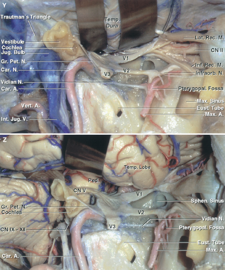
圖8.19。Y和Z,解剖基礎postauricular transtemporal方法。Y,概述之前打開硬腦膜。提供retrosigmoid postauricular方法提供了潛力,presigmoid, far-lateral曝光和可以用來訪問顳顬骨下的和pterygopalatine窩,軌道,subtemporal地區。在這種情況下,風險從retrosigmoid區域延伸到軌道。上頜竇已在軌道下麵打開地板上。Z,曝光後打開硬腦膜的概述。

點擊這裏查看這張圖片的交互模塊和相關內容。
圖8.20。f。Postauricular transtemporal方法。這種接觸包括transtemporal和顳顬骨下的方法結合顱骨切開術。向前,頭皮皮瓣已反映暴露胸鎖乳突肌,腮腺,耳大神經。B,外部運河已經分裂為腮腺反映皮瓣向前和頸部解剖暴露麵神經及其胴體、二腹肌後腹、頸內靜脈。C,乳突切開術完成公開presigmoid硬腦膜,乙狀竇,半規管。在下頜髁切除提供訪問顳顬骨下的窩。D, temporo-occipital顱骨切開術已經完成,顴弓打開,顳肌反映暴露上頜動脈翼肌顳顬骨下的窩。E、時間和顳顬骨下的風險敞口的放大圖。 The posterior wall of the external canal has been removed. The auriculotemporal branch of the mandibular nerve is often split into two rootlets by the middle meningeal artery. F, enlarged view of the tympanic cavity. The anterior part of the lateral semicircular canal is located above the tympanic segment of the facial nerve. The promontory overlies the basal cochlear turn. A., artery; Ac., acoustic; Aur., auricular; Bas., basilar; Car., carotid; Chor., chorda; CN, cranial nerve; Cond., condyle; Ext., external; Gl., gland; Gr., greater; Inf., inferior; Int., internal; Jug., jugular; Lat., lateral; M., muscle; Mandib., mandibular; Mast., mastoid; Max., maxillary; Mid., middle; Men., meningeal; N., nerve; Pet., petrosal, petrous; Proc., process; Seg., segment; Semicirc., semicircular; Sig., sigmoid; Sternocleidomast., sternocleidomastoid; Sup., superior; Temp., temporal; Trans., transverse; Tymp., tympani, tympanic; V., vein; Vert., vertebral.

圖8.20。G-L。Postauricular transtemporal方法。G,外部運河已經在準備暴露岩石的頸動脈切除。H,宮頸的結和堅硬的頸動脈被暴露在該地區在海角。莖突乳突的的側緣和頸小孔被暴露頸靜脈球下麵的半規管。我,下頜神經已經暴露在卵圓孔未閉。更廣泛的暴露的岩石的頸動脈已經完成,這樣可以反映出頸動脈管轉發給提供的鑽井岩石的頂端。J,堅硬的頸動脈被反映,堅硬的頂端暴露斜坡偽劣堅硬的竇中刪除。K,麵神經麵神經管已經搬出去了,和一個迷宮和堅硬的apicectomy已經完成。 L, a segment of the sigmoid sinus and the jugular bulb have been removed to expose the nerves passing through the jugular foramen. The dura has been opened and the facial nerve displaced posteriorly. The temporal lobe has been elevated to expose the subtemporal area while preserving the vein of Labbe.
疾病可以在任何地方出現在岩斜區和經常不限於一個解剖室的顱基地。參與多個顱神經和動脈發生之前因為顱基腫瘤傾向於達到相當大的規模生產臨床表現(32)。良性或惡性腫瘤的區別在這個領域不是剛性的,因為許多良性腫瘤可以有一個非常侵入性的特點。最好的手術方法的選擇取決於位置、擴展,病理學的大小和性質。
這些方法的一個優勢直接通過顳骨岩斜區是他們到達區通過組織飛機在口咽。他們提供了另一條路線,前硬膜內的病變位於內側神經進入內部聲學道和頸靜脈孔可以接近沒有進入鼻咽。他們還提供了一種大道的接觸損傷涉及顳和蝶骨頭除了斜坡。一個或外側的組合方法是常用的公開intraor硬膜外的clival病變也涉及到時間和蝶的骨頭。他們還提供前中腦、腦橋和延髓構成和後顱窩橋小腦角和神經。他們也可以提供更好的訪問顳骨,頸靜脈孔,堅硬的段頸內動脈比其他前部或後部的方法。該地區可以直接從側乳突,迷宮,耳蝸,如translabyrinthine transcochlear方法;從上麵通過subtemporal中間窩路線;從後麵的retrosigmoid枕骨下的方法;或從多個方向使用presigmoid等超越和infratentorial方法相結合方法,translabyrinthine或transcochlear方法可能被添加。 Alternative or extended approaches, most of which include some route through the mastoid and petrous parts, include the anterior transpetrosal, the subtemporal preauricular infratemporal, and the far-lateral transcondylar approach.
章中描述的retrosigmoid枕骨下的方法,在小腦橋腦角,提供了一個廣泛的觀點背後的小腦橋腦角和硬膜內的結構的側低的斜坡,但硬鋁表麵堅硬的頂端,上斜坡,幕的切跡不從這個曝光(26歲,35歲,36歲,46)(無花果。8.15和8.16)。刪除後壁的內部聽覺運河穿過retrosigmoid提供的內容道橫向縱向和橫向波峰。門廳可以打開如果需要移除腫瘤擴展到迷宮。護理是必要的,以避免受傷後半規管、共同小腿如果有保護聽力(29)的可能性。retrosigmoid方法提供了容易獲得的硬膜內的部分顱神經V,第七,第八,第九到十二。它還提供了訪問神經相關性段動脈的後循環。vertebrobasilar結可以暴露在某些情況下,雖然低顱神經和頸靜脈結節是頻繁的障礙。收縮的腦橋和顱神經之間的工作必須達到AICA從基部的動脈的起源。retrosigmoid遠側修改的方法,在遠側章中描述的方法,被設計來提供更好的接觸的外側和前方麵cervicomedullary結(45)。
32 presigmoid方法(8日)結合不同程度的超越和infratentorial曝光petrousectomy,同時保留靜脈交界處的拉貝風橫竇(無花果。8.14 - -8.17)。堅硬的骨頭的切除量可以從retrolabyrinthine變化最小的乳突切開術translabyrinthine或transcochlear暴露麵神經後位移。在選定的情況下,血管攝影顯示了兩個橫向之間的通信的開放鼻竇在中線,乙狀竇可以結紮提高曝光(24)。保存的引流靜脈的拉貝風和避免過度顳葉收縮是這種方法的主要目標上clival地區。接近結構下岩斜空間可能會限製頸靜脈球,可克服由乙狀竇的部門或工作後(36)。這種方法的主要優點是縮短工作距離clival損傷和解剖所提供的不同的角度。方法提供了訪問第三側顱神經通過十二和後循環的主要動脈。這種接觸的一個主要缺點是所提供的解剖變異,下麵描述,限製的大小通過特羅特曼接觸的三角形和迷宮。
translabyrinthine方法提供了訪問從它的起源在麵神經腦幹莖突乳突的孔,和接觸的內聽道的內容(圖8.6)(12、14)。腦橋外側表麵,下三叉神經的起源方麵,和麵部和蝸神經複合體可視化,但暴露的地區差的燈泡,在三叉神經,前內部聲學道通常是可憐的。暴露的程度達到translabyrinthine方法依賴於幾個解剖因素。高頸靜脈球,一個在前麵放置或大乙狀竇,或較低的中間凹板可能嚴重限製接觸(22日27)。
transcochlear方法相似的局限性與translabyrinthine曝光,盡管後換位的麵神經transcochlear方法允許更好地可視化結構前內部聽覺運河(15、16)。接觸的麵積非常狹窄,限製骨性外耳道的維護,但可以增加成就後運河的一部分。換位的麵部神經可能緊隨其後的是一個臨時或永久的麵部麻痹。
前subtemporal經岩方法使用硬膜外的開顱切除前岩石的金字塔的通過時間(無花果。8.12和8.13)。可能是結合顴骨切除增加獲得的地板中間窩(20)。岩石的頂端的麵積去除從內側延伸到內部聽覺運河和耳蝸堅硬的頂端和岩斜結,從岩石的脊頸動脈管前方後方。可能需要很大程度的顳葉收縮。這可能是減少使用與顴開顱額顳葉切除術。雖然隻有一扇小窗在堅硬的骨頭,接觸可以擴展除以相鄰小腦幕的一部分。外側和腦橋的前表麵上斜坡和相鄰的海綿竇的一部分可以通過這條路線接近(圖8.13)。蝸,麵部三叉神經和外展神經可以被識別。岩石的頸動脈可能限製醫生的視線和限製下岩斜地區的一部分,但這一限製可能克服與動脈的前動員(39歲,41)。方法提供了訪問前的腦幹和基底動脈之間的三叉神經上麵和下麵的麵部和蝸神經。 In approaching the basilar artery through this route, the size and location of the lesion in relation to the petrous ridge is critical. The trigeminal nerve can be mobilized to improve the exposure, although this may result in postoperative facial hypesthesia (19, 20). The anterior transpetrosal approach can be used alone for extradural pathologies restricted to the petrous apex or as a surgical step to approaching intradural pathologies in the petroclival region. It provides a route for resecting extradural lesions that extend from the level of the trigeminal nerve to the foramen magnum.
切除後岩石的金字塔的一部分已被用於聽神經瘤切除的擴展方法直接通過中間窩(42歲,21日,28日43)(圖8.12)。擴展方法結合不同程度的切除骨迷路的subtemporal transtentorial路線。擴大切除的堅硬的骨後方乳突和骨迷路使整個intrapetrous麵部神經,並提供訪問的小腦橋腦角subtemporal、translabyrinthine, presigmoid路線(無花果。8.12和8.13)(9)。
的subtemporal preauricular顳顬骨下的方法達到顱底的前外側的方向(無花果。8.10、8.13和8.18)。顴弓的部門,下頜髁切除或位移,和廣泛的切除外側部分中間窩層暴露顳顬骨下的窩,鼻咽,para -和咽後的區域,篩骨,蝶和上頜竇。方法還提供了訪問頸上部和堅硬的頸動脈。海綿竇也可以通過其橫向和基底接觸方麵。堅硬的頸動脈的前位移允許直接訪問斜坡和堅硬的骨頭的廣泛切除內側耳蝸。這暴露了硬膜外的clival地區從三叉神經的水平到枕骨大孔(33歲,36歲,38歲,39)。這種方法還可以提供對腦幹腹側硬膜內的空間(41)。小腦橋腦角的曝光和枕骨大孔是有限的,因為方法進行前內側通過第十二第七顱神經和耳蝸不是切除(36)。岩石的頸動脈的前換位允許不暴露的起源AICA vertebrobasilar結。可以作為一個替代的方法橫向路線midbasilar動脈的血管病變或vertebrobasilar結,當這些病變不能通過retromastoid或公開subtemporal transtentorial方法。
的postauricular transtemporal方法結合transcochlear曝光顳顬骨下的方法,可以作為其他preauricular顳顬骨下的方法當病理學涉及乳突顳顬骨下的窩和延伸到麵部休會,hypotympanic區域和頸靜脈球(5、6、34)(無花果。8.19和8.20)。的結構中低斜坡可以公開而不需要大腦收縮。麵部神經前上的流離失所,乙狀竇的結紮和分裂。位移的麵神經骨管嚴重幹擾其血管供應和臨時或永久喪失功能是將(33)。切除頸靜脈球允許接觸低顱神經的頸靜脈孔。動員的神經內側頸靜脈孔的一部分是極其困難和神經損傷可能發生未遂。腦橋外側和前表麵的低,髓質,cervicomedullary結很暴露。暴露的主要動脈的程度依賴於不同的血管解剖變異和方向的位移。暴露斜坡中間需要後麵部神經的結構位移和鑽井的迷宮順向銷毀任何殘餘聽力。外側和腦橋的前表麵的一部分可以接觸到的三叉神經的出現。 Exposure of the superior petroclival space requires that the transtemporal exposure be combined with a subtemporal exposure. The transtemporal approach can easily be extended to the infratemporal fossa, and the same exposure provided by the preauricular approach can be achieved. When this approach is combined with an infratemporal fossa exposure and anterior displacement of the intrapetrous carotid artery, the petrous part of the temporal bone can be completely removed, providing the widest possible exposure of the petroclival region (Figs. 8.19 and 8.20). The retrosigmoid, far-lateral, and transcondylar exposures can be obtained at the posterior margin of the exposure, and the anterior limit can be extended to include the pterygopalatine fossa and lateral part of the maxilla, orbit, and anterior cranial fossa.
廣泛切除病變涉及顱底經常需要合成骨的重建,神經,和硬腦膜的缺陷。腦脊液漏和奧羅的靠近受汙染的空間——或者鼻咽增加腦膜炎的風險。開放鼻竇應該消失,硬腦膜的切口和開口縫合和密封,應reanastomosed神經或嫁接,devascularized移植骨或硬腦膜應該盡可能地覆蓋著血管組織。
貢獻:阿爾伯特·l·Rhoton Jr
內容從Rhoton AL, Jr .)幕上的顱空間:顯微外科解剖和手術方法。神經外科2002年,51:S1-iii-S1-vi。doi.org/10.1097/00006123 - 200210001 - 00001。牛津大學出版社的許可的國會代表的神經外科醫生。©國會的神經外科醫生。
神經外科188bet手机app圖譜榮幸維護的遺產阿爾伯特·l·Rhoton Jr。
請登錄發布評論。
一定要按照我們在社交媒體上激動人心的內容,保持更新生活科恩博士與手術技術問題,和更多!


你必須登錄查看這些材料。
的188bet手机app幾乎完全取決於你的捐款。
我們不能繼續阿特拉斯沒有重大捐贈。
請提交至少每年250美元捐贈到阿特拉斯。沒有這一承諾,阿特拉斯將很快需要付費訂閱,將成為訪問世界各地許多外科醫生的病人的護理依賴於它。
現在請捐!
我們不能繼續阿特拉斯沒有重大捐贈。請提交至少每年250美元捐贈到阿特拉斯。
沒有這一承諾,阿特拉斯將很快需要付費訂閱並將成為訪問世界各地許多外科醫生的病人的護理依賴於它。現在請捐!