

你可以有所作為。
的188bet手机app這幾乎完全取決於你的捐款。
如果沒有你們的大量捐贈,我們就無法繼續開展地圖集。
請承諾每年至少捐贈250美元給Atlas。如果沒有這種承諾,Atlas將很快需要付費訂閱,世界各地的許多外科醫生將無法獲得它,他們的病人的護理依賴於它。
現在請捐!

最後更新:2021年4月8日
背景:目前尚無基於纖維解剖結合三維攝影的腦幹結構和安全手術進入區研究。
客觀的:研究進入中腦、橋腦和延髓的安全入口區域的三維內部結構和關係。
方法: 15個福爾馬林和酒精固定的人腦幹,采用纖維解剖技術,×6到×40放大,三維攝影,以確定解剖結構和安全進入區域。評估的進入區域是中腦的周動眼區、外側中腦溝和球上區和球下區;腦橋麵丘上方的腹膜周圍區、麵上和麵下入路、聽覺區和正中溝;以及髓質的前外側,橄欖後和背側髓溝。
結果對於位於表麵以下的病變,最安全的方法通常是最短和最直接的路線。以前的研究通常集中在表麵結構上。在這項研究中,定義了每個擬議的安全入口區域內可能處於危險中的深層結構以及每個入口區域的邊界。這項研究包括小腦梗、長束、顱神經軸內段和腦幹重要核與建議的安全進入區之間的關係的檢查。
結論纖維解剖技術結合三維攝影技術是一種有用的補充,使進入腦幹的目標更準確和安全。
改進的成像技術和電生理監測,以及更精確的顯微外科技術,增加了腦幹病變(如海綿狀畸形和膠質瘤)的手術數量,並導致了幾個擬議的安全進入區域的定義。1 - 4這些安全進入區域與腦幹內部結構之間的關係主要通過組織學和神經生理學研究來探討。5 - 81 .使用纖維解剖技術檢查安全進入區域的研究集中在腦幹前外側,大多數研究集中在表麵而不是內部解剖。9 - 12這是第一個使用纖維束解剖和三維(3-D)攝影相結合的方法檢查腦幹複雜內部結構的研究,以確定中腦、腦橋和髓質的安全進入區域邊界內和沿邊界的結構。顯微外科解剖輔以選擇性彌散張量成像(DTI)研究。
15例屍體腦幹在5%酒精溶液中保存1周後解剖並附小腦。摘除蛛網膜和表麵血管,然後使用蔡司手術顯微鏡(Carl Zeiss AG, Oberkochen,德國)提供的×6至×40放大率下的1- 3mm尖端的微解剖器解剖纖維束。暴露纖維束和顱神經及其核後,用電子秤測量10個腦幹(20側)(表)。該實驗室在之前的一篇文章中報道了解剖的每個階段都被記錄在3-D攝影中,以製備3-D浮雕圖像。13每個3-D插圖都顯示在標有相同標簽的2-D插圖的旁邊或上麵。浮雕圖像是使用Adobe Photoshop CS5, Version 12.0X 64 (Adobe,聖何塞,加利福尼亞州)組裝的。

在考慮中腦、腦橋和髓質的安全入口區之間的關係之前,我們回顧了小腦梗的纖維束解剖學,小腦梗包裹並穿過腦幹、長腦幹束和顱神經軸內段。
這是最大的小腦梗。它位於腦橋和小腦內的上小腦梗和下小腦梗的外側(圖1)。橋橫向纖維起源於腦橋腹側分散的核,通過中小腦梗到達小腦皮層。橋突橫向纖維聚集在一起形成小腦中梗在三叉神經離開橋突的位置。小腦中梗的纖維分布在整個小腦新皮層。14小腦中梗的纖維起源於橋腦核,在進入小腦之前,向外側和尾側斜移動,形成橋小腦角的底部。根據其相對於皮質脊髓束的位置和在小腦內的分布,小腦中梗的橋橫纖維分為淺組和深組。深層橋突橫向纖維位於皮質脊髓束的背側,淺層橋突橫向纖維位於皮質脊髓束的腹側。根據其相對於三叉神經腦幹入口部位的位置,將橋橋淺橫纖維分為上、下兩類(圖1A和B)。14深深的橋突橫向纖維位於小腦下梗的外側並覆蓋它(圖1C)。小腦中梗外側麵麵對橋小腦角池,但不到達或沿著第四腦室麵。

圖1 (f)。小腦總花梗。A,腦幹的前視圖。每張3-D插圖都附有標簽的2-D插圖。橋橫纖維在三叉神經與橋前外側交界處聚集形成小腦中梗。小腦中梗向背側略向尾側進入小腦,位於小腦和腦橋之間的角裂處,稱為橋小腦裂。位於皮質脊髓束前方的橋橫纖維稱為橋淺橫纖維,分為兩部分:位於三叉神經吻側的是橋淺上橫纖維,位於三叉神經尾側的是橋淺下橫纖維。B,左側淺層橋突橫向纖維被切除,露出橋突深層橫向纖維。淺層上、下層橋突橫纖維在右半腦橋皮質脊髓束的前方,深層橋突橫纖維在左半腦橋皮質脊髓束的後方。C,兩邊的橋橋麵橫向纖維都被切除了。 The middle cerebellar peduncle is situated lateral to the inferior cerebellar peduncle. D, posterior view of the inferior cerebellar peduncle in another specimen. Part of the cerebellum and the cuneate and gracile fasciculi in the left dorsal medulla have been removed. The inferior cerebellar peduncle ascends in the dorsolateral medulla, dorsal to the olive, lateral to the gracile and cuneate tubercles, and dorsolateral to the trigeminal spinal tract. At the level of the lateral recess, the inferior cerebellar peduncle ascends deep to the stria medullaris and dorsal cochlear nucleus and turns dorsally to reach the cerebellum. It is crossed ventromedially by the intrapontine segment of the facial nerve and dorsolaterally by the vestibulocochlear nerve. E, lateral view of the left inferior cerebellar peduncle in another specimen. Some left transverse pontine and middle cerebellar peduncle fibers have been removed to expose the inferior cerebellar peduncle. The inferior cerebellar peduncle passes dorsal to the olive and medial to the flocculus to reach the cerebellum. It surrounds the trigeminal nerve within the pons and mixes with fibers of the middle cerebellar peduncle. The inferior cerebellar peduncle within the cerebellum courses in a dorsomedial direction and passes dorsal to the upper two-thirds of the dentate nucleus and its hilus to reach the cerebellar vermis. F, left lateral view of a DTI showing the inferior cerebellar peduncle connecting the spinal cord and cerebellum. Cent., central; Cer., cerebellar; Cer. Pont., cerebellopontine; Coch., cochlear; Coll., colliculus; Cort., cortico; Decuss., decussation; Dent., dentate; Dors., dorsal; DTI, diffusion tensor imaging; Fiss., fissure; Flocc., flocculus; Gl., gland; Inf., inferior; Junc., junction; Lat., lateral; Lemn., lemniscus; Lob., lobule; Med., medial; Mid., middle; Nucl., nucleus; Ped., peduncle; Pon. Mes., pontomesencephalic; Preculm., preculminate; Rec., recess; Spin., spinal; STN, subthalamic nucleus; Sup., superior; Superf., superficial; TPF, transpontine fibers; Tr., tract; TST, trigeminal spinal tract; Tub., tubercle; Vent., ventral. (Images courtesy of AL Rhoton, Jr.)

圖1 (G-K)。小腦總花梗。G,另一個標本的後視圖。右小腦被切除,隻剩下齒狀核。左小腦下梗正好穿過左齒狀核的外側。齒狀核和其他小腦深部核的門位於小腦下梗的內側和下方。H,另一個標本的左側側麵圖。切除部分小腦下梗,顯露小腦上梗與齒狀核的關係。小腦下足梗的吻側緣延伸到小腦前裂的最深處,下緣延伸到小腦初級裂的最深處。鎖門和中央小葉位於小腦上梗的背側。 The superior cerebellar peduncle is located medial to the middle and inferior cerebellar peduncles. The lateral lemniscus, which ascends lateral to the superior cerebellar peduncle, is exposed above the trigeminal nerve. I, left lateral view of another dissection. The inferior cerebellar peduncle has been removed to expose the superior cerebellar peduncle, which connects the dentate nucleus to the red nucleus and thalamus. J, the left superior cerebellar peduncle and dentate nucleus have been removed to expose the medial surface of the superior cerebellar peduncle and dentate nucleus in the right lateral wall of the fourth ventricle. The superior cerebellar peduncle forms the lateral wall of the superior half of the fourth ventricle. K, posterior surface of another specimen showing the decussation of the superior cerebellar peduncle. The superior and inferior colliculi, cerebral aqueduct, and left thalamus have been removed to expose the decussation of the superior cerebellar peduncle and the left subthalamic and red nuclei. The decussation of the superior cerebellar peduncle is located at the level of the inferior colliculus. The ventral fibers of the superior cerebellar peduncle cross first and are located in the caudal part in the decussation. The dorsal fibers cross last and are located in the rostral part of the decussation. The most dorsal fibers continue without crossing. The most ventral fibers of the superior cerebellar peduncle are located at the caudal-most point of the decussation. After decussating, the superior cerebellar peduncle fibers turn laterally to reach the red nucleus, where they form the dorsal capsule of the red nucleus before reaching the thalamus. Cent., central; Cer., cerebellar; Cer. Pont., cerebellopontine; Coch., cochlear; Coll., colliculus; Cort., cortico; Decuss., decussation; Dent., dentate; Dors., dorsal; DTI, diffusion tensor imaging; Fiss., fissure; Flocc., flocculus; Gl., gland; Inf., inferior; Junc., junction; Lat., lateral; Lemn., lemniscus; Lob., lobule; Med., medial; Mid., middle; Nucl., nucleus; Ped., peduncle; Pon. Mes., pontomesencephalic; Preculm., preculminate; Rec., recess; Spin., spinal; STN, subthalamic nucleus; Sup., superior; Superf., superficial; TPF, transpontine fibers; Tr., tract; TST, trigeminal spinal tract; Tub., tubercle; Vent., ventral. (Images courtesy of AL Rhoton, Jr.)
小腦下梗連接脊髓、髓質和小腦,主要由脊髓小腦纖維形成(圖1)。小腦下梗在髓質背外側上升,橄欖背側,楔狀結節和纖細結節外側,三叉神經脊髓束背外側,三叉神經脊髓束與小腦下梗在喙部方向相同。它向上深入到髓紋和外側隱窩水平的耳蝸背核。它的外側被小葉覆蓋,腹側被下橄欖核覆蓋,腹內側被麵神經的橋膜內段覆蓋,背外側被前庭耳蝸神經覆蓋。它沿著三叉神經的腦橋內段向背部上升並與腦橋纖維混合形成小腦中梗。下小腦和中小腦梗可根據其軌跡識別。小腦下梗通過背內側繞齒狀核的上三分之二和門部到達小腦蚓部。齒狀核門和其他小腦深部核位於小腦下梗的正內側和下方(圖1E-H)。小腦下小腦梗的吻側邊界在小腦上小腦梗和齒狀核的連接處向背麵延伸,並在小腦幕麵四角小葉深的區域從外側向內側延伸。小腦下足梗上緣延伸至前裂最深點,下緣延伸至主裂最深點(圖1H)。
小腦的主要傳出通路源自齒狀核,並通往紅核和丘腦(圖1G-J)。15小腦上梗在中線處被小腦喙部和小腦中央小葉覆蓋,外側被小腦中央小葉的四角小葉和翼部覆蓋。小腦上足梗起源於齒狀核,位於小腦中、下足梗內側,形成第四腦室上半部的外側壁。它上升到中腦並在下丘水平交叉。這種交叉與視交叉類似,在視神經交叉中,中腦水平的外側纖維最多,也是最背側的纖維,在同側上升而不交叉,而中腦的內側纖維,即足梗的腹側纖維,則交叉到另一側(圖1K)。腹側纖維先交叉,位於交叉的尾部。背側纖維最後交叉,位於交叉的吻側。交交後,部分纖維進入紅核,但大部分纖維在紅核周圍側向轉動,形成紅核的背囊。然後向外側靠近內側丘,在丘腦腹外側部結束。16
內側係索將腦幹和安全進入區分為腹側和背側。
內側係索從纖細結節和楔形結節上升至丘腦,並將腦幹分為腹側和背側(圖2)。16在髓質中,在楔形和纖細的結節中產生後,向腹側彎曲。內側基板,當從側麵看,上升通過腦橋形成凹形麵向腹側。在中腦,它向背側上升至腦梗和黑質,腹外側上升至紅核,外側上升至丘腦下核,終止於丘腦(圖2B-G和3Q)。外側丘由耳蝸核向下丘延伸。15它上升到內側基板和小腦上梗的外側(圖1H和2A)。

圖2。腦幹的纖維束。A,左側側視圖。橋突橫向纖維和部分橋突皮質束已被切除,露出內側和外側lemmase。外側係索位於內側係索和小腦上梗的外側。B,另一個標本的側視圖。暴露內側係索和內側縱束。內側密管起於纖細結節和楔形結節,向上將腦幹分為腹側和背側部分,並在丘腦中傳遞。在髓質中,內側丘係位於髓質腹側的皮質脊髓束形成的金字塔的後麵。橄欖位於內側基板外側。 In the pons, the medial lemniscus in the lateral view is concave ventrally. In the midbrain, it ascends dorsal to the cerebral peduncle where its fibers intermingle with the substantia nigra. The medial longitudinal fasciculus curves ventrally at the lower edge of the facial colliculus and passes ventral to the hypoglossal triangle. It crosses the medial lemniscus at the level of the gracile and cuneate tubercles and descends in the ventral funiculus of the spinal cord. C, anterior view showing the relationships of the cerebral peduncle, medial lemniscus, and corticospinal tract in the pons. The ventral fiber tracts in the left half of the pons have been removed to expose the medial lemniscus. In the midbrain, the frontopontine fibers (green) are in the medial one-third, the temporoparieto-occipitopontine fibers (blue) are in the lateral one-third, and the corticospinal and corticobulbar tracts (red) are in the middle one-third of the cerebral peduncle. D, posterior view of the medial longitudinal fasciculus and the trigeminal mesencephalic and spinal tracts. Part of the dorsal pons and midbrain have been removed to expose the left medial lemniscus. The medial longitudinal fasciculus courses adjacent the midline near the floor of the fourth ventricle and connects to the trochlear nucleus at the level of the lower half of the inferior colliculus. The medial longitudinal fasciculus passes medial to the abducens nucleus and intrapontine segment of the facial nerve. The trigeminal nerve, after reaching its motor and main sensory nuclei, divides into the rostrally directed trigeminal mesencephalic tract and the caudally directed spinal tract. E, DTI, lateral view, showing the corticospinal tract, middle cerebellar peduncle, and medial lemniscus. F, DTI, lateral view of the medial longitudinal fasciculus. G, posterior view of the central tegmental tract, medial lemniscus, and medial longitudinal fasciculus. Parts of the dorsal pons and midbrain have been removed leaving the right central tegmental tract that connects the red nucleus and the olive. In the midbrain, the central tegmental tract originates from the dorsomedial part of the red nucleus and descends ipsilaterally between the superior cerebellar peduncle laterally, medial longitudinal fasciculus medially, and medial lemniscus ventrally. At the level of the facial colliculus, the central tegmental tract courses medial to the intrapontine segment of the facial nerve and lateral to the intrapontine segment of the abducens nerve to terminate in the olive. Cer., cerebellar, cerebral; Coll., colliculus; Cort., cortico; CTT, central tegmental tract; DTI, diffusion tensor imaging; Flocc., flocculus; Fr. Pon., frontopontine; Hypo., hypoglossal; Inf., inferior; Junc., junction; Lat., lateral; Lemn., lemniscus; Lim., limitans; Mam., mammillary; Med., medial; Mid., middle; MLF, medial longitudinal fasciculus; Nucl., nucleus; Ped., peduncle; Pon. Med., pontomedullary; Pon. Mes., pontomesencephalic; Seg., segment; Spin., spinal; Subst., substantia; Sulc., sulcus; Sup., superior; TMT, trigeminal mesencephalic tract; TPO Pon., temporoparieto-occipitopontine; Tr., tract; TST, trigeminal spinal tract; Tub., tubercle. (Images courtesy of AL Rhoton, Jr.)

圖3 (a e)。中腦。A,左側側視圖。中腦與上麵的間腦由視束和腦梗之間的溝隔開,與下麵的腦橋由橋腦腦溝隔開。B,內側係索將中腦分為腹側和背側。被蓋(紅色核)和頂蓋(四頭板)位於中腦背側,腦梗位於中腦腹側。外側中腦溝沿內側係索外側邊緣延伸(中斷的綠線)。C,外側中腦溝在中腦表麵,位於腦梗和外側腦丘之間,從上方內側膝狀體延伸到下方的橋腦溝。外側中腦溝沿內側腦丘外側邊緣延伸。D,當以與表麵成直角的角度進入外側中腦溝時,遇到的結構是三叉神經中腦和中央被蓋束、紅核、小腦上梗交叉、中線的動眼核和滑車核。 The red nucleus extends upward from the midlevel of the inferior colliculus to the level of the lateral wall of the third ventricle. The superior cerebellar peduncle decussates caudal to the red nucleus. E, left lateral view of the ventral and dorsal midbrain. Part of the cerebral peduncle and the left thalamus have been removed to expose the subthalamic nucleus and substantia nigra. The cerebral peduncle is in the ventral midbrain and the tegmentum (red nucleus) and tectum (quadrigeminal plate) are in the dorsal midbrain. The substantia nigra is located just dorsal the cerebral peduncle and along the ventral surface of the medial lemniscus. The medial lemniscus passes ventrolateral to the red nucleus to reach the thalamus. Ant., anterior; Cap., capsule; Cer., cerebellar, cerebral; Coll., colliculus; Cort., cortico; CTT, central tegmental tract; Decuss., decussation; Dors., dorsal; Fac., facial; Fasc., fasciculus; Fr. Pon., frontopontine; Gen., geniculate; Gl., gland; Inf., inferior; Int., internal; Interped., interpeduncular; Lat., lateral; Lemn., lemniscus; Mam., mammillary; Med., medial, medullaris; Mes., mesencephalic; Mid., middle; MLF, medial longitudinal fasciculus; Nucl., nucleus; Ped., peduncle; Perioc., perioculomotor; Pon., ponto; Quad., quadrigeminal; Retro., retroflexus; Rt., right; Seg., segment; Spin., spinal; STN, subthalamic nucleus; Str., stria; Subst., substantia; Sulc., sulcus; Sup., superior; Teg., tegmentum; Tent., tentorium; Thal., thalami, thalamic; TMT, trigeminal mesencephalic tract; Tr., tract; Vent., ventral. (Images courtesy of AL Rhoton, Jr.)

圖3 (f - k)。中腦。F,中腦被蓋左側側麵圖。切除左側中腦背側結構,露出右側被蓋中腦。動眼肌核位於上丘下半部分和下丘上半部分的中線附近,滑車核位於下丘下半部分的水平。紅色核從下丘的中段延伸到第三腦室的側壁。小腦上梗交叉位於紅核的尾部,位於下丘的水平。G,側麵圖。外側中腦溝位於內側腦丘外側緣外側(綠線中斷)。圖示中腦背側(藍色箭頭)和腹側(黃色箭頭)的入路。 H, midbrain axial section. The lateral mesencephalic sulcus is located lateral to the lateral edge of the medial lemniscus, the border between the ventral and dorsal midbrain. Entry into the lateral mesencephalic sulcus at a right angle (blue interrupted line) to the tectal surface (white interrupted line) reaches the dorsal midbrain. Angling 45° forward (green interrupted line) will reach the medial lemniscus. The substantia nigra is located along the anterior surface of the medial lemniscus. I, enlarged view of the midbrain. The medial longitudinal fasciculus located ventrolateral to the oculomotor nucleus ascends to mix with the intramesencephalic segment of the oculomotor nerve, and ends in the interstitial nucleus located rostral to the cerebral aqueduct. The oculomotor and trochlear nuclei in the midbrain are located between the cerebral aqueduct dorsally and the decussation of the superior cerebellar peduncle ventrally. The trochlear nerve is the only cranial nerve originating from the dorsal surface of the brainstem. J, left retrosigmoid surgical view of the brainstem. The lateral mesencephalic sulcus is located between the cerebral peduncle and lateral lemniscus. K, the same brainstem shown in J. The lateral mesencephalic sulcus has been preserved. The long tracts in the midbrain have been exposed. Ant., anterior; Cap., capsule; Cer., cerebellar, cerebral; Coll., colliculus; Cort., cortico; CTT, central tegmental tract; Decuss., decussation; Dors., dorsal; Fac., facial; Fasc., fasciculus; Fr. Pon., frontopontine; Gen., geniculate; Gl., gland; Inf., inferior; Int., internal; Interped., interpeduncular; Lat., lateral; Lemn., lemniscus; Mam., mammillary; Med., medial, medullaris; Mes., mesencephalic; Mid., middle; MLF, medial longitudinal fasciculus; Nucl., nucleus; Ped., peduncle; Perioc., perioculomotor; Pon., ponto; Quad., quadrigeminal; Retro., retroflexus; Rt., right; Seg., segment; Spin., spinal; STN, subthalamic nucleus; Str., stria; Subst., substantia; Sulc., sulcus; Sup., superior; Teg., tegmentum; Tent., tentorium; Thal., thalami, thalamic; TMT, trigeminal mesencephalic tract; Tr., tract; Vent., ventral. (Images courtesy of AL Rhoton, Jr.)
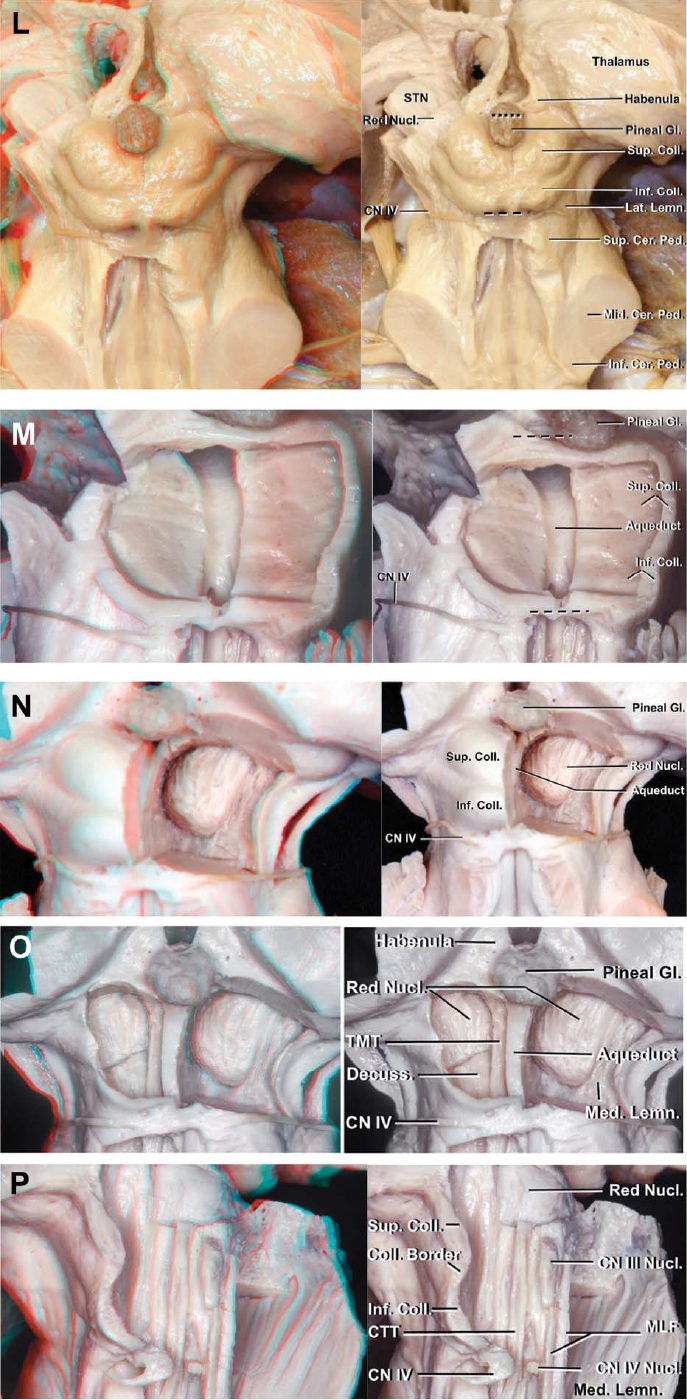
圖3(幫)。中腦。左,頂板後視圖,以及球上和球下安全進入區(斷續線)。小腦和左丘腦被切除了。橫丘上切口位於上丘上緣上方。枕下切口橫切於滑車神經和下丘下緣之間。如果橫切口向外側延伸過遠,小腦上梗和外側丘將被分割。M,後視圖,進一步剝離頂板。部分上丘和下丘被切除以顯露導槽,導槽是位於上丘和下丘入路腹側邊界的一個重要標誌。N,進一步剝離頂板。 The right red nucleus is exposed while preserving the superior and inferior colliculi on the left side. O, posterior view. The red nucleus extends from the midlevel of the inferior colliculus to the lateral wall of the third ventricle. An incision extending laterally from the midline encounters the habenula, trigeminal mesencephalic tract, central tegmental tract, and red nucleus as the incision is extended from dorsal to ventral. The decussation of the superior cerebellar peduncle is positioned caudal to the red nucleus and at the level of the inferior colliculus. P, posterior view of the midbrain tegmentum on the left side. The oculomotor nucleus is located at the level of the lower half of the superior colliculus and the upper half of the inferior colliculus. The trochlear nucleus is located caudal to the oculomotor nucleus at the level of the lower half of the inferior colliculus. The medial longitudinal fasciculus connects these cranial nuclei. Extending the infracollicular incision deeper than the aqueduct from dorsal to ventral will cross the trochlear nuclei, medial longitudinal fasciculus, and decussation of the superior cerebellar peduncle. Ant., anterior; Cap., capsule; Cer., cerebellar, cerebral; Coll., colliculus; Cort., cortico; CTT, central tegmental tract; Decuss., decussation; Dors., dorsal; Fac., facial; Fasc., fasciculus; Fr. Pon., frontopontine; Gen., geniculate; Gl., gland; Inf., inferior; Int., internal; Interped., interpeduncular; Lat., lateral; Lemn., lemniscus; Mam., mammillary; Med., medial, medullaris; Mes., mesencephalic; Mid., middle; MLF, medial longitudinal fasciculus; Nucl., nucleus; Ped., peduncle; Perioc., perioculomotor; Pon., ponto; Quad., quadrigeminal; Retro., retroflexus; Rt., right; Seg., segment; Spin., spinal; STN, subthalamic nucleus; Str., stria; Subst., substantia; Sulc., sulcus; Sup., superior; Teg., tegmentum; Tent., tentorium; Thal., thalami, thalamic; TMT, trigeminal mesencephalic tract; Tr., tract; Vent., ventral. (Images courtesy of AL Rhoton, Jr.)

圖3 q - t)(。中腦。Q,丘腦切除後的後視圖。在中腦,內側係索向腹外側上升至紅核,向外側上升至丘腦下核,進入丘腦。紅色核從下丘的中段延伸到第三腦室的側壁。丘腦下核位於紅色核的腹側和內囊的背內側。其他標記的結構是後屈束,它沿著紅色核的背內側延伸並連接著韁肌和踝間核,乳丘腦束,連接著乳體和丘腦前核,以及連接韁肌和中隔區域的丘腦髓質紋。R,踝間窩前視圖。周動眼神經安全進入區位於外側皮質脊髓束內側邊緣和內側動眼神經出口點之間。切口向內側傾斜過大會損傷動眼神經的腦內段,向外側傾斜過會進入皮質脊髓束。 S, further dissection. The perioculomotor entry will encounter the red nucleus just behind the medial lemniscus. T, left lateral view. The intramesencephalic segment of the oculomotor nerve passes medial to the red nucleus and exits the interpeduncular fossa. The red nucleus is located dorsal to the exit point of the oculomotor nerve from the brainstem. The surgical routes through the perioculomotor zone to the ventral (yellow arrow) and dorsal (blue arrow) midbrain are shown. Ant., anterior; Cap., capsule; Cer., cerebellar, cerebral; Coll., colliculus; Cort., cortico; CTT, central tegmental tract; Decuss., decussation; Dors., dorsal; Fac., facial; Fasc., fasciculus; Fr. Pon., frontopontine; Gen., geniculate; Gl., gland; Inf., inferior; Int., internal; Interped., interpeduncular; Lat., lateral; Lemn., lemniscus; Mam., mammillary; Med., medial, medullaris; Mes., mesencephalic; Mid., middle; MLF, medial longitudinal fasciculus; Nucl., nucleus; Ped., peduncle; Perioc., perioculomotor; Pon., ponto; Quad., quadrigeminal; Retro., retroflexus; Rt., right; Seg., segment; Spin., spinal; STN, subthalamic nucleus; Str., stria; Subst., substantia; Sulc., sulcus; Sup., superior; Teg., tegmentum; Tent., tentorium; Thal., thalami, thalamic; TMT, trigeminal mesencephalic tract; Tr., tract; Vent., ventral. (Images courtesy of AL Rhoton, Jr.)
腹側中腦和橋腦包含皮質脊髓束、皮質球束和皮質橋腦束(圖2)。尾髓隻包含皮質脊髓束。在中腦中,額橋纖維位於內側,皮質脊髓和皮質球束位於中部,顳頂-枕橋束位於腦梗外側(圖2C)。1、17 - 19從中腦開始的解剖很難定義橋腦的皮質脊髓束,但當從髓質金字塔下方解剖時,它可以遵循到腦梗的中間三分之一,這與其他解剖和臨床研究一致。1、15、17 - 19在腦橋,皮質脊髓束向前正中方向伸展。皮質球束向皮質脊髓束的背側下降與相關的腦神經核相連。橋腦皮質纖維在橋腦核處結束,橋腦核分布在皮質脊髓束和皮質球束的前後。14
背束包括內側縱束、中央被蓋束、三叉中腦束和三叉脊髓束。
內側縱束。這個有髓鞘的神經束從中腦延伸到上胸脊髓,並將視覺中樞和前庭中樞與控製眼睛、頭頸運動的神經核連接起來(圖2B和2D-G)。在中腦水平,與腦導水管背側與小腦上梗腹側交叉的中線相鄰。內側縱束向外側和腹側延伸至動眼神經核,在那裏與動眼神經的腦內段混合,並終止於位於大腦導水管吻側的間質核(圖2F和3I)。15在下丘下半部分水平,內側縱束與滑車核相連,並在第四腦室底部附近移動,並與腦橋中線相鄰。中間溝兩側的寬度為1毫米(圖2D)。它在麵丘的下緣向腹側彎曲,接近內側丘,並通過舌下三角的腹側。它在纖細結節和楔形結節水平穿過內側係索,並在脊髓腹側小索處繼續延伸(圖2B)。
三叉中腦和脊髓束。三叉神經在中腦橋進入腦幹。它通過小腦中梗向第四腦室延伸,到達三叉神經運動核和主感覺核,並在那裏分成三叉神經中腦束和脊髓束(圖2D)。三叉中腦束向上延伸至第四腦室底部的上半部分,位於小腦上梗外側、限製溝內側、中央被蓋束腹側和藍斑背側之間。三叉中腦束與正中溝之間的距離因中腦束的彎曲路線而變化。三叉中腦束和中溝之間的距離在三叉運動核和主感覺核水平平均5.9毫米,這些核位於麵丘的前外側。在上層的中層,這一距離平均為5.4 mm,在滑車神經水平,這一距離平均為3.7 mm(圖4I)。
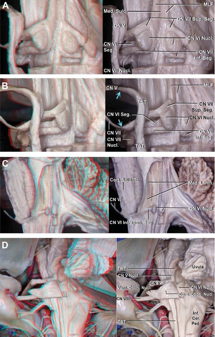
圖4(模擬)。腦橋。A,麵丘的後視圖。第四腦室室管膜和麵丘周圍的白質被切除。麵丘覆蓋在麵神經的外展核和橋突內段。外展核位於地板表麵腹側的中位。麵神經中間溝和外展神經核之間的內側縱束和橋膜內段。B,麵丘後外側視圖。藍色箭頭表示的是麵神經和三叉神經變成腦橋內神經的地方。麵核位於外展核的腹外側和三叉神經脊髓束的腹內側。 The initial intrapontine segment of the facial nerve extends dorsomedially toward the floor of the fourth ventricle, curves around the lower, medial, and upper edges of the abducens nucleus, and continues ventrolaterally along the medial side of the trigeminal spinal tract to exit the pons. C, left posterolateral view of another specimen after removing the left facial nerve. The intrapontine segment of the abducens nerve originates from the ventral face of its nucleus and proceeds anteriorly through the medial lemniscus and lateral to the corticospinal tract to exit the ventral pons. D, posterior view of another specimen. The dorsal cochlear nucleus sits on the dorsal surface of the inferior cerebellar peduncle, where it forms a smooth prominence called the auditory tubercle. The ventral cochlear nucleus is located on the lateral surface of the inferior cerebellar peduncle. The intrapontine segment of the vestibulocochlear nerve passes dorsal to the trigeminal spinal tract. Amb., ambiguus; Attach., attachment; Cer., cerebellar; CN, cranial nerve; Coch., cochlear; Coe., coeruleus; Coll., colliculus; Cort., cortico; CTT, central tegmental tract; Cun., cuneate; Dors., dorsal; Fac., facial; Flocc., flocculus; Fren., frenulum; Hypo., hypoglossal; Inf., inferior; Intrapon., intrapontine; Junct., junction; Lat., lateral; Lemn., lemniscus; Limit., limitans; Med., medial, median; MLF, medial longitudinal fasciculus; Nucl., nucleus; Ped., peduncle; Pon., pontine; Post., postrema; Rec., recess; Seg., segment; Spin., spinal; Sulc., sulcus; Sup., superior; Tent., tentorium; TMT, trigeminal mesencephalic tract; Tr., tract; Triang., triangle; TST, trigeminal spinal tract; Tub., tubercle; Vent., ventral; Vest., vestibular. (Images courtesy of AL Rhoton, Jr.)

圖4 (eg)。腦橋。E,另一個標本的前外側視圖。三叉神經進入區(斷線)位於三叉神經和麵神經之間。F,另一個標本的腋窩周圍區斜視。左側小腦和部分腦幹被切除,以顯示三叉神經脊髓束與顱神經之間的關係。藍色箭頭表示三叉神經離開腦幹的點,藍色斷線表示垂直的三叉神經周圍切口。三叉神經脊髓束在腦橋中段向尾側彎曲,向下延伸至脊髓。三叉神經脊髓束向脊神經7、9、10、11和12的腦橋內節段背側下行,並向脊神經8的腹側下行。G,另一個標本的乙狀結腸後切口的左側視圖。 The blue arrow shows the point where the facial and trigeminal nerves become intrapontine. The important structures in the peritrigeminal zone are the corticospinal tract anteromedially, the intrapontine segment of the posterior trigeminal root superiorly, the trigeminal motor and main sensory nuclei, intrapontine segment of the facial nerve, and trigeminal spinal tract posteromedially, and the ventral cochlear nucleus, and intrapontine segment of the abducens nerve inferiorly. Amb., ambiguus; Attach., attachment; Cer., cerebellar; CN, cranial nerve; Coch., cochlear; Coe., coeruleus; Coll., colliculus; Cort., cortico; CTT, central tegmental tract; Cun., cuneate; Dors., dorsal; Fac., facial; Flocc., flocculus; Fren., frenulum; Hypo., hypoglossal; Inf., inferior; Intrapon., intrapontine; Junct., junction; Lat., lateral; Lemn., lemniscus; Limit., limitans; Med., medial, median; MLF, medial longitudinal fasciculus; Nucl., nucleus; Ped., peduncle; Pon., pontine; Post., postrema; Rec., recess; Seg., segment; Spin., spinal; Sulc., sulcus; Sup., superior; Tent., tentorium; TMT, trigeminal mesencephalic tract; Tr., tract; Triang., triangle; TST, trigeminal spinal tract; Tub., tubercle; Vent., ventral; Vest., vestibular. (Images courtesy of AL Rhoton, Jr.)

圖4(設定h)。腦橋。H腦幹後表麵和第四腦室底。第四腦室的底部在中線被中間溝隔開。溝界限,另一個縱向溝,沿地板延伸至中溝外側。地板分為三個部分:上部或橋狀部分,中間或連接部分,下部或髓狀部分。上中央凹位於麵丘的外側,下中央凹位於舌下三角的外側。中腦,腦橋和髓質後視圖。三叉神經有3個感覺核:主要感覺核位於運動核的外側,三叉中腦核隨其神經束上升至中腦,三叉脊髓核隨其神經束下降至上脊髓。三叉中腦束在外側小腦上梗和內側溝界線之間上升,並在橋上端和中腦向中線彎曲。 The central tegmental descends deep to the trigeminal mesencephalic tract. The trigeminal mesencephalic and central tegmental tracts are located deep to the locus coeruleus. The inferior cerebellar peduncle ascends just lateral to the cuneate fasciculus. Amb., ambiguus; Attach., attachment; Cer., cerebellar; CN, cranial nerve; Coch., cochlear; Coe., coeruleus; Coll., colliculus; Cort., cortico; CTT, central tegmental tract; Cun., cuneate; Dors., dorsal; Fac., facial; Flocc., flocculus; Fren., frenulum; Hypo., hypoglossal; Inf., inferior; Intrapon., intrapontine; Junct., junction; Lat., lateral; Lemn., lemniscus; Limit., limitans; Med., medial, median; MLF, medial longitudinal fasciculus; Nucl., nucleus; Ped., peduncle; Pon., pontine; Post., postrema; Rec., recess; Seg., segment; Spin., spinal; Sulc., sulcus; Sup., superior; Tent., tentorium; TMT, trigeminal mesencephalic tract; Tr., tract; Triang., triangle; TST, trigeminal spinal tract; Tub., tubercle; Vent., ventral; Vest., vestibular. (Images courtesy of AL Rhoton, Jr.)

圖4 (J-L)。腦橋。J,後視圖。上中央凹呈三角形。三角形的頂點位於地板上部最外側的位置。它的上外側邊緣由小腦上梗構成,下外側邊緣由前庭區構成,內側底部由溝限肌構成。上中央凹三角形的上外側邊緣是三叉神經運動核和主要感覺核(黃圈)的深部位置的標誌。上中央凹三角形的頂端與麵丘的吻側邊緣位於同一橫向水平麵上。麵丘的外側邊界由溝限肌構成,內側邊界由內側縱束構成。K,麵表麵後視圖(綠色間斷線)。 The suprafacial approach is limited rostrally by the frenulum veli through which the trochlear nerve passes, caudally by the superior intrapontine segment of the facial nerve forming the upper edge of the facial colliculus, medially by the medial longitudinal fasciculus, and laterally by the sulcus limitans. L, enlarged posterior view of the infrafacial approach. The rostral border of this approach is the inferior intrapontine segment of the facial nerve forming the lower edge of the facial colliculus and corresponding to the level of a transverse line crossing the upper edges of the lateral recesses. The caudal border of the infrafacial approach, which is the site of horizontal attachment of the tela choroidea along the lower margin of the lateral recess, is positioned at the same transverse level as the upper margin of the hypoglossal triangle. The lateral edge of the ipsilateral medial longitudinal fasciculus forms the medial border and the facial nucleus and nucleus ambiguus are positioned below the surface of the floor at the lateral border of the infrafacial approach. The facial nucleus and nucleus ambiguus are found deep and just lateral to the most medial point of the attachment of the tela along the lower edge of the lateral recess (blue star). The facial nucleus is located at the level of the pontomedullary junction. Amb., ambiguus; Attach., attachment; Cer., cerebellar; CN, cranial nerve; Coch., cochlear; Coe., coeruleus; Coll., colliculus; Cort., cortico; CTT, central tegmental tract; Cun., cuneate; Dors., dorsal; Fac., facial; Flocc., flocculus; Fren., frenulum; Hypo., hypoglossal; Inf., inferior; Intrapon., intrapontine; Junct., junction; Lat., lateral; Lemn., lemniscus; Limit., limitans; Med., medial, median; MLF, medial longitudinal fasciculus; Nucl., nucleus; Ped., peduncle; Pon., pontine; Post., postrema; Rec., recess; Seg., segment; Spin., spinal; Sulc., sulcus; Sup., superior; Tent., tentorium; TMT, trigeminal mesencephalic tract; Tr., tract; Triang., triangle; TST, trigeminal spinal tract; Tub., tubercle; Vent., ventral; Vest., vestibular. (Images courtesy of AL Rhoton, Jr.)
三叉神經脊髓束在三叉神經運動和感覺核的水平向尾側彎曲,並在前庭耳蝸神經背膜內段和麵神經、舌咽神經、迷走神經、副神經和舌下神經腹側的橋膜內段之間下行,到達脊髓(圖4F)。
中央蓋的束.該束是錐體外係的一部分,連接著紅核和下橄欖核(圖2D, E, G, 3K,和4I)。在中腦水平,中央被蓋束起源於紅核的背內側部分,向同側下降,通過小腦上梗交叉,向內側縱束外側延伸。16中央被蓋束深入到第四腦室底部的上半部分,位於外側的限製溝和內側的小腦上梗之間。中央被蓋束位於藍斑斑和三叉中腦束的深處和內側係索的背側。在麵丘水平,中央被蓋束向外側在麵神經的橋膜內段和外展神經的橋膜內段之間,向內側延伸至下橄欖核的背內側。
中腦通過視束和腦梗之間的溝與上麵的間腦分開,通過橋腦後腦溝與下麵的橋腦分開(圖3)。內側係索將中腦分為腹側和背側兩部分。被蓋(紅色核)和頂蓋(四頭板)位於中腦背側,腦梗位於中腦腹側(圖3B和E)。中腦包含動眼神經和滑車神經的背腦段及其核。
動眼神經和核.該核位於上丘下半部分和下丘上半部分的中線附近,位於大腦導水管背側和小腦上梗腹側交叉之間(圖3D, F, I, T)。動眼器核的喙側長度平均5.3 mm。動眼神經的腦內段向內穿過,沿紅色核內側,在足間窩外側壁出中腦(圖3T)。
滑車神經和核.滑車核位於中腦中線附近,位於下丘下半部分水平(圖3D, F, I, P)。和動眼肌核一樣,滑車核位於腦導水管背側和小腦上梗腹側交叉之間。滑車纖維出核後向背側旋轉,繞導水管彎曲,在下丘下側出腦幹背麵,在小腦後腦裂內交叉至對側(圖3I)。
建議的中腦安全進入區域是沿著外側中腦溝、丘上區和丘下區以及眼周運動區(圖3)。
側中腦的溝。該溝位於中腦表麵,位於腦梗和外側丘之間,從橋腦腦溝向下延伸至上方內側膝狀體(圖3A-D)。17它位於中腦內側丘的腹側和中腦腹側和背側交界處的黑質的外側。將切口向前傾斜45°進入外側中腦溝,可到達中腦腹側和背側交界處的內側基板(圖3H)。將切口通過溝進一步向前傾斜,就會碰到腦梗。與頂蓋表麵成直角的切口將進一步向後側進入中腦背側(圖3H)。這個切口,根據進入點的水平,可以從外側到內側依次相遇:三叉中腦和中央被蓋束位於小腦上丘交叉的背側,紅核位於下丘上半部分靠近中線的水平沿第三腦室側壁向上延伸,小腦上丘交叉位於紅核位於下丘水平的尾部,動眼核位於上丘下半部分和下丘上半部分,滑車核位於下丘下半部分。紅色核位於中外側腦溝深度平均4.3 mm處,動眼器核和滑車核位於中外側腦溝表麵內側平均9.5 mm處的中線附近(圖3D-G)。
球上區和球下區.這些是導水管背側頂蓋(四邊形板)病變的建議安全進入區。1渡槽是確定引道深度的重要中線標誌(圖3L和M)。
對於球丘上入路,在上球丘上緣上方做一個橫向切口。比導水管深的切口會損傷動眼神經的腦內段和內側縱束,這兩者都位於導水管中線的腹側。當腦丘上切口從中線向外側延伸時,它將依次遇到韁肌、三叉中腦束、中央被蓋束和紅核。從上丘表麵到紅色核的深度平均5.5 mm(圖3N和O)。
對於下丘入路,在滑車神經和下丘下緣之間做一個橫向切口。一個比導水管深的切口,從淺到深,會碰到滑車核、內側縱束和小腦上梗交叉。橫向的球下切口向外側延伸,首先切開小腦上梗,然後切開外側係索。隨著外側切口從背側向腹側加深,它將遇到三叉中腦束、中央被蓋束和小腦上梗交叉(圖3P和圖T)。
Perioculomotor區.中腦腹中位病變可通過動眼周區到達,動眼周區由皮質脊髓和皮質球束之間的腦梗向外側直達動眼神經的內側出口點(圖3R-T)。1、14、18周圍動眼區的寬度、動眼神經的出口點向內側與皮質脊髓和皮質球束內側邊緣之間的距離是靠近動眼神經的一個狹窄區域,接近腦梗內側的三分之一至四分之一。1、15、17 - 19然而,最好將入路限製在椎弓根內側的1 / 4處。緊靠動眼神經外側的腦梗纖維是額突纖維。在中腦背側首先會遇到的結構是紅核和穿過它的動眼神經的腦內段。紅色核位於內側係索背側,是中腦動眼神經的出口點。必須注意避開紅核、動眼神經的背腦段、腦梗中部三分之一的皮質脊髓和皮質球束(圖3S和T)。
橋腦位於上麵的橋腦腦溝和下麵的橋腦髓溝之間。橋腦被內側神經束分為腹側(基底)和背側(被蓋)部分(圖2B和圖4)。它包含三叉神經、外展神經、麵神經和前庭耳蝸神經的橋膜內段及其各自的神經核。
三叉神經及其核.三叉神經運動核和主要感覺核位於中腦橋水平和第四腦室底部外側邊緣的深處(圖4)。三叉神經運動核和主要感覺核位於正中溝外側6mm處,即吻側、外側和麵丘上邊緣的深度。三叉神經核位於上中央凹三角形上外側邊緣和小腦上踝內側邊緣的深處(圖4J)。三叉神經有3個感覺核:(1)主感覺核,位於運動核的外側;(2)三叉中腦核,隨其神經束上升至中腦;(3)三叉脊髓核,隨其神經束下降至上脊髓(圖4I)。
外展神經及其核.外展核位於第四腦室底腹側的中位(圖4A- c, J和K)。在正中溝和外展核之間的麵神經的內側縱束和橋膜內段(圖4A)。外展神經的橋腦膜內段起源於外展神經核的腹側麵,向腹方向通過內側係索,向外側延伸至皮質脊髓束,在皮質脊髓束外側邊緣出橋腦橋(圖4C)。
麵神經及其核.麵核位於橋髓連接處水平,內側丘係背側,歧核吻側,外展核腹外側,三叉神經脊髓束腹內側,下橄欖核最上邊緣背內側(圖4A, B和J-L)。從地板表麵到麵核的深度平均為5.5 mm。麵神經的腦膜內段起源於麵神經核後向背內側延伸至第四腦室底部,依次環繞外展神經核的下、內、上邊緣,並在內側縱束的外側通過三叉神經脊髓束的內側,在橋小腦角處離開橋腦。
前庭耳蝸神經及其核.前庭耳蝸神經在橋髓溝外側端進入腦幹(圖4D)。前庭部分位於前上,耳蝸部分位於後下。耳蝸背核位於小腦下梗的背表麵在側隱窩中形成一個光滑的突起稱為聽覺結節。耳蝸腹側核位於小腦下梗外側表麵。耳蝸背核內側邊緣到正中溝和同側溝界限的距離分別為9毫米和6毫米。
腦橋腹側的安全進入區是小腦周圍區。
Peritrigeminal區。腦橋腹側病變的入路通常通過三叉神經和麵神經之間的縱向切口(圖4E和F)。20.切口穿過橋橋麵橫向纖維。隨著切口的加深,切口到達橋腦核並穿過橋腦橫向纖維。皮質脊髓束穿過腹內側腦橋。皮質脊髓束後外側緣到三叉神經和麵神經腦幹交界處的距離平均分別為9.2 mm和8 mm。這個切口不能從尾部延伸到麵神經出口區域的吻側邊緣,因為有損傷外展神經和麵神經的橋突內節段的風險。在三叉神經背側做切口有損傷三叉神經橋膜內段和耳蝸腹側核的風險。將切口的吻側延伸至三叉神經,尾側延伸至麵神經,可能會損傷三叉神經、外展神經和麵神經的橋突內段。三叉神經運動核位於三叉神經與腦橋交界處背內側平均10.1 mm處(圖4F和G)。
腦橋背側位於內側基肌腹側和第四腦室背側底部之間。
第四腦室的底部.第四腦室底部呈菱形(圖4H-L)。吻側三分之二的基底位於腦橋的後表麵尾側三分之一位於髓質的後表麵。腦導槽開口至腦尖,尾端位於腦obex。連接側隱窩吻側邊緣的一條線標誌著橋腦和第四腦室底髓質部分的交界處。基底分為上部(橋橋)、中間(連接)和下部(髓質)。上部有一個三角形的形狀:它的頂點是在渡槽,它的基礎是由橫向凹槽的吻側邊緣的一條線創建的。藍斑位於基底吻側邊緣的溝界線外側。中間部分是由橫向凹槽的上下邊緣之間的條狀物形成的。尾部呈三角形,橫向上受限於與脈絡膜背部相連的底的下外側邊緣,尾部受限於腹壁。
底板從吻端尖到尾端被中溝分成兩個對稱的半部分。另一個縱向的溝,沿室底延伸至正中溝外側。中位隆起是位於中溝和溝界限之間的縱向隆起。它是麵丘的位置,位於舌下核和迷走神經核上的突起,以及瘢痕後區域。這3個成對的三角形區域覆蓋舌下核和迷走神經核以及基底髓質部分的殘端區域,使該區域呈筆尖狀外觀,因此被稱為腳菖子。邊緣溝在兩點處加深,形成凹窩,稱為中央凹。上中央凹在麵丘的外側,下中央凹在舌下三角的外側。下中央凹位於地板的髓質部分,緊挨著舌下三角的外側,位於前庭上方區域和迷走神經下三角的上邊緣之間。髓紋在室底側隱窩處穿過。
優越的小窩.上中央凹呈三角形,位於麵丘的外側(圖4J)。三角的上外側邊緣由小腦上梗構成,下外側邊緣由前庭區構成,內側底部由溝極限肌構成。三角的頂點位於第四腦室吻側部的最外側點。上中央凹是估計麵丘位置和三叉神經運動核和主感覺核的深部位置的重要標誌。上中央凹三角形的頂端與麵丘的上邊緣位於同一橫向水平麵上。三角的上外側邊緣是估計三叉神經運動核和主要感覺核深度位置的一個標誌。如果麵丘的突出位置不清楚,可以用上中央凹三角形頂點的水平作為麵丘上緣的橫向水平。
麵丘.麵丘位於內側縱束和溝限肌之間的中線附近(圖4A、B和H-L)。麵丘的吻側緣是由麵神經的上腦膜內段形成的,與上中央凹三角形的外側尖位於同一橫向水平。它的尾緣是由麵神經的下腦膜內段形成的,位於橫線的水平,橫線穿過側隱窩的上邊緣。麵神經的下腦膜內段與麵神經核在同一橫平麵上。麵丘的喙側長度平均4.5 mm,寬度平均3.5 mm。
在橋腦背側,建議的安全進入區域分別是麵向麵丘上方和下方的麵表麵和麵下入路(圖4)。
同側的方法.麵上安全進入區在側麵受滑車神經穿過的velulum的限製,在尾部受位於麵丘上緣的麵神經的上部橋膜內段的限製,在內側受內側縱束的限製,在外側受溝限製肌的限製(圖4H-K)。這種入路的切口不應向內側延伸至內側縱束或向外側延伸至溝限,以避免損傷位於藍斑斑深部的三叉中腦束和中央被蓋束,或損傷位於上三角中心凹上外側邊緣深部的三叉運動核和主感覺核。三叉神經運動核和主要感覺核位於正中溝外側平均6毫米處。入口區喙部平均長度為12.7 mm。內側縱束寬度為1毫米。從地板表麵到內側基板的深度平均為4.5 mm。
Infrafacial方法.麵神經的下腦膜內段形成了麵丘的下緣,也形成了麵下入路的吻側邊界,位於穿過側隱窩上緣的橫線水平上(圖4L和5B)。尾緣位於舌下三角的上緣,位於脈絡膜尾部與側隱窩下緣的連接處。麵下安全進入區域的喙側長度等於側隱窩或地板中間(連接)部分上下邊界之間的距離。內側縱束形成內側邊界,從吻側至尾側深至底依次排列的麵核和麵核歧義形成外側邊界。麵核和模糊核位於脈絡膜背側窩下緣最內側附著點的外側深部。

圖5。A,左前外側觀。橄欖前溝位於橄欖和金字塔之間。橄欖口側凹陷,橄欖上窩,就在麵神經和前庭耳蝸神經與腦幹交界處的下方。舌咽神經、迷走神經和副神經出髓質,在橄欖後溝的背側,橄欖後溝位於橄欖和小腦下梗之間。舌下細根沿橄欖前溝出髓質。B,髓質,後視圖。岐核位於迷走神經三角的腹外側,三叉神經脊髓束的腹內側,楔形結節的腹側,橄欖的背側,麵核的尾側。舌咽神經、迷走神經和副神經的髓內段出岐核後向外側移動,在三叉神經脊髓束腹側,沿橄欖溝後出髓質。C, 3個背側髓質進入區分別位於中線obex以下的後正中溝、薄肌與楔形束之間的後中間溝和楔形束腹緣的後外側溝。 Amb., ambiguus; Cer., cerebellar; Hypo., hypoglossal; Inf., inferior; Inter., intermediate; Med., median; Nucl., nucleus; Ped., peduncle; Post., posterior; Posterolat., posterolateral; Sulc., sulcus; Triang., triangle; TST, trigeminal spinal tract; Tub., tubercle. (Images courtesy of AL Rhoton, Jr.)
內側髓係將髓質分為腹側和背側兩部分(圖5)。腹側髓質由覆蓋在皮質脊髓束上的金字塔組成(圖2B)。橄欖位於內側基板外側。橄欖前溝在錐體和橄欖之間縱向延伸。舌下神經髓內段起於舌下三角下方的舌下核,並向腹側延伸至內側縱束內側和橄欖側之間,沿橄欖前溝尾部的三分之二處出髓質。橄欖上窩是橄欖吻側的凹陷區,是麵神經和前庭耳蝸神經與腦幹連接的地方。橄欖後溝位於橄欖腹側和小腦下梗背側之間,位於舌咽神經、迷走神經和副神經出髓質的腹側。三叉神經脊髓束向腹內側下行,楔形束向內側上升至小腦下踝(圖4I)。
核Ambiguus.它位於迷走神經三角腹外側,三叉神經脊髓束腹內側,橄欖背側,麵核尾側(圖5B)。舌咽神經、迷走神經和副神經從岐核和三叉神經脊髓束的腹側向外側穿過,在橄欖後溝的背側出髓質。舌咽神經和迷走神經從橄欖後溝的上部的背側出,副細根沿著橄欖後溝的尾部出。
建議的髓質安全進入區沿著前外側(橄欖前)、橄欖後、後正中、後中間和後外側溝(圖5)。
前外側(橄欖前)溝.這個入口區位於舌下末梢根和C1吻側細根之間的橄欖前溝。由於它非常接近錐體束和錐體束交叉,因此僅外生病變首選此入口。2
的Postolivary溝.該區域通過位於橄欖和小腦下梗之間的橄欖後溝和舌咽根和迷走神經根腹側的橄欖後溝進入。9舌咽部和迷走神經細根在腦幹的背側與這個溝的上部相連。當切口變深時,它會碰到距溝表麵平均4毫米深的岐核。
背髓質.背側髓質病變的三個進入區已經被定義。這些是位於中線obex下方的後正中溝,位於薄肌和楔形束之間的後中間溝,以及位於楔形束外側的後外側溝。1三叉神經脊髓束腹向楔形束下延伸至上脊髓。
進入腦幹的外科手術可能導致嚴重的發病率,這需要了解腦幹的內部結構和安全進入區域邊界與內部結構之間的關係。2、21本研究采用纖維解剖技術檢查小腦和腦幹的白質,以評估進入中腦、橋腦和髓質的安全入口區邊界,以及它們在腦幹表麵的相應標記。通過選擇性3T磁共振(MR)束造影術支持的纖維束解剖確定每個安全入口區域的大小和位置。雖然已經有許多關於腦幹內部結構的組織學和放射學研究,但基於纖維解剖技術和3d攝影的安全進入區域的檢查還沒有。應用這些結果受到兩個事實的限製:第一,患者個體間存在差異;第二,很難知道通路的確切路徑,因為病變扭曲了解剖結構,因此正常解剖不再是異常解剖的充分指南。術中刺激有一定的幫助,特別是在麵核或錐體束區域,但它不能告訴我們腦幹內其他束的路線。
在腦幹病變切除的手術計劃中,目的應該是通過盡可能少的正常組織,並避免幹擾關鍵的核和束。22這些方法中最重要的問題是確定從表麵到病灶的最短距離和造成最小神經損傷的路徑。對於位於表麵以下的病變,最安全的方法通常是最短和最直接的方法。23對於到達腦幹表麵的病變,最安全的進入區是顯而易見的。24然而,為了保護關鍵的神經組織,可能需要其他方法。22對於深部病變,已經描述了一些安全進入區域。1、3一般來說,最好從最靠近病灶的表麵進入病灶。
三叉神經和麵神經之間的三叉神經周圍區域已被確定為腦橋腹外側部內側丘係腹側病變的安全進入區。20.對於是否需要垂直(縱向)或橫向切口,意見不一。4日,25日,26日橫向切口的目的是使其平行於橋突橫向纖維,從而減少對這些纖維的損傷。9在本研究中,三叉神經出口區到皮質脊髓束的距離平均為9毫米。三叉神經和麵神經橫切時應避免的關鍵神經結構是皮質脊髓束、三叉神經脊髓束、麵神經橋突內段以及三叉神經運動和感覺核。三叉神經和麵神經之間的垂直切口應避免的結構是麵神經和外展神經的橋突內段。當縱向切口向前深入時,會碰到皮質脊髓束。
第四腦室底麵丘的上、下區域已被報道為安全的手術進入區識別第四腦室底部安全進入區域邊界的一個重要問題是識別麵部丘。關於麵丘在地板中的位置,已有許多基於數值測量的研究。我們的研究與其他研究的不同之處在於,在解剖中暴露了麵神經、神經核、腦膜內節段以及外展神經核。這樣就可以更詳細地描述麵部上方和麵部下方以及其他提議的安全進入區域的邊界。在這個區域要避免的最重要的結構是麵丘。Bogucki et al,10在一項對40個標本的研究中,發現37%的麵丘在第四腦室底部沒有形成明顯的突出。他試圖根據其他永久性的地標來估計它的位置,比如bex和veli係帶,其他人則是基於數值測量。5、12我們的測量結果與其他研究結果一致。在這項研究中,解剖了麵神經的腦膜內段和產生麵神經丘的外展核,並詳細描述了與腦室底標記相關的麵上和麵下安全進入區邊界。
據報道麵上入路的邊界是內側縱束,尾部的麵丘,外側的小腦上梗內側邊緣。3.內側縱束在正中溝兩側向外側延伸1毫米。Bogucki等人認為包含滑車神經的絲狀帶是吻側界限。我們的結果與這個吻側界限一致,但與先前的報告不同,發現溝限製是一個比小腦上梗更安全的外側界限。使用限製溝而不是小腦上梗作為外側限製,將避免損傷三叉神經運動核、中腦束和位於限製溝外側的中央被蓋束。中央被蓋束損傷可引起眼球震顫和意圖性震顫。17日,27日28三叉神經運動核位於麵丘略上方,位於溝限肌外側。如果在地麵上看不到麵丘,上中央凹三角形的頂點與麵丘的上邊緣在同一水平麵上,是一個有用的標記。內側丘係距地麵平均4.5毫米深,是前界。如果在麵表麵區深部找不到病變,則很可能在腹側橋腦比內側丘係深。皮質脊髓束距地麵平均20毫米深。對於位於內側神經丘腹側的病變,應考慮采用脛束周圍入路。
這個入路的方向是在麵神經丘之間,由麵神經的腹膜內下段,舌下三角和迷走神經三角,內側縱束和外側的麵神經核形成。10在本研究中,我們發現麵下安全進入區域的吻側和尾側邊界分別位於側隱窩的上邊緣和下邊緣。在本研究中,這個入口區域的平均長度為6毫米,盡管也有其他長度的報道。5, 10, 12模糊核的上極,位於麵核的尾部,形成了麵下入路的外側邊界。麵核和模糊核位於脈絡膜底部沿側隱窩下緣最內側附著點的外側。切口應保持在這一點的內側,以避免損傷麵部核和核模糊。
髓紋被認為是麵下三角的尾緣。3.然而,這一發現受到了挑戰,因為髓紋有許多變化,而且在30%的第四心室底無法看到髓紋。10其他更可預測的地標也被探索過。5、6我們發現舌下三角的上邊緣與obex之間的距離平均為10.6毫米。舌下三角的上邊緣總是在迷走神經三角的吻側,並且與沿著側隱窩下緣的脈絡膜水平附著在同一橫向水平線上。5
綜上所述,建議麵下三角邊界的標誌為:側隱窩上下緣的橫線,內側縱束,外側隱窩下緣絨毛膜附著最內側點的側腹水平。腹側界限是中央被蓋束,距地麵平均4.9毫米深。
該入路用於橋腦上半部的中心病變,通過沿麵丘上緣上方正中溝的縱向切口。4, 18它有損傷內側縱束的風險,該束沿著中溝和滑車核的邊緣,位於麵丘上方15mm,靠近中線。如果位於中線附近的網狀結構在術後期間被雙側破壞,可能會觀察到改變或意識喪失。29
這個區域被建議用於位於中線較遠處的病變。1該方法所遇到的結構從淺到深依次為耳蝸背核、小腦下梗、三叉神經脊髓束、模糊核和麵核。
一個重要的限製是,在術前計算機斷層掃描和MRI上看到的安全進入區域的內部結構和邊界,可能會在顱骨和硬腦膜打開後,由於病變的扭曲作用而移位。因此,術中神經生理監測和繪圖的使用是至關重要的。6DTI顯示腦幹結構在術前計劃中也很有用。在這個和其他形態測量學研究中,測量可能會受到與固定相關的標本收縮的影響。5、30為了盡量減少這種收縮,我們隻使用5%的酒精進行固定,沒有像克林勒技術那樣冷凍標本。31我們沒有發現冷凍腦幹和未冷凍腦幹在纖維剝離難度上有任何顯著差異。這可能是因為腦幹的致密組織在冷凍時不允許水的擴散來分離纖維。我們希望這些知識的應用將使進入腦幹的手術更加準確和安全。
貢獻者:Kaan Yagmurlu, MD, Albert L. Rhoton, Jr, MD, Necmettin Tanriover, MD,和Jeffrey A. Bennett, MD
內容來自Yagmurlu K, Rhoton AL, Jr, Tanriover N, Bennett JA。三維顯微外科解剖和腦幹安全進入區。神經外科2014; 10:602 - 619。doi.org/10.1227/NEU.0000000000000466.經牛津大學出版社代表神經外科醫師協會批準。©神經外科醫生協會。
神經外科188bet手机app圖譜很榮幸能夠繼承Albert L. Rhoton, Jr . MD的遺產。
請登錄發表評論。
一定要在社交媒體上關注我們,獲取精彩內容並保持更新生活科恩醫生的會議,關於手術技術的問題,以及更多!


您必須登錄才能查看此材料。
的188bet手机app這幾乎完全取決於你的捐款。
如果沒有你們的大量捐贈,我們就無法繼續開展地圖集。
請承諾每年至少捐贈250美元給Atlas。如果沒有這種承諾,Atlas將很快需要付費訂閱,世界各地的許多外科醫生將無法獲得它,他們的病人的護理依賴於它。
現在請捐!
如果沒有你們的大量捐贈,我們就無法繼續開展地圖集。請承諾每年至少捐贈250美元給Atlas。
如果沒有這個承諾,Atlas將很快需要付費訂閱世界上許多病人的護理都依賴於它的外科醫生將無法使用它。現在請捐!