

你可以有所作為。
的188bet手机app這幾乎完全取決於你的捐款。
如果沒有你們的大量捐贈,我們就無法繼續開展地圖集。
請承諾每年至少捐贈250美元給Atlas。如果沒有這種承諾,Atlas將很快需要付費訂閱,世界各地的許多外科醫生將無法獲得它,他們的病人的護理依賴於它。
現在請捐!

最後更新:2021年4月7日
背景:大腦的纖維束可能是比皮層更重要的切除限度決定因素。更好地了解纖維通路的三維解剖組織對規劃安全、準確的大腦病變手術非常重要。
客觀的:研究人類大腦纖維束和皮層下灰質的地形解剖及其與一致的皮層、心室和核標誌物的關係。
方法: 25例福爾馬林固定人腦和4例整屍頭顱,采用纖維解剖技術,×6 ~ ×40放大檢查。纖維束和中央核結構,包括腦島和基底神經節,被檢查和他們的關係捕獲在3-D攝影。測量皮層回表麵與所選纖維束之間的深度。
結果:確定了大腦內重要的聯想、投射和束連與皮層淺表標誌的地形關係。與纖維束關係一致的重要標誌是皮質回和腦島溝、腦島的限製溝、中央核的核塊和側腦室。纖維束彼此之間也以一致的模式組織起來。將解剖結果與臨床放射學分析和術中纖維束刺激的功能數據進行簡要比較。
結論了解大腦纖維束的三維解剖組織對於計劃安全準確的腦部手術是至關重要的。

大腦纖維束的損傷可能導致比皮層損傷更嚴重和不可逆的缺陷。1 - 4皮質區域的可塑性似乎也高於皮層下區域。皮層下纖維束(白質)不僅在維持大腦連接方麵很重要,而且在確定切除範圍時也是一個必要的考慮因素。因此,纖維束在手術計劃中起著重要作用,同時還要考慮皮質、動脈和靜脈。4
大腦纖維束的解剖特征在以前的研究中被檢查過。5 - 10這項研究利用三維(3d)攝影技術,聚焦於纖維束和皮層下灰質之間複雜的空間關係,以及它們與包括皮層回在內的表麵標誌的關係。這些解剖發現與臨床放射學分析和術中纖維束刺激的功能數據進行了簡單的比較。然而,主要關注的是三維解剖關係。
25個福爾馬林固定的大腦(50個半球)和4個完整的屍體頭部被檢查。蛛網膜和表麵血管被從大腦中移除。所有標本在216°C冷凍2周。11冷凍過程完成後,標本被解凍,並使用蔡司手術顯微鏡(Carl Zeiss AG, Oberkochen, Germany)提供的×6至×40放大率下的微型解剖器對纖維束進行解剖。標本在解剖過程中保存在5%福爾馬林溶液中。屍體頭部的纖維束通過額顳部開顱術檢查。解剖是逐步進行的。在這些解剖的每一個較深的步驟中發現的纖維束的位置是根據它們從覆蓋的皮層回的深度來記錄的。從外側到內側,檢查了島葉皮層和中線之間的中央核心結構。
在逐步解剖纖維束的暴露過程中,使用電子秤對10個大腦(20個半球)進行測量(表1)。根據該實驗室之前的一篇文章報道,每個階段的解剖都用3-D攝影記錄,以製備3-D圖像。12分析與上覆回或回相關的皮質下纖維束的地形位置和深度,特別是在額葉,以提供手術相關的3-D圖像。每個三維圖像都顯示在相應的標有標簽的二維圖像的旁邊或上麵。使用Adobe Photoshop CS5, Version 12.0X 64 (Adobe,聖何塞,加利福尼亞州)組裝的浮雕圖像,可以用紅色和藍色的眼鏡觀看。印刷頁麵上的浮雕圖像最好在有良好照明的情況下平躺時佩戴紅色和藍色眼鏡觀看;然而,一個光線充足、放大了3d圖像的電腦顯示器可以提供更好的觀看效果。

移除大腦皮層暴露出短的關聯纖維,即所謂的回間纖維或U纖維,它們連接著相鄰的腦回。較短的關聯纖維深的是較長的關聯纖維(圖1)。一般來說,關聯纖維通路的深度和長度成正比;連接遙遠區域的較長的連接纖維比較短的連接纖維位於更深(更中間)的位置。

圖1 (a - c)。佩裏絲纖維和長連接纖維。A,睫狀體周圍皮層由腹側運動前皮層(ba6)、額下回包括蓋部(ba44)、三角回(ba45)和眶回(ba47)、額中回(ba46 /9)、頂葉下小葉包括上緣回(ba40)和角回(ba39)、上回(ba22、ba41和ba42)、中回(ba21、ba37)和顳下回(ba20)組成。B,中央葉,由前腦回和後腦回組成,被保存了下來。移除皮質和鄰近的短纖維,暴露出長纖維。皮質和短纖維和長纖維是按橫向到中間的順序排列的。SLF II連接角回(ba39)或枕中回(ba19)的後部和額中回(ba10或ba46 /9),並深入到枕中回的前部、頂葉下小葉、中央前後回的中段和額中回。C,最淺表的長聯係纖維是額頂纖維和額顳纖維。額頂纖維由額頂連接和額顳連接的AF組成。SLF II發生於心房上部和側腦室體的表麵。位於庇護上區的腹側纖維通路是SLF III,它連接著邊緣上回和蓋部。 The AF is divided into ventral and dorsal segments. The ventral segment extends from the mid and posterior parts of the superior gyrus and mid part of the middle temporal gyrus, passes deep to the lower part of the supramarginal gyrus and medial to the SLF III in the frontoparietal operculum to reach the inferior frontal gyrus. The dorsal segment extends from the posterior part of the middle and inferior temporal gyri, passes through the angular gyrus and ventral to the SLF II to reach the inferior and middle frontal gyri. AF, arcuate fasciculus; Ang., angular; Ass., association; BA, Brodmann area; Cent., central; Dors., dorsal; Front., frontal; Gyr., gyrus; IFOF, inferior fronto-occipital fasciculus; ILF, inferior longitudinal fasciculus; Inf., inferior; Mid., middle; MdLF, middle longitudinal fasciculus; Occip., occipital; Operc., opercularis; PIP, posterior insular point; Preoccip., preoccipital; SLF, superior longitudinal fasciculus; Sulc., sulcus; Sup., superior; Supramarg., supramarginal; Temp., temporal; Triang., triangularis; Vent., ventral. (Images courtesy of AL Rhoton, Jr.)

圖1 (D-F)。D, SLF I, II, III和AF節段的顳葉皮層區已經暴露。由於SLF I位於腦半球的內側,所以在對側暴露。SLF I位於扣帶溝上岸,位於扣帶帶上方,胼胝體纖維內側,連接楔前葉與內側額回和前扣帶皮層。SLF III, SLF II和SLF I的位置依次從外側到內側,從腹側到背側。E,庇護上區AF和SLF節段的關係。AF腹段與SLF III一起移動並位於SLF III內側。AF背段與SLF II一起向腹移動。F,頂葉下小葉放大圖。AF腹側段穿過邊緣上回,與額頂頂蓋的SLF III一起向前投射。 The AF dorsal segment passes through the angular gyrus and projects forward just ventral to the SLF II. The AF dorsal segment passes through the lower part of the angular gyrus, whereas the SLF II passes through the upper part of the angular gyrus. AF, arcuate fasciculus; Ang., angular; Ass., association; BA, Brodmann area; Cent., central; Dors., dorsal; Front., frontal; Gyr., gyrus; IFOF, inferior fronto-occipital fasciculus; ILF, inferior longitudinal fasciculus; Inf., inferior; Mid., middle; MdLF, middle longitudinal fasciculus; Occip., occipital; Operc., opercularis; PIP, posterior insular point; Preoccip., preoccipital; SLF, superior longitudinal fasciculus; Sulc., sulcus; Sup., superior; Supramarg., supramarginal; Temp., temporal; Triang., triangularis; Vent., ventral. (Images courtesy of AL Rhoton, Jr.)

圖1(胃腸道)。G, AF腹段在三角部的末端和顳葉已經暴露。H,切除庇護下區的所有AF節段,暴露MdLF和ILF。後半回起於顳極,經過顳上回到達頂葉下小葉,尤其是角回。在島島後點(中斷的紅線)、上下限溝和下下限溝的連接處可分為前、後兩部分。中低層的前部位於島島後點的前方,位於IFOF的表麵,而中低層的後部與IFOF混雜在一起。ILF連接顳極和枕皮質背外側,但不到達鈣質皮質。在枕前切跡的水平,它向上旋轉45°到達枕後外側皮層。我,MdLF的放大視圖。房顫、弓狀纖維束; Ang., angular; Ass., association; BA, Brodmann area; Cent., central; Dors., dorsal; Front., frontal; Gyr., gyrus; IFOF, inferior fronto-occipital fasciculus; ILF, inferior longitudinal fasciculus; Inf., inferior; Mid., middle; MdLF, middle longitudinal fasciculus; Occip., occipital; Operc., opercularis; PIP, posterior insular point; Preoccip., preoccipital; SLF, superior longitudinal fasciculus; Sulc., sulcus; Sup., superior; Supramarg., supramarginal; Temp., temporal; Triang., triangularis; Vent., ventral. (Images courtesy of AL Rhoton, Jr.)
最淺的長連接纖維是上縱束(SLF)和弓狀束(AF)。SLF由額頂葉連接纖維和額顳葉連接纖維AF組成。SLF有3個部分:SLF I、II和III。SLF I位於額上回,SLF II位於額中回,SLF III位於額下回。從外側表麵開始,向內側移動,SLF的組成以相反的順序遇到:SLF III, SLF II,然後SLF I(圖1)。13SLF I,背側纖維通路,位於扣帶溝、楔前葉(頂葉內側表麵)和額上回(額內側回)的上端,在扣帶上方和胼胝體纖維內側移動(圖1D)。9日,13 - 15一項纖維解剖研究報告SLF I是一個短的關聯通路;然而,我們發現SLF I的前部與後部是連續的。16位於顱上區的長連接纖維通路由於在凹處繞溝偏轉而呈波浪形。在SLF II中可以看到同樣的波浪狀外觀。SLF I被認為與以身體為中心的動作的高階控製和運動活動的啟動有關。13日,17歲,18歲SLF II,中間通路,在角額回和中額回之間,位於心房上邊緣和側腦室體之間。在功能上,SLF II與空間意識的調節有關。13反過來,SLF II的損傷可能導致空間工作記憶和注意力障礙,如忽視綜合征。14、19術中對SLF II的直接電抑製導致對側視覺刺激欣賞的短暫下降。14、19位於額頂葉蓋內的腹側纖維通路是SLF III,它連接著邊緣上回和蓋部。13在功能上,左側(顯性)SLF III參與語音處理(工作記憶)。13、20、21刺激左側SLF III可引起關節紊亂,如構音障礙/構音障礙。13、20、21而右側(非顯性)SLF III則與視覺空間注意、韻律和音樂處理有關。13、14、22 - 24
另一個表麵的長關聯纖維通路是AF,它連接運動(布洛卡區)和感覺(韋尼克區)語言中心。房顫分為腹側段和背側段(圖1)。腹側段在庇護上區位於背側段的腹側,但在庇護下區位於背側段的前方。腹側段從顳上回的中後部和顳中回的中部經過邊緣上回的下部和額頂蓋SLF III的內側到達額下回。背段從顳中回和顳下回的後部穿過角回的下部,然後向腹側輕微移動到SLF II到達額下回和額中回。根據我們的解剖和來自神經成像、基於損傷任務和彌散張量成像(DTI)研究的文獻,AF腹側段與語音語言處理相關,而AF背側段與詞彙和語義語言處理相關。AF的腹側段與經典的AF模型相當。25日,26日
在顱下區,主要的長連接纖維通路是中、下縱束(MdLF和ILF)(圖1H和1I)。中下回位於顳上回內,內下回位於顳下回內。顳下回發源於顳極,經顳上回到達頂葉下小葉,尤其是角回。MdLF最初是由薩爾茨和潘迪亞描述的27在獼猴身上使用放射自顯影技術。他們的發現首次適用於人腦,顯示患有DTI的人腦中存在MdLF。28
在島島後點水平,上、下界限溝交界處和前顳橫回後內側端,中顳橫回可分為前後兩部分。前半段位於額枕下束表麵,後半段與額枕下束混雜在一起。29使用纖維解剖技術或成像技術都不可能將MdLF纖維與角回後方的IFOF和矢狀層纖維區分開來,這與先前報道的枕葉MdLF終止的一些研究相反。30、31日
MdLF的功能還不清楚。Makris等28表明MdLF可能在語言和注意力方麵起著重要作用。然而,德威特哈默等人298例患者在切除及電刺激後未發現語言障礙。因此,他們得出結論,中顳葉的前部與語言處理無關。目前還沒有關於中下丘腦後部的數據,但有人認為它與聲音的定位有關,因為它在角回(大腦導航中心的一部分)和海氏回(Heschl’s gyrus)上的聽覺接收區之間穿行。31
下縱束(ILF)連接顳極和枕皮質背外側,但不到達鈣質皮質(圖1H、2G和2K)。在枕前切跡水平,向上彎曲45°角,分布於枕皮質背外側。DTI研究顯示,ILF是一種枕顳連接,由起源於紋狀外的短纖維和長纖維組成,吻端末端位於顳中回和下回、顳極、海馬旁回、海馬和杏仁核(也稱為杏仁體)。32ILF涉及到物體的識別和識別。17日,33雖然術中對ILF的刺激沒有引起任何語言障礙,但由於它與梭狀回(枕顳回)和顳極等語義和句法處理相關區域的連接,ILF被認為是一種間接的語義語言通路。34也有人認為,作為腹側語義通路的一部分,ILF與MdLF和極端囊一起在語言理解中發揮作用。35、36ILF位於側腦室顳角軸位以下。切除顳下回和潛在的ILF,即使是在顯性半球,也不會導致嚴重的缺陷。6

圖2 (a - c)。A,穿過大腦中樞的軸向切麵。中央核心區位於外側島葉皮層和內側腦室之間。中央核包括島葉皮層、極囊、閉囊和外囊、殼核、蒼白球、內囊、尾狀核和丘腦。B,從外側到內側,暴露的中央核心結構包括島葉皮層、極囊、閉鎖、外囊的腹側和背側、殼核、蒼白球和內囊的後肢。C,中心核心結構的上外側視圖。尾狀核、蒼白球和丘腦已暴露。島葉皮層形成中央核心的外側邊界,上麵的側腦室和下麵的第三腦室形成內側邊界。內囊膝位於前連合的後麵,與後島短回和Monro孔在同一冠狀麵。Acc。, accumbens; AIP, anterior insular point; Amyg., amygdala; Ant., anterior; Call., callosal, callosum.; Caps., capsule; Caud., caudate; Cent., central, centrum; Claust., claustrum; Claustrocort., claustrocortical; CN, cranial nerve; Comm., commissure; Cor., corona; Corp., corpus; Dia., diagonal; Dors., dorsal; Dorsolat., dorsolateral; Ext., extension, external; Extr., extreme; FLP, frontal limen point; For., foramen; Front., frontal; Gl., gland; Glob., globus; Gyr., gyrus; Hippo., hippocampus; Hypoth., hypothalamus; IFOF, inferior fronto-occipital fasciculus; ILF, inferior longitudinal fasciculus; Inf., inferior; Innom., innominata; Ins., insula, insular; Int., internal; Lat., lateral; Lent., lenticular; LGB, lateral geniculate body; Lim., limiting; Mam., mammillary, mammillo; Med., medial, medullaris; MLF, medial longitudinal fasciculus; Nucl., nucleus; Occip., occipital; Orb. Front., orbitofrontal; Pall., pallidus; Par., parietal, parieto; PIP, posterior insular point; Post., posterior; Rad., radiata, radiations; Sag., sagittal; Sept., septal; SLF, superior longitudinal fasciculus; Str., stria; Strat., stratum; Subst., substantia; Subthal., subthalamic; Sulc., sulcus; Sup., superior; Temp., temporal; Thal., thalami, thalamic; TLP, temporal limen point; Tr., tract; UF, uncinate fasciculus; Vent., ventral, ventricle; Ventromed., ventromedial. (Images courtesy of AL Rhoton, Jr.)

圖2 (D-F)。D,額頂葉和顳葉蓋被切除露出腦島。島中央溝在凸麵上深入並幾乎平行於島中央溝,將島皮質分為短島回和長島回。腦島被上、下、前限製溝包圍。前、上界限溝的交界處稱為島前點,上、下界限溝的交界處稱為島後點。另外兩個重要的點是顳部和額部閾點,分別位於下溝和前溝與閾島的交界處。E,去除島葉皮層暴露極囊,極囊由短的聯係纖維組成,提供島葉回和腦白區之間的連接。F,切除極囊暴露外囊和隱窩。外囊和閉囊分為腹側和背側兩部分。背側外囊由幽閉皮質纖維形成,相互連接皮層和背側幽閉。 The dorsal claustrum is located between the extreme and external capsules. The ventral claustrum is formed by scattered clumps of gray matter in the ventral external capsule that reach the amygdala. The green interrupted line marks the border between the ventral and dorsal external capsule. Acc., accumbens; AIP, anterior insular point; Amyg., amygdala; Ant., anterior; Call., callosal, callosum.; Caps., capsule; Caud., caudate; Cent., central, centrum; Claust., claustrum; Claustrocort., claustrocortical; CN, cranial nerve; Comm., commissure; Cor., corona; Corp., corpus; Dia., diagonal; Dors., dorsal; Dorsolat., dorsolateral; Ext., extension, external; Extr., extreme; FLP, frontal limen point; For., foramen; Front., frontal; Gl., gland; Glob., globus; Gyr., gyrus; Hippo., hippocampus; Hypoth., hypothalamus; IFOF, inferior fronto-occipital fasciculus; ILF, inferior longitudinal fasciculus; Inf., inferior; Innom., innominata; Ins., insula, insular; Int., internal; Lat., lateral; Lent., lenticular; LGB, lateral geniculate body; Lim., limiting; Mam., mammillary, mammillo; Med., medial, medullaris; MLF, medial longitudinal fasciculus; Nucl., nucleus; Occip., occipital; Orb. Front., orbitofrontal; Pall., pallidus; Par., parietal, parieto; PIP, posterior insular point; Post., posterior; Rad., radiata, radiations; Sag., sagittal; Sept., septal; SLF, superior longitudinal fasciculus; Str., stria; Strat., stratum; Subst., substantia; Subthal., subthalamic; Sulc., sulcus; Sup., superior; Temp., temporal; Thal., thalami, thalamic; TLP, temporal limen point; Tr., tract; UF, uncinate fasciculus; Vent., ventral, ventricle; Ventromed., ventromedial. (Images courtesy of AL Rhoton, Jr.)

圖2(胃腸道)。G,背側外囊和幽閉皮質纖維連接背側幽閉和前部輔助運動區和後部頂葉後部之間的皮層。在枕前切跡的水平,可以看到髂內f以45°角向上彎曲。H,切除一些幽閉皮質纖維帶,露出硬殼核。第一,硬殼核的後半部分被切除露出蒼白球和內囊。蒼白球位於硬殼核的內側,顏色比硬殼核淺。外囊位於硬殼核外側,內囊位於硬殼核和蒼白球內側,在硬殼核上方聚集形成輻射冕。外囊由IFOF和UF形成。Acc。,核; AIP, anterior insular point; Amyg., amygdala; Ant., anterior; Call., callosal, callosum.; Caps., capsule; Caud., caudate; Cent., central, centrum; Claust., claustrum; Claustrocort., claustrocortical; CN, cranial nerve; Comm., commissure; Cor., corona; Corp., corpus; Dia., diagonal; Dors., dorsal; Dorsolat., dorsolateral; Ext., extension, external; Extr., extreme; FLP, frontal limen point; For., foramen; Front., frontal; Gl., gland; Glob., globus; Gyr., gyrus; Hippo., hippocampus; Hypoth., hypothalamus; IFOF, inferior fronto-occipital fasciculus; ILF, inferior longitudinal fasciculus; Inf., inferior; Innom., innominata; Ins., insula, insular; Int., internal; Lat., lateral; Lent., lenticular; LGB, lateral geniculate body; Lim., limiting; Mam., mammillary, mammillo; Med., medial, medullaris; MLF, medial longitudinal fasciculus; Nucl., nucleus; Occip., occipital; Orb. Front., orbitofrontal; Pall., pallidus; Par., parietal, parieto; PIP, posterior insular point; Post., posterior; Rad., radiata, radiations; Sag., sagittal; Sept., septal; SLF, superior longitudinal fasciculus; Str., stria; Strat., stratum; Subst., substantia; Subthal., subthalamic; Sulc., sulcus; Sup., superior; Temp., temporal; Thal., thalami, thalamic; TLP, temporal limen point; Tr., tract; UF, uncinate fasciculus; Vent., ventral, ventricle; Ventromed., ventromedial. (Images courtesy of AL Rhoton, Jr.)

圖2 (j - k)。J, IFOF在額葉,頂葉和枕葉之間。在中額回水平,IFOF向輻射冠外側和SLF II內側移動,並深入到上限製溝的前三分之一(藍線)和前限製溝的上一半(藍線)。它的纖維剛好在UF上方的皮層島水平處變窄,深入到下極限溝的中間三分之一處(藍線),並在上顳回和中顳回內向後延伸,到達頂葉和枕葉的後部。在深入到下極限溝後,頂枕皮層IFOF皮層分布的上限沿一條線(黃色虛線)連接著大腦皮層島前緣的中點和頂枕溝的上端。這條線也是背側(幽閉皮質纖維)和腹側(IFOF)外囊之間的邊界。UF深入到前限製溝的下半部分和下限製溝的大部分前部,連接眶額區和間隔區到顳極。ILF已經暴露在顳下回的水平。K,另一個標本。示出了IFOF和UF的分離。 The IFOF and UF are positioned deeper than the extreme capsule in the central core area. The IFOF passes deep to the middle third of the inferior limiting sulcus (blue line) to reach the occipital lobe while the UF courses deep to the most anterior part of the inferior limiting sulcus to reach the temporal pole. The ILF passes deep to the inferior temporal gyrus and dorsolateral occipital cortex. Acc., accumbens; AIP, anterior insular point; Amyg., amygdala; Ant., anterior; Call., callosal, callosum.; Caps., capsule; Caud., caudate; Cent., central, centrum; Claust., claustrum; Claustrocort., claustrocortical; CN, cranial nerve; Comm., commissure; Cor., corona; Corp., corpus; Dia., diagonal; Dors., dorsal; Dorsolat., dorsolateral; Ext., extension, external; Extr., extreme; FLP, frontal limen point; For., foramen; Front., frontal; Gl., gland; Glob., globus; Gyr., gyrus; Hippo., hippocampus; Hypoth., hypothalamus; IFOF, inferior fronto-occipital fasciculus; ILF, inferior longitudinal fasciculus; Inf., inferior; Innom., innominata; Ins., insula, insular; Int., internal; Lat., lateral; Lent., lenticular; LGB, lateral geniculate body; Lim., limiting; Mam., mammillary, mammillo; Med., medial, medullaris; MLF, medial longitudinal fasciculus; Nucl., nucleus; Occip., occipital; Orb. Front., orbitofrontal; Pall., pallidus; Par., parietal, parieto; PIP, posterior insular point; Post., posterior; Rad., radiata, radiations; Sag., sagittal; Sept., septal; SLF, superior longitudinal fasciculus; Str., stria; Strat., stratum; Subst., substantia; Subthal., subthalamic; Sulc., sulcus; Sup., superior; Temp., temporal; Thal., thalami, thalamic; TLP, temporal limen point; Tr., tract; UF, uncinate fasciculus; Vent., ventral, ventricle; Ventromed., ventromedial. (Images courtesy of AL Rhoton, Jr.)
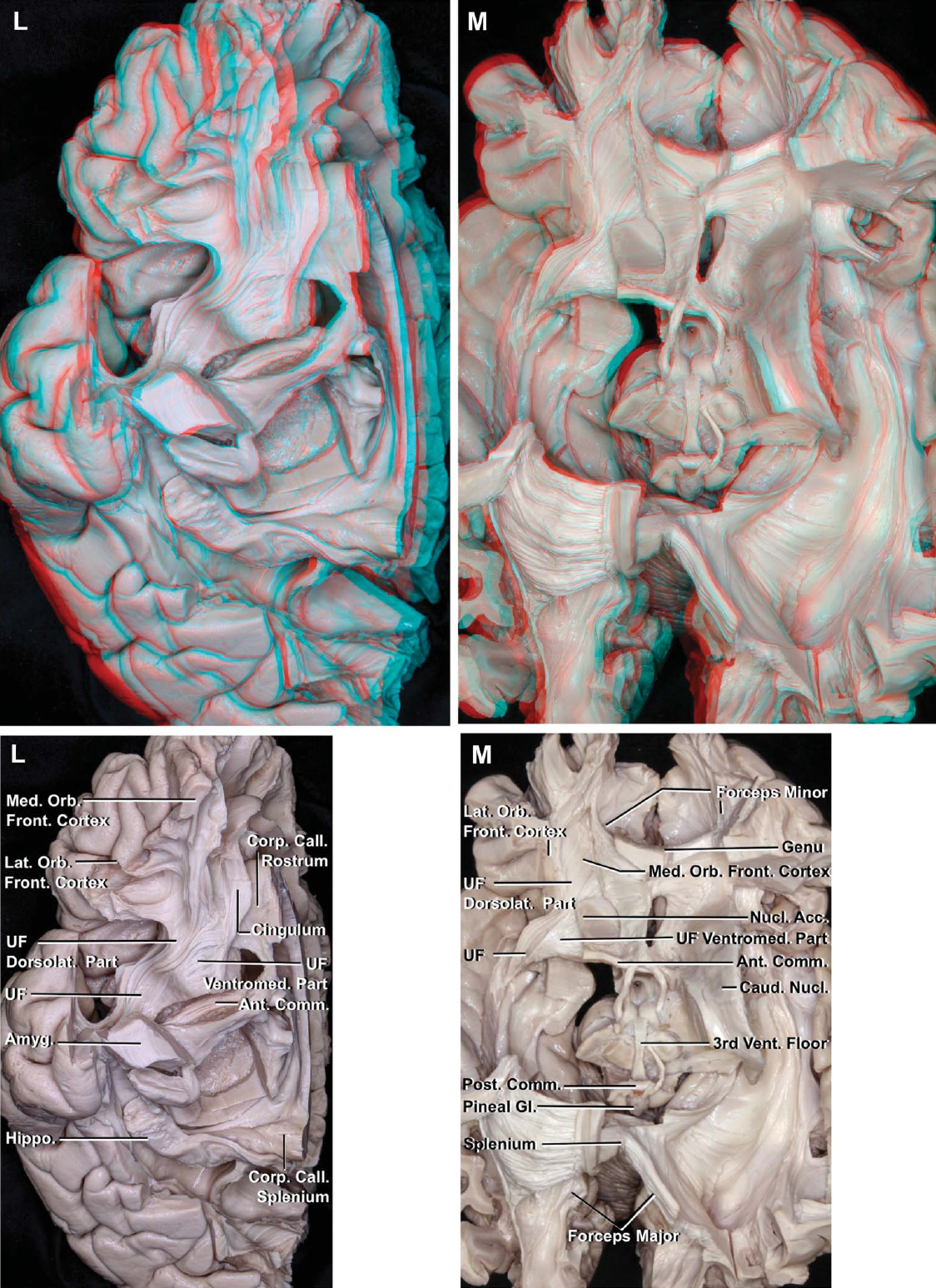
圖2 (l m)。左,UF的兩個部分的下視圖。背外側部分投射到眶側區,腹內側部分投射到伏隔核、眶隔區和內側眶額區。腹內側部分覆蓋伏隔核的下壁和內壁,在胼胝體膝下的皮質區與扣帶相交。M,腓脛纖維和UF的背外側和腹內側部的上視圖。腹內側部分連接伏隔核和眼窩前區。伏隔核位於尾狀核的下方。連合纖維是前連合,構成第三腦室前壁和胼胝體的一部分。後連合處位於大腦導水管上端的背側。在膝上交叉的胼胝體纖維,稱為小鉗,向前轉動連接額部區域。 The fibers crossing in the splenium, called the forceps major, turn posteriorly to interconnect the parieto-occipital regions. Acc., accumbens; AIP, anterior insular point; Amyg., amygdala; Ant., anterior; Call., callosal, callosum.; Caps., capsule; Caud., caudate; Cent., central, centrum; Claust., claustrum; Claustrocort., claustrocortical; CN, cranial nerve; Comm., commissure; Cor., corona; Corp., corpus; Dia., diagonal; Dors., dorsal; Dorsolat., dorsolateral; Ext., extension, external; Extr., extreme; FLP, frontal limen point; For., foramen; Front., frontal; Gl., gland; Glob., globus; Gyr., gyrus; Hippo., hippocampus; Hypoth., hypothalamus; IFOF, inferior fronto-occipital fasciculus; ILF, inferior longitudinal fasciculus; Inf., inferior; Innom., innominata; Ins., insula, insular; Int., internal; Lat., lateral; Lent., lenticular; LGB, lateral geniculate body; Lim., limiting; Mam., mammillary, mammillo; Med., medial, medullaris; MLF, medial longitudinal fasciculus; Nucl., nucleus; Occip., occipital; Orb. Front., orbitofrontal; Pall., pallidus; Par., parietal, parieto; PIP, posterior insular point; Post., posterior; Rad., radiata, radiations; Sag., sagittal; Sept., septal; SLF, superior longitudinal fasciculus; Str., stria; Strat., stratum; Subst., substantia; Subthal., subthalamic; Sulc., sulcus; Sup., superior; Temp., temporal; Thal., thalami, thalamic; TLP, temporal limen point; Tr., tract; UF, uncinate fasciculus; Vent., ventral, ventricle; Ventromed., ventromedial. (Images courtesy of AL Rhoton, Jr.)
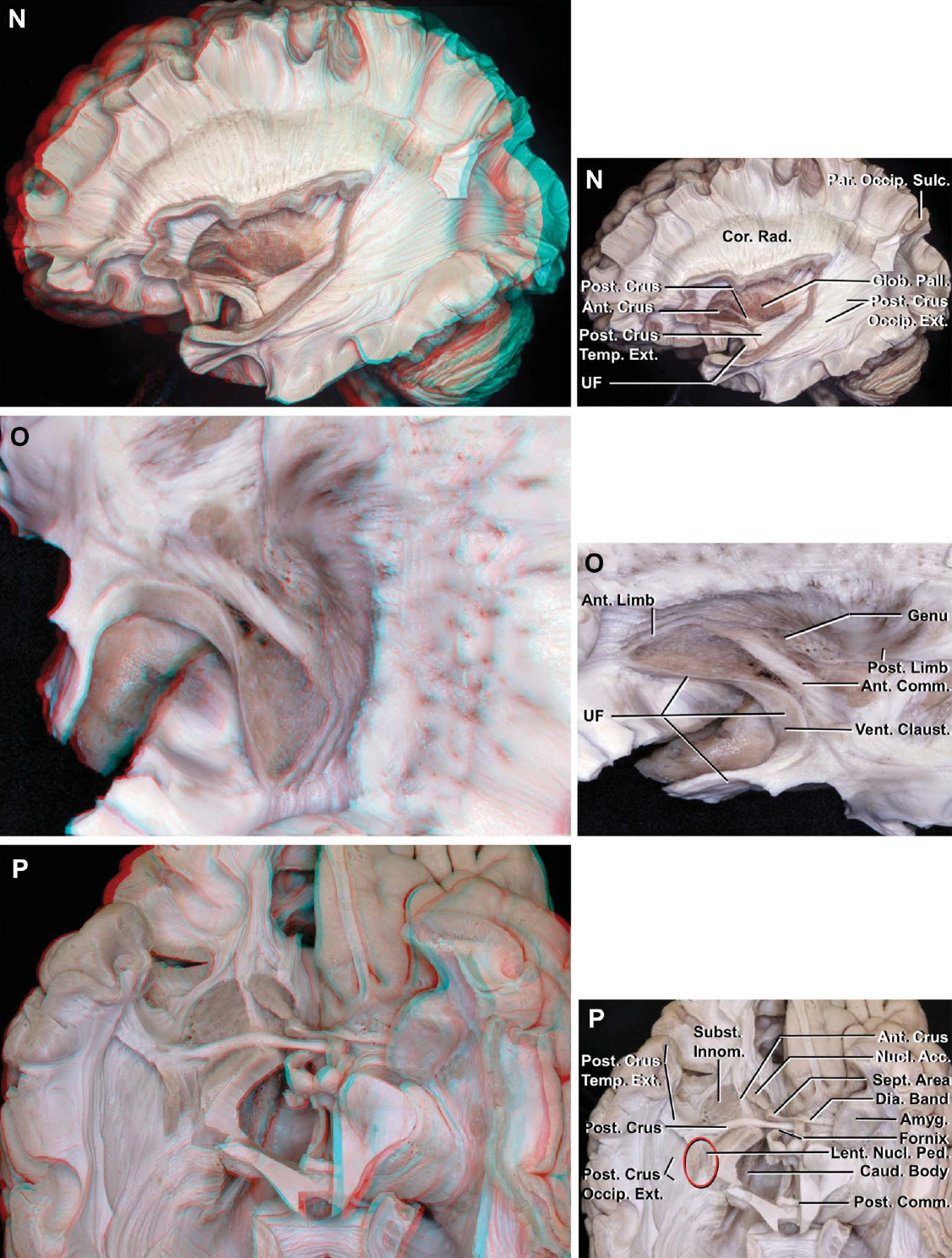
圖2(阻燃劑)。N,側麵圖。取出IFOF和殼核暴露前連合和蒼白球。前連合位於內囊前肢的下方,形成前小腿,凸出眶前區和嗅核。前連合的後腳向尾側和外側穿過蒼白球的腹側,位於葛提奧雷管,並有顳部和枕部的延伸。顳延伸向下延伸至顳極和UF後麵的杏仁核,枕部延伸向後延伸至枕葉。O,前合角的後腳在葛提奧管內橫向運行並發生扭轉,使其上纖維沿合角內側下降到達顳葉,下纖維沿合角外側上升到達枕葉。內囊的扭轉與前連合方向一致,使後肢的下部比前肢更側向外側。P,劣質的觀點。扁形核通過扁形核的下延伸與杏仁核相連,稱為扁形核梗。 The anterior commissure resembles bicycle handlebars located immediately in front of the columns of the fornix and forms part of the anterior wall of the third ventricle. The temporal and occipital extensions of the posterior crus are exposed. Structures that sit below and anterior to the anterior commissure include the substantia innominata, nuclei accumbens and basalis, diagonal band of Broca, and medial septal nuclei. The substantia innominata is located lateral to the anterior crus of the anterior commissure and anterior limb of the internal capsule. The nucleus accumbens is situated medial to the anterior crus of the anterior commissure and anterior limb of the internal capsule, and below the level of posterior crus of the anterior commissure. The septal area is located in front of the anterior commissure next to the midline. Acc., accumbens; AIP, anterior insular point; Amyg., amygdala; Ant., anterior; Call., callosal, callosum.; Caps., capsule; Caud., caudate; Cent., central, centrum; Claust., claustrum; Claustrocort., claustrocortical; CN, cranial nerve; Comm., commissure; Cor., corona; Corp., corpus; Dia., diagonal; Dors., dorsal; Dorsolat., dorsolateral; Ext., extension, external; Extr., extreme; FLP, frontal limen point; For., foramen; Front., frontal; Gl., gland; Glob., globus; Gyr., gyrus; Hippo., hippocampus; Hypoth., hypothalamus; IFOF, inferior fronto-occipital fasciculus; ILF, inferior longitudinal fasciculus; Inf., inferior; Innom., innominata; Ins., insula, insular; Int., internal; Lat., lateral; Lent., lenticular; LGB, lateral geniculate body; Lim., limiting; Mam., mammillary, mammillo; Med., medial, medullaris; MLF, medial longitudinal fasciculus; Nucl., nucleus; Occip., occipital; Orb. Front., orbitofrontal; Pall., pallidus; Par., parietal, parieto; PIP, posterior insular point; Post., posterior; Rad., radiata, radiations; Sag., sagittal; Sept., septal; SLF, superior longitudinal fasciculus; Str., stria; Strat., stratum; Subst., substantia; Subthal., subthalamic; Sulc., sulcus; Sup., superior; Temp., temporal; Thal., thalami, thalamic; TLP, temporal limen point; Tr., tract; UF, uncinate fasciculus; Vent., ventral, ventricle; Ventromed., ventromedial. (Images courtesy of AL Rhoton, Jr.)

圖2 (q s)。Q,劣質的觀點。前小腿和前連合後小腿的枕部和顳部延伸部分都被切除了。內囊膝位於前連合後方。中斷的綠線標記了內囊的前後邊界。內囊膝與Monro孔在同一冠狀麵。內囊部分的下緣是前肢的前連合,膝的下丘腦和後肢的外側膝狀體。R,前、後毛細血管的內側視圖。後連合起源於後連合的核,位於動眼肌核的前麵。S,稱為輻射狀冠的額頂投射纖維由內外囊形成,位於SLF II的內側。 The occipital and temporal projection fibers, called the sagittal stratum, are positioned medial to the occipital extension of the anterior commissure and the IFOF and ILF. The border between the corona radiata and sagittal stratum is positioned at a line (broken line) connecting the upper end of the parieto-occipital sulcus and the midpoint of the anterior edge of the limen insulae. The sagittal stratum is composed of occipitothalamic fibers, also called the optic radiations, and temporo- and occipitopontine fibers. Acc., accumbens; AIP, anterior insular point; Amyg., amygdala; Ant., anterior; Call., callosal, callosum.; Caps., capsule; Caud., caudate; Cent., central, centrum; Claust., claustrum; Claustrocort., claustrocortical; CN, cranial nerve; Comm., commissure; Cor., corona; Corp., corpus; Dia., diagonal; Dors., dorsal; Dorsolat., dorsolateral; Ext., extension, external; Extr., extreme; FLP, frontal limen point; For., foramen; Front., frontal; Gl., gland; Glob., globus; Gyr., gyrus; Hippo., hippocampus; Hypoth., hypothalamus; IFOF, inferior fronto-occipital fasciculus; ILF, inferior longitudinal fasciculus; Inf., inferior; Innom., innominata; Ins., insula, insular; Int., internal; Lat., lateral; Lent., lenticular; LGB, lateral geniculate body; Lim., limiting; Mam., mammillary, mammillo; Med., medial, medullaris; MLF, medial longitudinal fasciculus; Nucl., nucleus; Occip., occipital; Orb. Front., orbitofrontal; Pall., pallidus; Par., parietal, parieto; PIP, posterior insular point; Post., posterior; Rad., radiata, radiations; Sag., sagittal; Sept., septal; SLF, superior longitudinal fasciculus; Str., stria; Strat., stratum; Subst., substantia; Subthal., subthalamic; Sulc., sulcus; Sup., superior; Temp., temporal; Thal., thalami, thalamic; TLP, temporal limen point; Tr., tract; UF, uncinate fasciculus; Vent., ventral, ventricle; Ventromed., ventromedial. (Images courtesy of AL Rhoton, Jr.)

圖2(你不)。T與G相比,切除了部分幽閉皮質纖維和內囊,暴露了連接皮層和丘腦不同部位的丘腦輻射。額前和枕丘腦輻射水平方向。額上側和頂側丘腦輻射從其丘腦端斜向上。U,與S相比,去掉前連合的IFOF和枕部延伸,暴露矢狀層。半卵圓體位於胼胝體上方,由SLF II聯係、輻射冠投影和胼胝體纖維組成。Acc。,核;AIP,島前點;Amyg。, amygdala; Ant., anterior; Call., callosal, callosum.; Caps., capsule; Caud., caudate; Cent., central, centrum; Claust., claustrum; Claustrocort., claustrocortical; CN, cranial nerve; Comm., commissure; Cor., corona; Corp., corpus; Dia., diagonal; Dors., dorsal; Dorsolat., dorsolateral; Ext., extension, external; Extr., extreme; FLP, frontal limen point; For., foramen; Front., frontal; Gl., gland; Glob., globus; Gyr., gyrus; Hippo., hippocampus; Hypoth., hypothalamus; IFOF, inferior fronto-occipital fasciculus; ILF, inferior longitudinal fasciculus; Inf., inferior; Innom., innominata; Ins., insula, insular; Int., internal; Lat., lateral; Lent., lenticular; LGB, lateral geniculate body; Lim., limiting; Mam., mammillary, mammillo; Med., medial, medullaris; MLF, medial longitudinal fasciculus; Nucl., nucleus; Occip., occipital; Orb. Front., orbitofrontal; Pall., pallidus; Par., parietal, parieto; PIP, posterior insular point; Post., posterior; Rad., radiata, radiations; Sag., sagittal; Sept., septal; SLF, superior longitudinal fasciculus; Str., stria; Strat., stratum; Subst., substantia; Subthal., subthalamic; Sulc., sulcus; Sup., superior; Temp., temporal; Thal., thalami, thalamic; TLP, temporal limen point; Tr., tract; UF, uncinate fasciculus; Vent., ventral, ventricle; Ventromed., ventromedial. (Images courtesy of AL Rhoton, Jr.)

圖2 (V-W)。V,前額丘腦輻射,連接丘腦和前額葉皮層和額極,在內囊內水平移動。額丘上輻射,連接丘腦和額葉內側區和扣帶回前部,通過側腦室管膜的外側,是內囊內最內側的纖維。穿過脾的絨氈層纖維下降,將來自心房側壁的光輻射和側腦室的顳角和枕角分開。W,切除放射狀冠暴露額角室管膜和側腦室體。在脾中交叉的絨氈層纖維沿著心房的頂部和側壁以及側腦室的顳角和枕角走行。光輻射從外側膝狀體發出,經過顳角的頂部和側壁、心房的側壁和側腦室的枕角,到達鈣質溝。島前穴位於側腦室額角外側,島後穴位於體心房交界處。Acc。,核; AIP, anterior insular point; Amyg., amygdala; Ant., anterior; Call., callosal, callosum.; Caps., capsule; Caud., caudate; Cent., central, centrum; Claust., claustrum; Claustrocort., claustrocortical; CN, cranial nerve; Comm., commissure; Cor., corona; Corp., corpus; Dia., diagonal; Dors., dorsal; Dorsolat., dorsolateral; Ext., extension, external; Extr., extreme; FLP, frontal limen point; For., foramen; Front., frontal; Gl., gland; Glob., globus; Gyr., gyrus; Hippo., hippocampus; Hypoth., hypothalamus; IFOF, inferior fronto-occipital fasciculus; ILF, inferior longitudinal fasciculus; Inf., inferior; Innom., innominata; Ins., insula, insular; Int., internal; Lat., lateral; Lent., lenticular; LGB, lateral geniculate body; Lim., limiting; Mam., mammillary, mammillo; Med., medial, medullaris; MLF, medial longitudinal fasciculus; Nucl., nucleus; Occip., occipital; Orb. Front., orbitofrontal; Pall., pallidus; Par., parietal, parieto; PIP, posterior insular point; Post., posterior; Rad., radiata, radiations; Sag., sagittal; Sept., septal; SLF, superior longitudinal fasciculus; Str., stria; Strat., stratum; Subst., substantia; Subthal., subthalamic; Sulc., sulcus; Sup., superior; Temp., temporal; Thal., thalami, thalamic; TLP, temporal limen point; Tr., tract; UF, uncinate fasciculus; Vent., ventral, ventricle; Ventromed., ventromedial. (Images courtesy of AL Rhoton, Jr.)
大腦的中央核位於島葉皮層外側和腦室內側之間。該區域包括極囊、隱窩、外囊、慢狀體(殼核+蒼白球)和尾狀核、內囊、基底前腦和丘腦(圖2)。37
第一個出現在中樞的結構是島葉皮層(圖2A-2D),向外側向內側移動。腦島被限製溝(圓形)包圍,並被前、上、下限製溝以三角形的形狀包圍著腦島,與額葉、頂葉和顳蓋分開。373個限製性島溝及其連接處的4個點是島骨手術的重要標誌,因為它們很容易在裂裂後被發現。這些標誌是島葉前後點和額葉和顳葉limen點。島前穴位於前、上界限溝的交界處,島後穴位於上、下界限溝的交界處。38另外兩個重要的點是顳部和額部閾點,分別位於閾島與下限溝和前限溝的交界處。
另一個重要的定向標誌是島中央溝,它向深部延伸,幾乎與島中央溝在凸起處平行,並將島皮質劃分為短島回和長島回(圖2D)。短島腦回位於島中央溝的前麵,長島腦回位於島中央溝的後麵。37後島短回位於內囊膝和Monro室間孔的同一冠狀麵外側(圖2C)。
位於額下回三角部內側的從淺到深的結構是島前點、IFOF、來自內囊前肢的輻射冠纖維和側腦室的額角。位於後島點內側的結構,從淺到深依次為外包膜、內包膜後肢的放射冠纖維、尾狀核的尾部以及側腦室體和心房的交界處。腦回的後內側邊緣位於腦島後點。限製溝,島島前、後、額葉和顳葉閾點,是規劃島島手術最一致的外科標誌(圖2D)。38
移除島葉皮層暴露極囊,它水平連接相鄰的島葉回,垂直突出,深入到極限腦溝,直至腦蓋區(圖2E)。有人認為,極囊是由連接鄰近島腦回的短連接纖維組成的。39對非人靈長類動物損傷的實驗研究表明,短的關聯纖維沿著極囊連接額葉、顳葉和頂葉。40相反,對獼猴的放射自顯影研究和對人類的DTI研究表明,極囊是連接枕葉和額葉的長連接纖維通路。17歲,35歲,40歲這些纖維通路也被描述為與人類的IFOF相對應,因為IFOF在獼猴體內不存在。有研究表明,極端囊可能參與語言的句法處理和腹側語義功能,通常歸因於相鄰的IFOF和鉤束。41極端囊的解剖和功能仍有爭議。在纖維解剖研究中,極囊似乎含有連接島腦回彼此和額頂葉和顳葉蓋的短關聯纖維。
切除極端囊暴露隱窩和外囊(圖2F)。隱窩是位於外側的極囊和內側的外囊之間的灰質的薄集合。外囊和隱窩均由腹側和背側兩部分組成。8背側外囊是由幽閉皮質投射纖維形成的,它在頂葉前部的輔助運動區和後部的頂葉後部之間連接著幽閉和皮層(圖2F-2H)。幽閉皮質係統參與視覺、體感和運動信息的整合。8該係統的雙側損傷已導致嚴重的腦病。8背側隱窩位於極端和外部囊之間。腹側外囊是由上麵的IFOF和下麵的UF形成的。腹側隱窩由分散在腹側外囊內的灰質島嶼組成,向外側延伸至杏仁核(圖2H和2I)。
IFOF是額枕聯合纖維通路,連接額中回和額下回到頂葉和枕葉的後部(圖2I-2K)。在額葉,它到達背外側前額葉皮層(額中回的中部部分),眶部和三角部。IFOF位於輻射冠纖維外側(淺)和額葉SLF II和AF節內側(深)。IFOF深入到上限製溝的前三分之一和前限製溝的上一半。它在下丘腦間葉的上方變窄,深入到下極限溝的中間三分之一處,並在上顳回和中顳回內繼續向後延伸,到達枕葉。在深入到下極限溝後,頂枕皮層IFOF皮層分布的上限位於連接皮層島中點和頂枕溝上端的一條線下。這條線也位於幽閉皮質纖維的後邊緣(圖2G、2J和2K)。IFOF是腹側語義通路的主要組成部分。34雖然無法在獼猴中鑒定到IFOF,但我們認為人類的IFOF與獼猴的極端囊相當。已經注意到,IFOF通過上顳回、中顳回和顳幹,將枕內側下葉和可能的內側頂葉(楔前葉)連接到額下回、額中回中部(前額葉皮層背外側)和額極。32在我們的觀察中,IFOF也投射到頂葉的後部。IFOF被認為在語義處理、視覺識別、多模態感覺輸入和運動規劃的整合、閱讀和寫作,以及理解和產生有意義的講話中發揮著重要作用。6日,34歲的42、43有人注意到,術中電刺激整個IFOF會產生語義錯語(對目標詞的意思有錯誤)。6日,44IFOF也被建議作為左顳葉切除術的內側界限,以避免術後語言障礙。6IFOF覆蓋了光輻射纖維,當它們深入到上顳回和中顳回和枕葉,並在側腦室的顳角、心房和枕角的外側。手術中避免進入IFOF的深度有助於防止視神經輻射損傷。
UF是額顳聯合纖維通路,通過顳極的背外側分支連接眶外側區,通過腹內側分支連接眶內側區和眶間隔區(圖2J-2M)。45
UF位於前穿孔物質的前方,覆蓋伏隔核的下側和內側,到達胼胝體膝下區域。在亞屬區,UF被認為是腹側邊緣通路的一部分,它與扣帶的纖維連接在一起,被認為是背側邊緣通路。33斷開聯係可能會導致行為障礙。33歲的45伏隔核與眶額區之間的連接是由UF的腹內側支提供的。UF深通至前限製溝的下半部分和下限製溝的大部分前部,形成鉤狀。
外囊內側是由外側的殼核和內側的蒼白球形成的慢狀核(圖2H和2I)。通過橫跨內囊前肢的分散的灰質束與尾狀核連接,也稱為灰質囊橋(尾狀豆狀灰質橋),通過被稱為殼核底(殼核的前腹部)的延伸到達基底前腦,通過被稱為小核梗的小核的下延伸到達杏仁核。37慢狀核的基底和核梗都是跨越慢狀核和相鄰核之間區域的灰質集合(圖2P和3D)。去除殼核後暴露蒼白球和前連合。蒼白球位於前連合後方,參與自主運動的調節。46蒼白球的意思是“蒼白的球體”,在我們的檢查中,它的顏色比所有標本的殼核的顏色都要淺(圖2I, 2N,和2Q)。蒼白球有位於外側的外部部分和位於內側的內部部分。外(外側)部分向內側延伸至內囊膝,但內(內側)部分僅麵向內囊後肢(圖3F)。蒼白球主要對運動有抑製控製。蒼白球損傷可引起不自主震顫,這就是蒼白球切開術用於治療某些運動障礙的原因。46

圖3 (a - c)。圖2中分解的逐步繼續。A,身體外側壁室管膜和側腦室額角以及絨氈層和光學輻射纖維的間斷帶被切除。視輻射與顳角、枕角和側腦室的心房由絨氈層隔開。絨氈層纖維以垂直方向運動,而光輻射纖維則以水平方向運動。B,優越的觀點。額頂骨(輻射狀冠)和枕骨(矢狀層)凸出。側腦室的心房和枕角在外側與矢狀層和絨氈層纖維交界,在內側與大鉗交界。額角外側壁的上緣和側腦室體被放射狀纖維包圍。C,絨氈層纖維被移除以暴露胼胝體球部覆蓋在大鉗纖維上,以及胼胝體覆蓋在側腦室心房內側壁的鈣質溝深端。 After arising in the lateral geniculate body, the optic radiation fibers pass between the inferior limiting sulcus and tail of the caudate nucleus. AIP, anterior insular point; Amyg., amygdala; Ant., anterior; Call., callosal, callosum; Caps., capsule; Caud., caudate; Cing., cingulate; Claust., claustrum; Comm., commissure; Cor., corona; Corp., corpus; Front., frontal; Glob., globus; Gyr., gyrus; Hippo., hippocampus; ILF, inferior longitudinal fasciculus; Inf., inferior; Innom., innominata; Int., interna, internal; Lat., lateral; Lent., lenticular; LGB, lateral geniculate body; Lim., limiting; Med., medullaris; Nucl., nucleus; Occip., occipital; Pall., pallidus; Par., parietal, parieto; Ped., peduncle; PIP, posterior insular point; Rad., radiata, radiations; Sag., sagittal; Sept., septal; SLF, superior longitudinal fasciculus; Str., stria; Strat., stratum; Subst., substantia; Sulc., sulcus; Sup., superior; Term., terminalis; Thal., thalami; Vent., ventral, ventricle. (Images courtesy of AL Rhoton, Jr.)
圖3 (D-F)。D,尾狀核的尾部深入到下極限溝並融入杏仁核。扁形核的核梗與杏仁核混合在一起,兩個核之間沒有明確的分界線。無名質位於前連的後小腿的下麵和前麵,在前小腿的外側。E,島前點位於側腦室額角的外側。島後點位於尾狀核尾部的外側,位於側腦室體與心房的交界處。尾狀核的頭部位於島區邊界的深處和內部。島前點位於尾狀核的上方。尾狀核的主體延伸到上極限溝的上方並深入到島島後點。尾狀核的尾部開始於島後點深處,並深入到下極限溝的後部下方。 F, removal of the globus pallidus externus (lateral segment) exposes the globus pallidus internus (medial segment). AIP, anterior insular point; Amyg., amygdala; Ant., anterior; Call., callosal, callosum; Caps., capsule; Caud., caudate; Cing., cingulate; Claust., claustrum; Comm., commissure; Cor., corona; Corp., corpus; Front., frontal; Glob., globus; Gyr., gyrus; Hippo., hippocampus; ILF, inferior longitudinal fasciculus; Inf., inferior; Innom., innominata; Int., interna, internal; Lat., lateral; Lent., lenticular; LGB, lateral geniculate body; Lim., limiting; Med., medullaris; Nucl., nucleus; Occip., occipital; Pall., pallidus; Par., parietal, parieto; Ped., peduncle; PIP, posterior insular point; Rad., radiata, radiations; Sag., sagittal; Sept., septal; SLF, superior longitudinal fasciculus; Str., stria; Strat., stratum; Subst., substantia; Sulc., sulcus; Sup., superior; Term., terminalis; Thal., thalami; Vent., ventral, ventricle. (Images courtesy of AL Rhoton, Jr.)

圖3(胃腸道)。G, SLF的側麵圖I位於胼胝體纖維內側,在扣帶上方。H, SLF的內側視圖。它位於扣帶溝的上岸,位於胼胝體纖維的內側,位於扣帶的上方。我,側麵圖。切除限製溝和丘腦,露出丘腦終紋和髓質紋。終紋起源於終紋的床核,位於中隔區域的前連合附近,環繞丘腦並與杏仁核融合。丘腦髓紋連接隔區和韁肌,並將背側與丘腦內側表麵分開。AIP,島前點;Amyg。杏仁核; Ant., anterior; Call., callosal, callosum; Caps., capsule; Caud., caudate; Cing., cingulate; Claust., claustrum; Comm., commissure; Cor., corona; Corp., corpus; Front., frontal; Glob., globus; Gyr., gyrus; Hippo., hippocampus; ILF, inferior longitudinal fasciculus; Inf., inferior; Innom., innominata; Int., interna, internal; Lat., lateral; Lent., lenticular; LGB, lateral geniculate body; Lim., limiting; Med., medullaris; Nucl., nucleus; Occip., occipital; Pall., pallidus; Par., parietal, parieto; Ped., peduncle; PIP, posterior insular point; Rad., radiata, radiations; Sag., sagittal; Sept., septal; SLF, superior longitudinal fasciculus; Str., stria; Strat., stratum; Subst., substantia; Sulc., sulcus; Sup., superior; Term., terminalis; Thal., thalami; Vent., ventral, ventricle. (Images courtesy of AL Rhoton, Jr.)
前連合穿過殼核基部的中線,連接眶額葉、枕葉和顳葉,特別是杏仁核(圖2N-2R)。33前連合位於穹窿柱的正前方,類似於自行車的車把。它是第三腦室前壁的一部分(圖2M和2P-2R)。前聯合有一個前腳,向前延伸到嗅覺核並到達內側眼窩前額區,還有一個後腳,向外側延伸並分為顳和枕部。47前小腿形成內側伏隔核和外側無名質之間的邊界(圖2P和2Q)。33歲的45它向前延伸到內囊前肢腹表麵的下方。前連合的後腳在脛管內橫向伸展並發生扭轉,使其上纖維延伸至顳葉,下纖維延伸至枕葉(圖2O)。48後小腿深入到下界限溝的前三分之二處和IFOF內側。後小腿的顳部延伸向下延伸至顳極和UF後麵的杏仁核,枕部延伸深入至上顳回和中顳回,到達枕葉。前連合是癲癇手術中重要的半腦間連接。它的顳延伸被認為是癲癇活動在內側顳葉之間快速傳播的途徑,在半球切開術或功能性半球切除術中應該被分割。33在天生沒有胼胝體的患者中,它的枕部延伸也被證明能夠傳遞半球間的視覺信息。33視覺信息部分是由前連合的枕部延伸和胼胝體的脾處理的。33在前連合後腳的前麵和下麵是無名質,由Meynert基底核形成,為皮質提供膽堿能輸入。33歲,49歲,50歲供應中央核心結構的莢狀紋狀動脈穿過前穿孔物質,並通過相鄰的前連合的前後邊緣。
基底前腦位於前連連後腳的前方和下方,前連連包括無名質、基底核和伏隔核、布羅卡對角帶、內側隔核和延伸的杏仁核,杏仁核複體延伸至終紋床核(圖2P)。45歲,47包含基底核的無名質位於後小腿前方、前連合和內囊前肢的前小腿外側和前穿孔物質上方。形成殼核底(前腹部)的灰質與無名質混合,位於前穿孔物質的上方,兩者之間沒有明確的邊界。伏隔核位於前連合前小腿內側和內囊前肢內側,在前連合下方。中隔區位於前連合前的中線附近。在神經生理學上,無名質和伏隔核之間、隔核和尾狀核頭部之間沒有明確的界限。33歲,47歲,49歲,50歲
由投射纖維組成的內囊位於慢狀核的正中(圖2I和2Q)。軸向切片內囊的前外側和後外側邊緣分別與慢狀核的前內側和後內側邊緣成近直角連接。內囊有三個部分:前肢、後肢和膝。內囊的前肢穿過硬殼核和尾狀核的頭部之間。37內囊膝位於前肢和後肢的交界處,位於前連合的後麵,與Monro孔和後島短回位於同一冠狀平麵上。37內囊的扭轉方向與前連合方向一致,使後肢下部比前肢更側向外側(圖2O和2Q)。17日,33
內囊膝包括皮質球纖維,它連接中央前回的下三分之一和腦幹中相關的顱核,以及丘腦前部和前部輻射的纖維。33歲的51內囊的前肢包含前、上額丘腦輻射纖維和額脊束(圖2T-2V)。33歲的51歲,52歲內囊前肢最外側的纖維起源於額葉蓋。內囊前肢中部的纖維起於包括額極在內的前額皮質,在內囊內水平運行,向後通過內囊前肢和膝到達丘腦。來自內側額葉區和扣帶皮層前部的纖維位於前肢最內側的位置。17日,53歲,54
內囊的後肢位於慢狀核和丘腦之間。後肢包括皮質脊髓、皮質橋突、皮質被蓋和頂葉丘腦輻射(文獻中為正中後或上丘腦輻射)。51、52它還包含來自Brodmann區4和6的額突纖維和額骨纖維。皮質脊髓纖維起源於鄰近的中央溝,穿過內囊後肢的前部。
內包膜的前、上、後邊界位於硬殼核的前內側邊緣和尾狀核的上外側邊緣之間。外囊與內囊在慢狀核的上邊緣連接,形成輻射冕(圖2I和2N)。17內囊的膝和後肢的內側緣麵對丘腦的網狀核,前肢的內側緣麵對尾狀核的頭部。內囊部分的下界是前肢的前連合、膝的下丘腦和後肢的膝狀外側體的水平(圖2Q)。17
位於殼核上緣上方的額頂投射纖維稱為輻射冠,但輻射冠的確切位置和纖維類型在文獻中尚未明確。51它們包括囊內纖維和囊背外纖維,並對應於蓮形前纖維和蓮形上纖維(圖2T、2U和3B)。輻射冠包括內側囊內的皮質脊髓、皮質橋質和丘腦皮質纖維,外側囊外的幽閉皮質纖維。額頂投射纖維穿過膝和內囊的前肢和後肢。皮質丘腦或丘腦皮質纖維與輻射冠和內囊分離進入丘腦,形成丘腦輻射。47歲,49
連接頂枕溝上端和閾島中點的一條線將上麵的輻射狀冠與下麵由枕部和顳部投射纖維組成的矢狀層分開(圖2S)。這條線也對應於幽閉皮質(背側外囊)和頂骨投射纖維的後邊界和IFOF(腹側外囊)的上邊界(圖2J和2S)。枕部投射纖維由枕橋突和枕丘腦纖維組成,外側與IFOF、MdLF和ILF等相關纖維相鄰(圖2S-2U和3B)。枕丘腦輻射是由從外側膝狀體和枕骨到鈣質皮層的視輻射形成的(圖3A-3C)。47
丘腦輻射是根據其相關的皮質區域命名的,如上額和前額、頂葉和枕葉丘腦輻射,而不是根據它們在丘腦中的連接,如前、後、上(中央)和下丘腦輻射(圖2T-2V和3A-3D)。根據丘腦而不是皮質的連接來命名丘腦纖維,導致有兩種下丘腦輻射:來自眼窩前額皮質的纖維或來自杏仁核(踝袢的分支)的纖維。17日,55皮質丘腦纖維位於內囊內皮質脊髓和皮質橋質纖維的內側。47丘腦額前放射斜向下和向後,通過內囊和膝的前肢到達丘腦背內側。17上麵的額葉上丘腦輻射首先垂直,然後向後方向通過額葉前丘腦輻射的內側,通過膝和內囊的前肢和後肢到達丘腦。頂丘腦輻射通過內囊的後肢到達丘腦。枕丘腦輻射(視輻射)分為三個波段:前、中、後。視輻射的前束向前外側延伸,通常到達側腦室的顳角尖端,就在纖維向後轉成一個環的前連合的顳延伸處(邁耶氏環)的後麵。這條帶覆蓋顳角的頂部和側壁以及側腦室心房的下表麵,延伸到鈣質溝的下唇。中央視神經輻射束從外側膝狀體和枕骨向下延伸,然後轉回到達鈣質皮層。後束的視輻射,從外側膝狀體和枕骨發出後,直接向後延伸到上唇的鈣質溝。
額葉切除術的皮質邊界在額極後方7 - 8厘米處,避開前中央回和額下回(圖4)。56安全額葉切除術的基本皮層和皮層下邊界是中央前回及其後麵的纖維,額下回和弓狀束,以及內側側腦室的尾狀核頭部和額角。57

圖4 (a - c)。A,左額顳側開顱術。側靜脈淺部額葉和顳葉已經暴露。B,深入到額上回和中回的纖維束被暴露,同時保留了額前中央回和額下回。移除皮質和短的關聯纖維,暴露SLF II,它深入中額回。輻射冕已經暴露出來了。C,從內側開始,SLF II的切除將IFOF深度暴露到額中回的中部和輻射冕。移除額上回水平的輻射冠纖維暴露胼胝體纖維。部分胼胝體纖維被切除,露出SLF i、AF、弓形束;AIP,島前點; Ant., anterior; Call., callosal, callosum; Caps., capsule; Caud., caudate; Cent., central; Cor., corona; Corp., corpus; Dors., dorsal; Front., frontal; Glob., globus; Gyr., gyrus; IFOF, inferior fronto-occipital fasciculus; Inf., inferior; Ins., insular; Int., internal; Lat., lateral; Lim., limiting; Mid., middle; Operc., opercularis; Orb., orbitalis; Pall., pallidus; Precent., precentral; Rad., radiata; SLF, superior longitudinal fasciculus; Sulc., sulcus; Sup., superior; Temp., temporal; Triang., triangularis; UF, uncinate fasciculus; Vent., ventral, ventricle. (Images courtesy of AL Rhoton, Jr.)
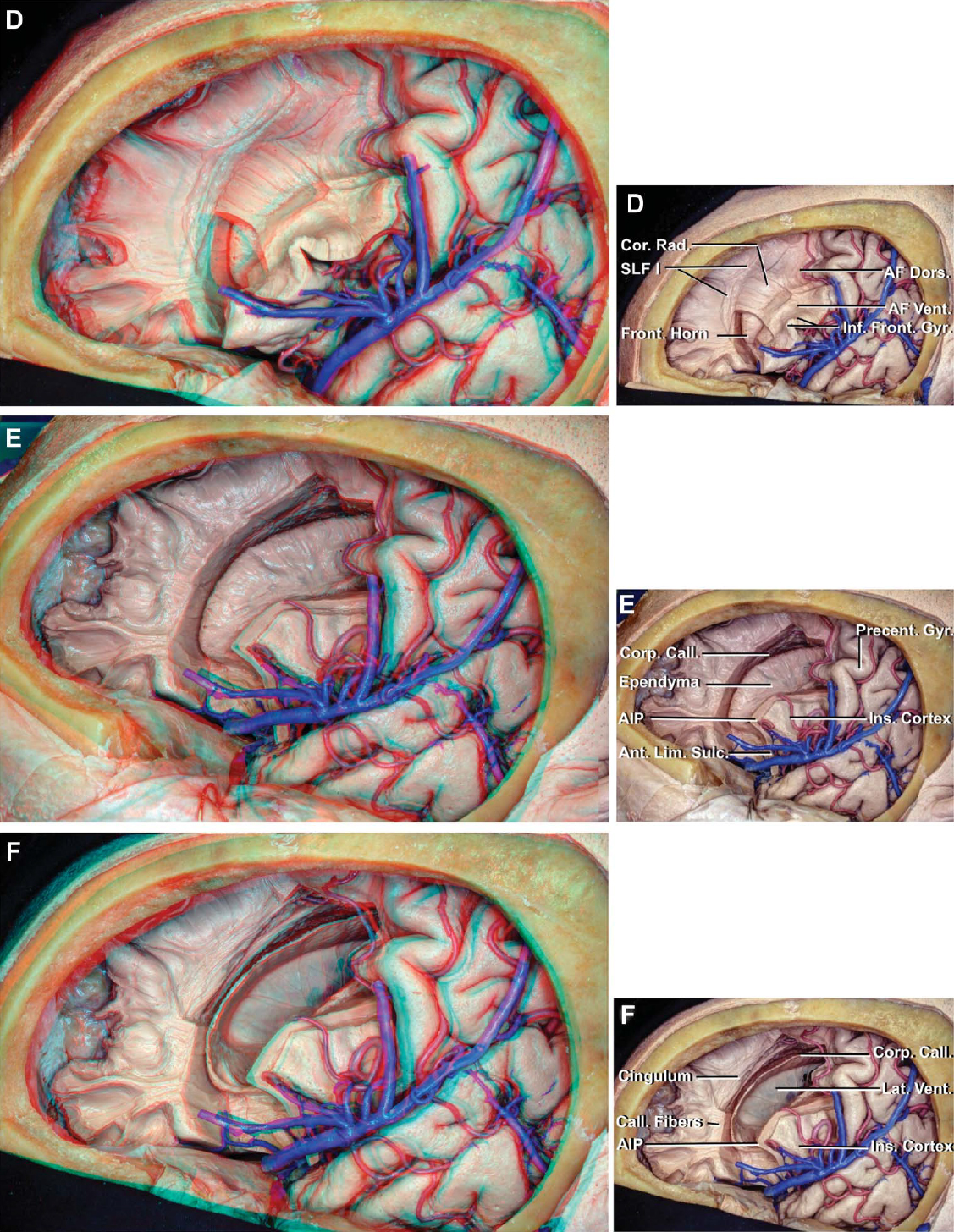
圖4 (D-F)。D,將暴露深度延伸至額中回和額下回,暴露側腦室額角和AF腹背節。側腦室上方暴露胼胝體和SLF I。E,額下回已經被移除露出島葉皮層。胼胝體暴露在側腦室管膜上方。F,側腦室,胼胝體和島葉表麵暴露。島前點位於側腦室額角的外側。進一步切除胼胝體纖維暴露扣帶。房顫、弓狀纖維束;AIP,島前點; Ant., anterior; Call., callosal, callosum; Caps., capsule; Caud., caudate; Cent., central; Cor., corona; Corp., corpus; Dors., dorsal; Front., frontal; Glob., globus; Gyr., gyrus; IFOF, inferior fronto-occipital fasciculus; Inf., inferior; Ins., insular; Int., internal; Lat., lateral; Lim., limiting; Mid., middle; Operc., opercularis; Orb., orbitalis; Pall., pallidus; Precent., precentral; Rad., radiata; SLF, superior longitudinal fasciculus; Sulc., sulcus; Sup., superior; Temp., temporal; Triang., triangularis; UF, uncinate fasciculus; Vent., ventral, ventricle. (Images courtesy of AL Rhoton, Jr.)

圖4 (G-H)。G,島葉皮層被切除了。IFOF深入到上限界溝的前三分之一和前限界溝的上半部,UF深入到前限界溝的下半部。H,硬殼核被移至IFOF的保留帶,露出內囊和蒼白球。顳葉的UF已經暴露。房顫、弓狀纖維束;AIP,島前點;螞蟻。前;調用。, callosal, callosum; Caps., capsule; Caud., caudate; Cent., central; Cor., corona; Corp., corpus; Dors., dorsal; Front., frontal; Glob., globus; Gyr., gyrus; IFOF, inferior fronto-occipital fasciculus; Inf., inferior; Ins., insular; Int., internal; Lat., lateral; Lim., limiting; Mid., middle; Operc., opercularis; Orb., orbitalis; Pall., pallidus; Precent., precentral; Rad., radiata; SLF, superior longitudinal fasciculus; Sulc., sulcus; Sup., superior; Temp., temporal; Triang., triangularis; UF, uncinate fasciculus; Vent., ventral, ventricle. (Images courtesy of AL Rhoton, Jr.)
在初步保留前中央和額下回的情況下,切除額葉。在額葉中回水平切除皮質層和短關聯纖維,使SLF II暴露於皮質表麵2厘米深。從內側開始,SLF II的切除暴露了位於中額回後部的AF背側段和位於中額回中部的IFOF,深度為3厘米,並在超過3厘米的深度暴露了輻射冠纖維,包括幽閉皮層和額toponine纖維和額丘腦輻射。額葉上丘腦輻射呈斜向分布,額葉前丘腦輻射水平分布,比額葉上丘腦輻射更淺。在額上回水平,輻射冠纖維暴露在皮質表麵以下1.5 cm處。去除放射狀冠,暴露出2厘米深的胼胝體纖維和2.2厘米深的SLF I。在剝離後期移除額下回,暴露出島葉皮層和距島葉前皮層0.6 cm深的IFOF。IFOF深入到前限製溝的前三分之一和前限製溝的上半部,而鉤突束深入到前限製溝的下半部。進一步在額中回和額下回深度進行解剖,露出距離表麵4cm處的側腦室額角和體,以及額下回的AF節段。胼胝體位於側腦室頂部。 The frontal horn and body of the lateral ventricle are positioned medial to the inferior frontal gyrus, but, if enlarged, they extend upward to the level of the middle frontal gyrus.
從地形上看,中額回可分為前半部分,包括額極皮層(Brodmann區[BA] 10),中半部分包括背外側前額葉皮層(BAs 9和46),後半部分包括前運動皮層(BAs 6和8)和運動皮層(BA 4)。1額下回包括腹外側前額葉皮層(BAs 44, 45, 47)。運動皮層(ba4)和皮質脊髓束的損傷會導致對側偏癱或麻痹。前中央回的前上方是額眼區,其損傷可能導致凝視異常。58歲的59中額回的中間部分,被稱為背外側前額葉皮層(BAs 9和46),與認知有關。它和它的底層纖維通路的損傷會導致以記憶缺陷、抑製增加、抽象思維和目標導向執行功能受損為特征的障礙。1、2、60 - 62背外側前額葉皮層也被認為是一個語義中心。電刺激這個皮層區域和潛在的IFOF會產生語義錯語。4, 60背外側前額葉皮層按橫向和內側順序通過短聯想纖維與額葉皮層的其餘部分連接,下頂葉由SLF II連接,枕葉和顳葉由IFOF連接,基底神經節和丘腦由額丘腦輻射連接,對側前額葉皮層由小鉗連接,海馬(或邊緣係統)由扣帶連接。1, 14日,63
額葉蓋(包含SLF III和AF的額節)的損傷可能分別產生構音障礙和失語障礙。20、21也有報道稱,單側電刺激或手術切除位於尾狀核頭部背側前運動皮層(ba6)水平的底層纖維通路可能導致雙側運動障礙(運動停止而不喪失任何張力),而刺激或損傷位於運動皮層下層的纖維束(錐體束)可能導致對側運動障礙。64年,65年腹側前運動皮層(下ba6)可塑性差,皮質和下層纖維束(SLF III)的損傷可能導致不可逆的語言障礙(構音障礙/構音障礙)。66
額下回(腹外側前額葉皮層)由眶部、蓋部和三角部組成,包括位於蓋部的布洛卡區和三角部的一小部分。刺激左額下葉皮層可誘發語錯(更多的後刺激誘發音位錯,更多的前刺激誘發語意錯),可能包括句法錯誤。67年,68年位於額下回下方的纖維束,從淺到深,是短的U連接纖維,SLF III, AF腹側和背側節,IFOF和輻射冠。纖維束的內側界限是尾狀核和側腦室的額角和體。
內側額葉,尤其是前扣帶皮層的病變,其特征是冷漠、缺乏動力、目標導向行動減少以及缺乏好奇心和興趣。69年,70年額葉上內側表麵更多的後側病變也可能導致自主運動的減少和沉默。71內側前額葉皮層通過扣帶連接到其他葉的內側表麵,通過小鉗連接到對側內側額葉皮層,通過SLF I連接到楔前葉(內側頂葉)。1、13、14、19
眼窩前表麵分為內側和外側兩部分。內側眶額皮質通過UF的腹內側部與內側顳葉結構、伏隔核、間隔區和嗅覺區相連,外側眶額皮質則分別通過UF的背外側部和IFOF與顳極和背外側前額葉皮層相連。與對側眼窩前區相連的是小鉗和前合角的前腳(圖2L、2M和2P)。眼窩額葉皮層與行為處理有關。損傷對皮質和底層纖維通路的影響因部位的不同而顯著不同,通常包括以去抑製、社交不當和性專注為特征的性格變化。1、72、73
額葉中傳統的功能區是額下回(布洛卡區)和中央前回(運動皮層)。57位於額下回下方的運動前纖維、SLF和IFOF也被認為是活躍的皮層下纖維束。57、74應保留位於額中回下方的IFOF和SLF II,以避免術後缺損。
在以前的出版物中,我們強調了基於大腦淺表標誌與深部結構之間的關係發展三維大腦概念的重要性。37這個概念在以前的出版物中通過顯微外科和內窺鏡解剖和方法的三維攝影得到了加強。33歲,75之前研究的關係包括淺表標記的位置,如大腦淺表和深部結構的縫合處,以及淺表大腦結構與深部甚至中線結構的關係,以在外科醫生的腦海中建立一條路線圖,以便在大腦的不同深度導航。37還有很多其他的例子,包括纖維束與腦幹安全進入區之間的關係。75
本研究中顯示的一些“首次”纖維解剖發現包括(1)上縱束的三個細分(圖1、3G和3H);(2) SLF的三段與弓狀肌束背段和腹側段的關係(圖1D);(3)中縱束與島島後點水平IFOF的關係(圖1H、1I);(4)枕前切跡水平下縱束45°向上彎曲(圖1H、2G、2K);(5)基底節區和中央核的纖維束與腦島上、前、下界限溝的關係(圖2、3);(6) UF的腹內側段和背外側段,以及UF(腹側邊緣通路)和扣帶(背側邊緣通路)在亞屬區的連接(圖2L和2M);(7)中樞基底神經節結構之間的關係(圖2B、2C、2M、2P、3D-3F);(8)前合角的旋轉及其纖維分布(圖2N-2P);(9)內囊各部分的核束和纖維束邊界(圖2Q);(10)輻射冠和矢狀層之間的邊界,幽閉皮質纖維(背側外囊)和IFOF(腹側外囊)之間的邊界,以及頂骨投影纖維和枕骨投影纖維之間的邊界,並在形成半卵圓體的纖維解剖中表現出來(圖2G, 2J, 2S,和2U); (11) fiber tracts composing the anterior limb of the internal capsule in order laterally to medially (Figures 2T-2V); (12) thalamic radiations based on the cortical connections of the thalamic fibers (Figures 2T-2W); (13) relationship of the optic radiations to the inferior limiting sulcus, atrium and temporal horn of the lateral ventricle, and adjacent fiber tracts (Figures 2T-2W and 3A-3D); (14) frontal lobe fiber tract anatomy in step-by-step dissections in the surgical view (Figure 4); and (15) topographic anatomy of the fiber tracts in relation to cortical gyri and their depth from the surface of the cortical gyri (Figures 5A-5E and 6A-6C and Tables 1 and 2).

圖5 (a - c)。第一,大腦中央核心的結構被疊加在皮層表麵。前溝(紅色)、上溝(黃色)和下溝(藍色)勾勒出腦島形成的中央核的外側表麵。前限製溝位於三角肌前部邊緣深處。上限溝的前部位於額下回的內側,後限溝的後部位於中央前腦回和中央後腦回的下三分之一和中三分之一與邊緣上回前部交界處的內側。下極限溝位於顳上溝的深處。尾狀體頭部的前部延伸到前限溝。身體上緣略高於上極限溝的水平,尾部的一段向後延伸,低於下極限溝的水平。尾狀核的尾部,在向前推進的過程中,經過下極限溝的前部與杏仁核融合。核心的其他結構包括殼核、蒼白球和丘腦。 B, long association pathways. The SLF I extends deep to the superior frontal gyrus and the superior parietal lobule. The SLF II passes deep to the middle frontal gyrus and midlevel of the pre- and postcentral gyri and deep to the upper part of the supramarginal and angular gyri. The SLF III passes deep to the inferior frontal gyrus, the lower part of the pre- and postcentral gyri, and the lower part of the supramarginal gyrus. The MdLF passes deep to or through the superior temporal and angular gyri and the ILF medial to the inferior temporal gyrus and dorsolateral occipital cortex. C, position of the UF, IFOF, claustrocortical fibers, and the dorsal and ventral segments of the AF in relation to the cortical surface. The UF passes medial to the temporal pole, anterior part of the superior and middle temporal gyri, and limen insulae and connects to the medial and lateral orbitofrontal areas. The IFOF passes deep to the mid part of the middle frontal gyrus and anterior part (pars orbitalis and triangularis) of the inferior frontal gyrus. In the insular area, the IFOF passes deep to the short insular gyri and limen insulae, and deep to the superior and middle temporal gyri, posterior part of the inferior parietal lobe, and occipital lobe. The claustrocortical fibers pass deep to the part of the cerebral cortex between the supplementary motor area anteriorly and posterior parietal lobe posteriorly. The claustrocortical fibers form the dorsal external capsule, and the IFOF and UF form the ventral external capsule. The AF ventral segment is positioned ventral to the AF dorsal segment in the area above the sylvian fissure, but anterior and dorsal to the dorsal segment below the fissure. The AF ventral segment passes deep to the mid part of the superior and middle temporal gyri, posterior part of the superior temporal gyrus, lower part of the supramarginal gyrus, and post- and precentral and inferior frontal gyri. The AF dorsal segment passes deep to the posterior part of the middle and inferior temporal gyri, lower part of the angular gyrus, post- and precentral gyri, and posterior part of the middle and inferior frontal gyri. AF, arcuate fasciculus; Ang., angular; Ant., anterior; Call., callosum; Caud., caudate; Claustrocort., claustrocortical; Comm., commissure; Corp., corpus; Dors., dorsal; Front., frontal; Glob., globus; Gyr., gyrus; IFOF, inferior fronto-occipital fasciculus; ILF, inferior longitudinal fasciculus; Inf., inferior; LGB, lateral geniculate body; Lim., limiting; Marg., marginal; MdLF, middle longitudinal fasciculus; Mid., middle; Pall., pallidus; Par., parietal; Postcent., postcentral; Precent., precentral; Rad., radiations; SLF, superior longitudinal fasciculus; Sulc., sulcus; Sup., superior; Temp., temporal; Thal., thalamic; UF, uncinate fasciculus; Vent., ventral. (Images courtesy of AL Rhoton, Jr.)

圖5 (d e)。D,胼胝體和胼胝體纖維的位置,包括小鉗和大鉗,絨氈層纖維和穿過前合角的纖維。胼胝體膝位於額下回前部深部,身體位於中央前後回深部和邊緣上回前部,脾位於邊緣上回深部。前連合的後腳深入到上顳回和中顳回和枕回。E,丘腦輻射部位,連接丘腦和各種皮層區域。額葉前丘腦和視神經輻射水平傳遞,額葉上丘腦和頂葉輻射斜向傳遞。光輻射深入到上、中顳和枕回。房顫、弓狀纖維束;Ang。角; Ant., anterior; Call., callosum; Caud., caudate; Claustrocort., claustrocortical; Comm., commissure; Corp., corpus; Dors., dorsal; Front., frontal; Glob., globus; Gyr., gyrus; IFOF, inferior fronto-occipital fasciculus; ILF, inferior longitudinal fasciculus; Inf., inferior; LGB, lateral geniculate body; Lim., limiting; Marg., marginal; MdLF, middle longitudinal fasciculus; Mid., middle; Pall., pallidus; Par., parietal; Postcent., postcentral; Precent., precentral; Rad., radiations; SLF, superior longitudinal fasciculus; Sulc., sulcus; Sup., superior; Temp., temporal; Thal., thalamic; UF, uncinate fasciculus; Vent., ventral. (Images courtesy of AL Rhoton, Jr.)
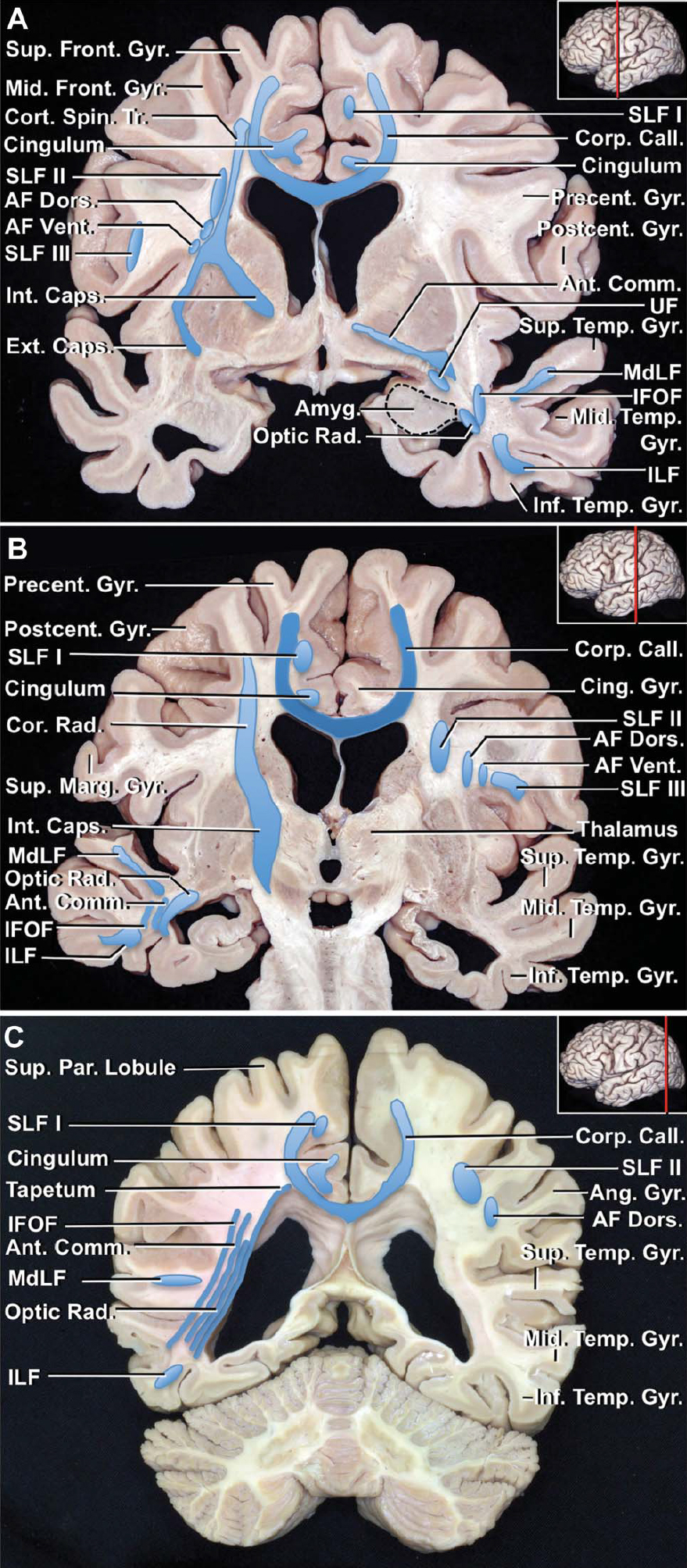
圖6。冠狀切片纖維束的位置。A,中央前回水平的冠狀麵。在庇護上區,為了從外側到內側,SLF III深入到額頂骨蓋,AF腹段位於SLF III的內側,AF背段的腹外側。SLF II位於AF背內側。放射狀冠由慢狀核上方的內外囊結合形成,位於AF節段和SLF II的內側。胼胝體之上的胼胝體纖維位於輻射狀冠內側。扣帶位於胼胝體上方,胼胝體纖維內側。SLF I位於扣帶上方。在庇護下區,中顳下回深通至顳上回表麵,內顳下回深通至顳下回。 The IFOF passes deep to the MdLF and ILF. The fibers in the posterior crus of the anterior commissure are located deep to the lower part of the precentral sulcus on the convexity. The optic radiations are located deep to the superior and middle temporal gyri and lateral to the temporal horn of the lateral ventricle. B, coronal section at the level of the postcentral gyrus. In the suprasylvian area, laterally to medially, are the SLF III, AF ventral and dorsal segments, and SLF II. The corticospinal tract, a part of the corona radiata, is located deep to the association fibers, where it projects vertically downward in the internal capsule. In the infrasylvian area, the IFOF, fibers crossing in the anterior commissure, and optic radiations pass deep to the superior and middle temporal gyri. The ILF passes medial to the inferior temporal gyrus. C, coronal section at the level of the angular gyrus. The AF dorsal segment passes deep to the lower part of the angular gyrus. The SLF II passes deep to the upper part of the angular gyrus. The MdLF passes deep to or through the superior temporal gyrus, and the ILF passes deep to or through the inferior temporal gyrus. Laterally to medially, the fibers of the IFOF, anterior commissure, optic radiations, and tapetum pass lateral to the atrium of the lateral ventricle. The optic radiations pass lateral to the tapetum and inferior two-thirds of the atrium of the lateral ventricle. AF, arcuate fasciculus; Amyg., amygdala; Ang., angular; Ant., anterior; Call., callosum; Caps., capsule; Cing., cingulate; Comm., commissure; Cor., corona; Corp., corpus; Cort. Spin., corticospinal; Dors., dorsal; Ext., external; Front., frontal; Gyr., gyrus; IFOF, inferior fronto-occipital fasciculus; ILF, inferior longitudinal fasciculus; Inf., inferior; Int., internal; Marg., marginal; MdLF, middle longitudinal fasciculus; Mid., middle; Par., parietal; Postcent., postcentral; Precent., precentral; Rad., radiata, radiations; SLF, superior longitudinal fasciculus; Sup., superior; Temp., temporal; Tr., tract; UF, uncinate fasciculus; Vent., ventral. (Images courtesy of AL Rhoton, Jr.)

本研究還關注了纖維束的關係,從淺到深,以及它們從皮層回表麵的深度(表1;圖5和圖6)。額葉回水平的纖維束,從淺到深依次為額上回(輻射冠、胼胝體纖維和SLF I)、額中回(SLF II, AF、IFOF的背側段、輻射冠和胼胝體纖維);和額下回(SLF III, AF, IFOF和輻射冕的腹側和背側段)。深入頂葉表麵的纖維束是頂葉上小葉(輻射狀冠、胼胝體纖維和SLF I)和頂葉下小葉(SLF III、AF背段和腹段、SLF II、MdLF、IFOF、輻射狀冠和絨氈層)。顳葉內從淺到深的纖維束分別是顳上回(MdLF、UF、IFOF、前連連、視神經輻射和絨氈層)、顳中回(AF、IFOF、UF、前連連、矢狀層和絨氈層的背段和腹段);以及顳下回(AF和ILF的背側段)。ILF位於側腦室顳角軸位以下。枕回下麵的纖維束是枕上回(ILF, IFOF,前連連,矢狀層,大鉗,和扣帶),枕中回(SLF II[有時],ILF, IFOF,前連連,矢狀層,和絨氈層);和枕下回(ILF, IFOF,前連合,矢狀層和大鉗)。9
DTI為纖維束的可視化提供了一個強大的工具,就像血管造影術為我們理解血管病理提供了一個極好的工具。然而,了解血管造影和DTI是一項技能,而獲得3d知識則是另一項技能,它允許外科醫生使用手眼和大腦導航技能通過手術訪問動脈或纖維束。我們正在進入一個纖維追蹤的階段,類似於腦血管造影引入時所發生的情況。隨著外科醫生了解血管造影並利用這些知識完成血管病理的顯微外科、內窺鏡和血管內治療,腦血管外科得到了發展。當DTI和腦內纖維束三維知識相結合時,纖維束解剖的技能將為筋束旁和筋束內手術的進步鋪平道路。本研究結合了顯微外科解剖技術和三維攝影技術,以幫助開發在處理大腦病理時在纖維束之間導航所必需的技能和知識。
DTI能夠追蹤大腦的主要纖維束。目前還沒有定量的DTI研究報告每個纖維通路與上覆腦回的確切位置和深度。然而,DTI與解剖解剖的比較分析顯示有良好的相關性。33、76、77DTI能夠在同一幅圖像中顯示多個纖維路徑,並可能更好地顯示它們之間的關係,而不像在纖維解剖中,一些路徑隻能以逐步的方式顯示。另一方麵,通過仔細的規劃,可以在一張纖維解剖圖像中顯示多個纖維束及其關係(圖2-4)。
本研究的解剖結果已與來自臨床放射學分析和術中纖維束刺激的功能數據進行了簡單的比較(表2)。更好地了解纖維通路的三維解剖組織對規劃安全、準確的大腦病變手術很重要。
貢獻者:Kaan Yagmurlu,醫學博士,Alexander L. Vlasak,和Albert L. Rhoton, Jr醫學博士
內容來自Yagmurlu K, Vlasak AL, Rhoton AL, Jr.大腦三維地形纖維束解剖。神經外科2015; 11:274 - 305。doi.org/10.1227/NEU.0000000000000704.經牛津大學出版社代表神經外科醫師協會批準。©神經外科醫生協會。
神經外科188bet手机app圖譜很榮幸能夠繼承Albert L. Rhoton, Jr . MD的遺產。
請登錄發表評論。
一定要在社交媒體上關注我們,獲取精彩內容並保持更新生活科恩醫生的會議,關於手術技術的問題,以及更多!


您必須登錄才能查看此材料。
的188bet手机app這幾乎完全取決於你的捐款。
如果沒有你們的大量捐贈,我們就無法繼續開展地圖集。
請承諾每年至少捐贈250美元給Atlas。如果沒有這種承諾,Atlas將很快需要付費訂閱,世界各地的許多外科醫生將無法獲得它,他們的病人的護理依賴於它。
現在請捐!
如果沒有你們的大量捐贈,我們就無法繼續開展地圖集。請承諾每年至少捐贈250美元給Atlas。
如果沒有這個承諾,Atlas將很快需要付費訂閱世界上許多病人的護理都依賴於它的外科醫生將無法使用它。現在請捐!