
![圖2.5。頸內動脈棘上部分的前、下視圖。A,前視圖。視神經進入前斜突內側的視神經管。漏鬥經視交叉下方至腦下垂體。頸動脈在視神經的後麵。蝶平麵在交叉溝和鞍結節的前麵。頸動脈的穿支在交叉下間隙向內側穿過。垂體上動脈發源於頸動脈並通至漏鬥。鐮狀突是硬腦膜的褶皺,它經過視神經上方,靠近視神經孔。 B, the right optic nerve has been divided at the optic foramen and elevated to show the perforating branches of the supraclinoid portion of the carotid arteries. The right anterior cerebral artery was divided at its origin so that the optic nerve and chiasm could be elevated. The carotid artery gives rise to multiple perforating branches as well as the ophthalmic, posterior communicating, anterior choroidal, and the middle cerebral arteries. The supraclinoid portion of the ICA is divided into three segments based on the origin of its major branches: the ophthalmic segment (C4-Op.) extends from the origin of the ophthalmic artery to the origin of the PComA, the communicating segment (C4-Co.) extends from the origin of the PComA to the origin of the AChA, and the choroidal segment (C4-Ch.) extends from the origin of the AChA to the bifurcation of the carotid artery. The perforating branches arising from the ophthalmic segment pass to the optic nerve, chiasm, infundibulum, and the floor of the third ventricle. The perforating branches arising from the communicating segment pass to the optic tract and the floor of the third ventricle. The perforating branches arising from the choroidal segment pass upward and enter the brain through the anterior perforated substance. The diaphragma sellae surrounds the infundibulum above the pituitary gland. The temporal lobe is below the middle cerebral artery. C, the left optic nerve has been divided at the optic foramen and the anterior cerebral artery divided near its origin so that both optic nerves and the chiasm and tract could be elevated to show the perforating branches of the carotid artery. The Liliequist membrane is posterior to the infundibulum and hides the basilar artery, but not the posterior cerebral artery. The perforating branches of the ophthalmic segment pass upward to the infundibulum and the optic nerve, chiasm, and tract. D, both optic nerves and both ACAs and the infundibulum have been divided to permit the optic nerves and chiasm to be elevated with a forceps for this view under the optic chiasm and across the diaphragma sellae and dorsum to the upper part of the basilar artery and the oculomotor nerves. The oculomotor nerves pass forward below the PCAs. The perforating branches of the supraclinoid segment of the carotid artery pass upward to supply the infundibulum, the optic chiasm and tracts, and the floor of the third ventricle; some enter the brain through the anterior perforated substance. The right AChA is very large. A., artery; A.C.A., anterior cerebral artery; A.Ch.A., anterior choroidal artery; Ant., anterior; B.A., basilar artery; C.A., carotid artery; Ch., choroidal; Ch., chiasm, chiasmatic; Co., communicating; Diaph., diaphragm; Falc., falciform; Hyp., hypophyseal; Infund., infundibulum; M.C.A., middle cerebral artery; N., nerve; O., optic; Op., Ophth., ophthalmic; P.C.A., posterior cerebral artery; P.Co.A., posterior communicating artery; Perf., perforated; Post., posterior; Subst., substance; Sulc., sulcus; Sup., superior; Temp., temporal; Tr., tract. (From, Gibo H, Lenkey C, Rhoton AL Jr: Microsurgical anatomy of the supraclinoid portion of the internal carotid artery. J Neurosurg 55:560–574, 1981 [15].)](https://assets.neurosurgicalatlas.com/neuroanatomy/Rhoton_-_Missed_Images/Supratentorial_2.5_edited.jpg)
![圖2.19。大腦中動脈的分支模式。這些從五個大腦半球解剖的MCAs圖顯示了主幹的不同分支模式。主幹在78%的半球上分叉,12%的半球上三分叉,10%的半球上多分支(四條或四條以上的主幹)。這些圖顯示了主、上、中、下主幹。這些主幹產生了透鏡紋狀動脈、眼窩前額動脈、前額動脈、中央動脈、中央動脈、前頂葉動脈、後頂葉動脈、角動脈、顳枕動脈、後顳動脈、中顳動脈、前顳動脈和顳極動脈。A、分岔:等量主幹(占半球的18%)。主幹分為大約相同直徑的上主幹和下主幹,並提供相似大小的皮層區域。上幹通過角動脈形成眶額動脈,下幹通過顳枕動脈形成顳極動脈。B,分岔:下主幹為主(占半球的32%)。 The inferior trunk has a larger diameter and area of supply than the superior trunk. The superior trunk supplies the orbitofrontal through the anterior parietal areas, and the inferior trunk supplies the posterior parietal through the temporopolar areas. C, bifurcation: superior trunk dominant (28% of hemispheres). The superior trunk has a larger diameter and area of supply than the inferior trunk. It supplies the orbitofrontal through the temporo-occipital areas, and the inferior trunk supplies the temporal areas except for the temporopolar area, which is supplied by an early branch (Early Br.) that arises from the main trunk. D, trifurcation pattern (12% of hemispheres). The main trunk of the MCA divides into three trunks. The superior trunk supplies the orbitofrontal and prefrontal areas, the middle trunk supplies the precentral through the posterior parietal areas, and the inferior trunk supplies the angular through the anterior temporal areas. The temporopolar artery arises from the main trunk as an early branch. E, multiple trunks (10% of hemispheres). The main trunk gives rise to more than three trunks. There are five trunks in the specimen shown. A., arteries, artery; Ang., angular; Ant., anterior; Br., branch; Cent., central; Inf., inferior; Len. Str., lenticulostriate; Mid., middle; Orb.Fr., orbitofrontal; Par., parietal; Post., posterior; Pre. Cent., precentral; Pre. Fr., prefrontal; Sup., superior; Temp., temporal; Temp. Occ., temporo-occipital; Temp. Pol., temporopolar; Tr., trunk. (From, Gibo H, Carver CC, Rhoton AL Jr, Lenkey C, Mitchell RJ: Microsurgical anatomy of the middle cerebral artery. J Neurosurg 54:151–169, 1981 [14].)](https://assets.neurosurgicalatlas.com/neuroanatomy/Rhoton_-_Missed_Images/Supratentorial_2.19_edited.jpg)
![圖2.20。莖動脈形態。主幹動脈起源於主幹,形成皮質動脈。中間的圖示顯示了左大腦半球的側麵,在額葉、頂葉和顳葉區域之間有一個空間。額葉由眼窩前額區、前額區、中央區和中央區組成;頂葉由前頂葉、後頂葉和角區組成;顳葉和枕葉由顳極、前顳區、中顳區、後顳區和顳枕區組成。中央區域的後部,實際上是頂葉的一部分,包括額葉。中心圖顯示了最常見的詞幹模式,外圍圖顯示了接下來三種最常見的模式。每一種顏色或顏色的深淺表示由一根莖動脈供血的區域。 The percentage of hemispheres having the stem pattern shown is listed on each diagram. The most common frontal lobe pattern involves two stem arteries: one gives rise to the branches to the orbitofrontal, prefrontal, and precentral areas, and the other supplies the central area. The most common parietal lobe pattern involves three stem arteries, one each for the anterior and posterior parietal and the angular areas. The most common temporal and occipital lobe pattern involves four stem arteries: one stem artery supplies both the temporopolar and the anterior temporal areas, and there is one stem each for the middle temporal, posterior temporal, and temporo occipital areas. The next three most common stem patterns for each lobe are shown on the peripheral diagrams. The four patterns shown for each lobe do not account for 100% of the hemispheres, but show only the four most common patterns for that lobe. Ang., angular; Ant., anterior; Cent., central; Mid., middle; Orb. Fr., orbitofrontal; Par., parietal; Post., posterior; Pre. Cent., precentral; Pre. Fr., prefrontal; Temp., temporal; Temp. Occ., temporo-occipital; Temp. Pol., temporopolar. (From, Gibo H, Carver CC, Rhoton AL Jr, Lenkey C, Mitchell RJ: Microsurgical anatomy of the middle cerebral artery. J Neurosurg 54:151–169, 1981 [14].)](https://assets.neurosurgicalatlas.com/neuroanatomy/Rhoton_-_Missed_Images/Supratentorial_2.20.jpg)
![圖2.26。從大腦半球剖開的大腦前動脈圖。可見胼胝體周圍動脈、胼胝體邊緣動脈、眼窩前額動脈、額極動脈、額內前動脈、中動脈和後動脈、旁中心動脈、頂上動脈和頂下動脈、短胼胝體動脈、下胼胝體動脈、複發動脈和胼胝體前動脈。A,這裏沒有通訊動脈ACA所有單獨的皮層分支都直接來自於胼胝體周圍動脈。有兩個後內額動脈和旁中心動脈。短小的胼胝體分支起源於胼胝體周圍動脈。B,胼胝體邊際動脈起源於兩個皮層分支:額極動脈和額內前動脈。其他皮層分支來自於胼胝體周圍動脈。可見胼胝體前動脈和下動脈。C,胼胝體邊際動脈是額內中動脈和額內後動脈的起源。 Short and inferior callosal arteries are present. D, four cortical branches arise from the callosomarginal artery. A., artery; A.I.F.A., anterior internal frontal artery; Cal., callosal; Cm., callosomarginal; Fp., frontopolar; I., inferior; Inf., inferior; M.I.F.A., middle internal frontal artery; Of., orbitofrontal; Par., parietal; Pce., paracentral; Perical., pericallosal; P.I.F.A., posterior internal frontal artery; Precal., precallosal; Rec., recurrent; S., superior; Sh., short. (From, Perlmutter D, Rhoton AL Jr: Microsurgical anatomy of the distal anterior cerebral artery. J Neurosurg 49:204–228, 1978 [27].)](https://assets.neurosurgicalatlas.com/eyJidWNrZXQiOiJuc2F0bGFzLWFzc2V0cyIsImtleSI6Im5ldXJvYW5hdG9teVwvUmhvdG9uXy1fTWlzc2VkX0ltYWdlc1wvU3VwcmF0ZW50b3JpYWxfMi4yNi5qcGciLCJlZGl0cyI6eyJyZXNpemUiOnsiZml0IjoiY292ZXIiLCJ3aWR0aCI6MTgzMH0sImpwZWciOnsicXVhbGl0eSI6NjV9fX0=)
![圖2.31。動脈進入前麵的穿孔物質。A,劣勢視角。左側的前穿孔物質向前延伸到內側和外側嗅紋,向後延伸到視神經束和顳葉,向外側延伸到外膜島,在視交叉上方的內側延伸到半球間裂。顳葉前部已被切除以暴露顳角。頸內動脈、脈絡膜前動脈、大腦前動脈和中動脈(M1和M2)形成了前穿孔物質的分支。PComA不為前部穿孔物質提供分支。大腦的中間分支,稱為紋狀透鏡體動脈,分為內側、中間和外側紋狀透鏡體組。外側透鏡狀紋狀動脈在M1段分叉附近外側出現。脈絡膜前支和頸動脈前支進入前穿孔物質的後部,靠近視神經束。 The branches from the anterior cerebral artery enter the narrow strip of the anterior perforated substance above the optic chiasm. The recurrent artery arises from the anterior cerebral artery, near the level of the AComA, and passes laterally above the carotid bifurcation to enter the anterior perforated substance anterior to the branches from the other sources. The M1 segment gives rise to an early branch. B, another specimen. The optic nerve and chiasm have been reflected inferiorly. The branches from the left A1 segment enter the narrow medial sector of the anterior perforated substance extending above the optic chiasm to the interhemispheric fissure. A perforating artery arises from an early branch of the M1. Some of the lateral lenticulostriate arteries arise near the M1 bifurcation. The intermediate lenticulostriate arteries have a candelabra appearance. The anterior choroidal artery sends branches to the posterior half of the anterior perforated substance. Two recurrent arteries arise near the anterior communicating artery. C, another specimen. The anterior choroidal branches to the anterior perforated substance arise near the origin of the anterior choroidal artery. The lateral lenticulostriate arteries arise near the M1 trifurcation and have a roughly S shaped course. The intermediate lenticulostriate arteries have a candelabra appearance. The medial lenticulostriate arteries pass near the perforating branches arising from the carotid artery and the medial half of the A1 segment. D, inferior view, right side. The intermediate lenticulostriate arteries have a candelabra appearance. The A1 branches enter the anterior perforated substance medial to those from the internal carotid, anterior choroidal, and middle cerebral arteries. The recurrent artery arises above the optic chiasm, passes laterally above the carotid bifurcation, and gives rise to branches that enter the anterior perforated substance in front of those from other sources. E, perforating branches of the anterior cerebral artery, anterior view. The recurrent artery arises above the optic chiasm near the level of the AComA. The A1 segment arises from the carotid artery and its perforating branches to enter the medial half of the anterior perforated substance in the narrow sector extending above the optic chiasm. A., arteries, artery; A.C.A., anterior cerebral artery; Ant., anterior; Bifurc., bifurcation; Car., carotid; Chor., choroid, choroidal; Comm., communicating; Br., branch; Fiss., fissure; Front., frontal; Gyr., gyrus; I.C.A., internal cerebral artery; Infund., infundibulum; Int., intermediate; Interhem., interhemispheric; Lat., lateral; Len. Str., lenticulostriate; Med., medial; N., nerve; Olf., olfactory; Orb., orbital; P.C.A., posterior cerebral artery; Ped., peduncle; Perf., perforated, perforating; Plex., plexus; Post., posterior; Rec., recurrent; Subst., substance, substantia; Temp., temporal; Tr., tract; Trifurc., trifurcation. (From, Rosner SS, Rhoton AL Jr, Ono M, Barry M: Microsurgical anatomy of the anterior perforating arteries. J Neurosurg 61:468–485, 1984 [36].)](https://assets.neurosurgicalatlas.com/neuroanatomy/Rhoton_-_Missed_Images/Supratentorial_2.31_edited.jpg)

你可以有所作為。
的188bet手机app幾乎完全取決於你的捐款。
如果沒有你們的大量捐贈我們無法繼續製作《地圖集》。
請承諾每年至少向Atlas捐贈250美元。如果沒有這種承諾,Atlas將很快需要付費訂閱,世界各地的許多外科醫生將無法使用它,他們的病人的護理依賴於它。
請立即捐款!

最後更新:2021年4月7日
幕上動脈包括頸內動脈的枕骨上部分及其大腦前、中、眼、後交通和脈絡膜前分支,威利斯圈的組成部分,在後中線包括基底尖,最後是大腦後動脈。所有這些動脈的起源都位於大腦中心下方的深處,它們的近端幹相對難以接近,因為它們在sylvian或半球間裂或腦幹和顳葉之間的基底池中行進(圖2.1)。在側凸處隻有較小的末梢分支可達,即使在側凸處,這些分支通常隱藏在皮質溝中,而不是在腦回表麵。沒有一個手術入路能到達大腦三大動脈的所有分支,因為它們的路線很長。因此,每一種手術入路都必須根據所涉及動脈段的關係精心定製。這些動脈與常見動脈瘤部位及其手術暴露的關係將在第3章中進行回顧。

點擊這裏查看此圖像的交互模塊和相關內容。
圖2.1 A-F。基底池的動脈。A,前視圖。幾乎相等大小的a1穿過終板的前部。右A2在左A2前麵進入半球間裂。左回動脈起於前交通動脈(AComA)附近,並在前穿孔物下方外側通過。穿支動脈起源於AComA。B,視野轉移到頸動脈分叉上方。返動脈在A1上方外側穿過,並與M1的透鏡狀紋狀分支交織。後交通動脈(PComA)指向內側,通過頸動脈、視神經和A1之間的視頸三角可見。 C, anterolateral view. The PComA is seen through the opticocarotid triangle. The M1 bifurcates into superior and inferior trunks at the limen insula. D, the basal cisterns have been opened and the temporal pole retracted to expose the oculomotor nerve. The PComA is directed backward above and medial to the oculomotor nerve. The superior cerebellar artery courses below the oculomotor nerve. E, the temporal lobe has been elevated. The anterior choroidal artery (AChA) ascends on the medial side of the uncus. The PComA and the P1 join to form the P2, which continues backward on the medial side of the posterior part of the uncus. A medial posterior choroidal artery (MPChA) passes backward around the brainstem. The superior cerebellar artery passes below the oculomotor and trochlear nerves. The branches forming the P3 course through the quadrigeminal cistern. The P2 courses through the ambient and crural cisterns. A MPChA encircles the brainstem. F, the tentorium has been divided to expose the upper part of the basilar artery. The trigeminal nerve is exposed in the lateral margin of the tentorial opening. The posterior cerebral artery (PCA) courses above and the superior cerebellar artery courses below the oculomotor nerve.

點擊這裏查看此圖像的交互模塊和相關內容。
圖2.1 G-L。G,另一個標本的顳下暴露。PComA比D和e所示的大。動眼神經在PCA和小腦上動脈之間向前傳遞。H,暴露沿腦幹一側進一步向後延伸至四叉神經池。小幕被分開,露出小腦的上部。主動脈和小腦上動脈環繞腦幹到達四叉神經池。P2分為位於鉤突和腦梗之間的腳側池的P2A和位於中腦海馬旁回之間的周圍池的P2P。P3位於四叉神經池。滑車神經起於下丘下方並在小腦上動脈分支上方交叉。I,暴露進一步向後方延伸,在幕上到四叉神經池的左半部分。 The tributaries of the vein of Galen have been retracted to expose the pineal. The PCA courses above the tentorium and the superior cerebellar artery below. The trochlear nerve arises below the inferior colliculus and passes around the brainstem. J, the exposure has been directed below the tentorium. The internal cerebral veins exit the roof of the third ventricle and the basal veins exit the basal cisterns to join and form the vein of Galen. The P3 courses through the quadrigeminal cistern. K, midline infratentorial exposure. The pineal is exposed between the posterior cerebral arteries and basal veins and below the internal cerebral veins. The exposure into the fissure between the cerebellum and midbrain is not as great as can be achieved when the exposure is directed off to the side of the vermian apex in a paramedian location as shown in J. L, enlarged view of the midline infratentorial exposure. A., artery, arteries; A.Co.A., anterior communicating artery; Bas., basilar; Bifurc., bifurcation; Br., branch; Car., carotid; Cer., cerebral; Cist., cistern; Clin., clinoid; CN, cranial nerve; Coll., colliculus; Front., frontal; Gl., gland; Inf., inferior; Int., internal; Lam., lamina; Lent. Str., lenticulostriate; M.P.Ch.A., medial posterior choroidal artery; Olf., olfactory; P.Co.A., posterior communicating artery; Perf., perforating; Pit., pituitary; Post., posterior; Quad., quadrigeminal; Rec., recurrent; S.C.A., superior cerebellar artery; Str., straight; Sup., superior; Temp., temporal; Tent., tentorial; Term., terminalis; Tr., tract, trunk; V., vein.
頸內動脈(ICA)的鞘上段是顱內動脈瘤的常見部位,其分支經常被拉伸、移位或被顱底腫瘤包圍。在Willis圈動脈瘤和蝶骨脊、前、中顱窩、鞍上區腫瘤手術中,常暴露ICA及其主支和穿支。內頸動脈發育不全或再生障礙性病變是罕見的。
ICA分為四個部分:C1或頸部分從它與頸總動脈的交界處延伸到頸動脈管的外口;C2或岩部在頸動脈管內並在動脈進入海綿竇處結束;C3或海綿狀部分在海綿竇內,並在動脈穿過硬腦膜形成海綿竇頂部的地方結束;C4或枕骨上部分開始於動脈進入蛛網膜下腔,並在分叉處終止於大腦前動脈(ACA)和大腦中動脈(MCA)(圖2.2)(25,36)。
C4開始於硬腦膜的動脈,形成海綿竇的頂部。它通過前斜突內側和視神經下方進入顱腔。它經過後、上、略外側,到達視交叉的外側,並在前穿孔物質下分叉,在sylvian裂的內側端形成ACA和MCA。C4段被定義為包括MCA和ACA產生的胯部,ACA和MCA起源之間的壁的頂端發源於分支被認為是ICA的分支,就像在這個頂端發源於ICA分叉的動脈瘤被認為是ICA的分支一樣。從側麵看,海綿(C3)和顱內(C4)部分有幾條曲線,形成S型,這些部分一起被稱為頸動脈虹吸。S的下半部分,主要由海綿體內部分形成,前部凸,而由枕骨上部分形成的上半部分,後部凸。前凸段和後凸段的交界處沿前斜突的內側。C4的分叉前分支包括眼動脈、脈絡膜前(AChA)、後交通動脈(PComA)、穿通動脈和垂體上動脈。
硬膜內暴露C4和Willis圓的前部沿同側蝶脊或眶頂指向前斜突。在暴露ICA時,入路通常從近端到遠端,從眼節開始,向遠端分支工作。眼動脈硬膜內長度較短,且位於視神經下,不易暴露。
在眼動脈起點之外暴露C4時,盡管AChA出現在PComA遠端,但外科醫生通常會在PComA之前看到AChA(圖2.1和2.3)。這是因為三組解剖環境。首先,C4向後外側向上傳遞,將AChA的起點置於比PComA的起點更靠近中線的外側。其次,AChA通常比PComA在C4部分後壁的外側更遠。在94%的半球中,AChA起源於C4部分後壁的部位位於PComA起始部位的外側(33)。第三,AChA比PComA更偏向橫向;前者沿外側繞過腦梗並進入顳角,而後者最常見的初始路徑是動眼神經上方的後內側方向,通往腳梗間窩。
![圖2.2。左側頸內動脈(ICA)的側位圖(左)和前方圖(右),以及甲、乙、棘上節段(C4)。A, C4部分的側麵圖。B, C4部分的前視圖。ICA分為四個部分。這些部分,從近端到遠端,是C1到C4部分。頸部分(C1,紅色)從ICA的起點延伸到顳骨岩頸動脈管的外孔。岩部(C2,橙色)從頸動脈管的外口延伸到動脈離開頸動脈管進入海綿竇的地方。海綿狀部分(C3,黃色)始於動脈進入海綿竇的地方,終止於前斜突內側硬腦膜進入顱內的地方。顱內(斜突上)部分(C4,米色)開始於動脈進入前斜突內側的顱腔,並在前穿孔物以下終止,在那裏動脈分叉進入大腦前動脈和中動脈。 The ICA gives rise to the ophthalmic, posterior communicating, anterior choroidal, anterior cerebral, and the middle cerebral arteries. The supraclinoid portion of the ICA is divided into three segments based on the origin of these branches. The ophthalmic segment (C4-Op., dark blue) extends from the origin of the ophthalmic artery to the origin of the PComA. The communicating segment (C4-Co., light green) extends from the origin of the PComA to the origin of the anterior choroidal artery. The choroidal segment (C4-Ch., dark green) extends from the origin of the anterior choroidal artery to the bifurcation of the internal carotid artery into the anterior and middle cerebral arteries. A., artery; A.C.A., anterior cerebral artery; A.Ch.A., anterior choroidal artery; Ch., choroidal; Co., communicating; M.C.A., middle cerebral artery; Op., ophthalmic; Ophth., ophthalmic; P.Co.A., posterior communicating artery. (From, Gibo H, Lenkey C, Rhoton AL Jr: Microsurgical anatomy of the supraclinoid portion of the internal carotid artery. J Neurosurg 55:560–574, 1981 [15].)](https://assets.neurosurgicalatlas.com/neuroanatomy/Rhoton_Book_-_Supratentorial/Figure_2.2.png)
點擊這裏查看此圖像的交互模塊和相關內容。
圖2.2。左側頸內動脈(ICA)的側位圖(左)和前方圖(右),以及甲、乙、棘上節段(C4)。A, C4部分的側麵圖。B, C4部分的前視圖。ICA分為四個部分。這些部分,從近端到遠端,是C1到C4部分。頸部分(C1,紅色)從ICA的起點延伸到顳骨岩頸動脈管的外孔。岩部(C2,橙色)從頸動脈管的外口延伸到動脈離開頸動脈管進入海綿竇的地方。海綿狀部分(C3,黃色)始於動脈進入海綿竇的地方,終止於前斜突內側硬腦膜進入顱內的地方。顱內(斜突上)部分(C4,米色)開始於動脈進入前斜突內側的顱腔,並在前穿孔物以下終止,在那裏動脈分叉進入大腦前動脈和中動脈。 The ICA gives rise to the ophthalmic, posterior communicating, anterior choroidal, anterior cerebral, and the middle cerebral arteries. The supraclinoid portion of the ICA is divided into three segments based on the origin of these branches. The ophthalmic segment (C4-Op., dark blue) extends from the origin of the ophthalmic artery to the origin of the PComA. The communicating segment (C4-Co., light green) extends from the origin of the PComA to the origin of the anterior choroidal artery. The choroidal segment (C4-Ch., dark green) extends from the origin of the anterior choroidal artery to the bifurcation of the internal carotid artery into the anterior and middle cerebral arteries. A., artery; A.C.A., anterior cerebral artery; A.Ch.A., anterior choroidal artery; Ch., choroidal; Co., communicating; M.C.A., middle cerebral artery; Op., ophthalmic; Ophth., ophthalmic; P.Co.A., posterior communicating artery. (From, Gibo H, Lenkey C, Rhoton AL Jr: Microsurgical anatomy of the supraclinoid portion of the internal carotid artery. J Neurosurg 55:560–574, 1981 [15].)

點擊這裏查看此圖像的交互模塊和相關內容。
圖2.3 A-F。翼麵暴露威利斯圈。第一,左額顳骨瓣被抬高硬腦膜被打開。左額葉和顳葉被收回以暴露頸動脈進入硬腦膜內側前床突。頸動脈分叉暴露出來了。透鏡狀紋狀動脈起源於M1。M1以三叉分叉的方式分裂。B,暴露已從交叉和額葉延伸到AComA和對側A1和A2s。在AComA附近出現的一根複發動脈在頸動脈分叉的外側通過。C,基底分叉通過位於內頸動脈A1和視神經之間的視頸三角暴露出來。 D, the carotid bifurcation has been depressed to expose the basilar apex in the interval between the carotid bifurcation and the lower margin of the optic tract. Perforating branches crossing the area can make the approach hazardous. A thalamoperforating artery arises from the ipsilateral P1. E, the temporal pole has been retracted posteriorly for a pretemporal exposure. The carotid and anterior choroidal arteries have been elevated to expose the PComA, which gives rise to a large perforating branch referred to as a premamillary artery. The M1 gives rise to an early branch proximal to the trifurcation. The P2 extends above and the superior cerebellar artery (SCA) extends below the oculomotor nerve. F, anterior subtemporal view. The temporal pole and the carotid artery have been elevated to the expose the origin of the normal-sized PComA. The AChA passes backward along the medial edge of the uncus. A large MPChA arises from the P1 and loops downward as it passes to the quadrigeminal cistern.

點擊這裏查看此圖像的交互模塊和相關內容。
圖2.3 G-J。G, AChA升高,暴露出PComA的一個大的穿支,稱為乳頭前動脈。H, PComA已經升高,以提供基底尖和p1的良好曝光。同側SCA為雙側動脈。在滑車神經進入邊緣的地方,幕被分開了。這增加了基底動脈暴露的長度。重複小腦上動脈的主幹向下延伸至三叉神經。J,切除岩尖以完成前路岩切入路,這增加了通往腦幹前部和基底動脈的通路。在本例中,包括耳蝸和半規管在內的迷路,以及內聽道中的神經已經暴露出來,以顯示前路岩石切開術的鑽孔與這些結構的關係。前路岩石切開術的鑽孔指向頸岩動脈後的迷宮內側,並向內側進入岩下竇和斜坡一側。 The abducens nerve and the ICA are in the lower margin of the exposure. A., arteries, artery; A.Ch.A., anterior choroidal artery; A.Co.A., anterior communicating artery; A.I.C.A., anteroinferior cerebellar artery; Ant., anterior; Bas., basilar; Br., branch; Car., carotid; Clin., clinoid; CN, cranial nerve; Contra., contralateral; Front., frontal; Gr., greater; Ipsi., ipsilateral; Lent. Str., lenticulostriate; M.C.A., middle cerebral artery; M.P.Ch.A., medial posterior choroidal artery; N., nerve; Olf., olfactory; P.Co.A., posterior communicating artery; Pet., petrosal; Post., posterior; Premam., premamillary; Rec., recurrent; S.C.A., superior cerebellar artery; Seg., segment; Semicirc., semicircular; Temp., temporal; Tent., tentorial; Thal. Perf., thalamoperforating; Tr., tract; Trifurc., trifurcation.
C4根據眼球、PComA和AChA的起源部位分為三個節段。眼段從海綿竇頂部和眼動脈起點延伸至PComA起點;所述通信段從所述PComA的原點延伸至所述AChA的原點;脈絡膜段從AChA的起點延伸到ICA的末端分叉。眼段最長,通訊段最短(15)。
C4射孔支
三個C4節段中的每一個都發出一係列具有相對恒定的終止位置的射孔分支。平均有8個(範圍3-12個)穿通動脈(不包括眼動脈、PComA和AChA)起源於C4(圖2.4-2.6)。
眼段平均有4個(範圍從1到7個)穿通動脈。大多數發生於動脈的後部或內側。這些分支最常分布於腦垂體漏鬥部(柄部)、視交叉,較少分布於視神經、第三腦室底前乳突部和視神經束(頻率由高到低)。少數血管終止於硬腦膜,覆蓋前斜突、蝶鞍和鞍結節。從這一節段起並通至垂體漏鬥的動脈稱為垂體上動脈(13,15)。
在超過一半的半球中,沒有從通信段產生的穿孔分支,即使存在,也隻有一到三個。它們起於後半壁,按頻率由高到低依次終止於視神經束、第三腦室底前乳狀部、視交叉和漏鬥部,偶爾也會進入前或後穿孔物質。分支常在後交通動脈瘤頸部周圍伸展。
脈絡膜段平均有4個分支(範圍從1到9個)。大多數分支起源於動脈壁的後半部分,按頻率由高到低依次終止於前穿孔物、視神經束和鉤結。
垂體上動脈是一組1 - 5個(平均為2個)小分支,起源於C4的眼段,終止於垂體柄和腺體,但也向視神經、交叉和第三腦室底發送分支(圖2.4-2.6)。最大的分支常被稱為垂體上動脈。大多數分支起源於動脈的後內側、內側或後部。漏鬥動脈是一組發源於PComA並分布於漏鬥的動脈。漏鬥動脈比垂體上動脈少。四分之一的半球有一條或兩條漏鬥動脈,其餘的半球沒有。
垂體上動脈和漏鬥動脈經交叉下方的內側到達灰塊瘤。它們混合在一起,在垂體柄周圍形成一個精細的吻合神經叢,稱為漏鬥周吻合。這些動脈和漏鬥周神經叢分布於垂體柄和前葉。海綿內頸動脈腦膜垂體幹的下垂體分支灌注於頸動脈後葉。囊狀動脈也起源於海綿內頸動脈並供應腦垂體囊(16)。
這個漏鬥周圍神經叢有升動脈和降動脈。降動脈包括短柄動脈和淺動脈。短柄動脈穿過漏鬥,形成毛細血管,通向沿柄而下的竇狀動脈。淺表動脈在蛛網膜下腔的莖部外側向下行,並穿透前葉。升動脈供應灰塊莖、中隆起和視交叉下表麵。垂體上動脈也有分支到達視神經的交叉和近端。

點擊這裏查看此圖像的交互模塊和相關內容。
圖2.4 a-b .ICA的射孔分支。A,劣勢視角。頸內動脈包括眼動脈、後交通動脈、脈絡膜前動脈、大腦前動脈和大腦中動脈。ICA的枕骨上部分根據這些分支的起源分為三個節段:眼節段(C4-Op。(藍色),從眼動脈的起點延伸到PComA的起點;通信段(C4-Co。(淺綠色),從PComA的原點延伸到AChA的原點;和脈絡膜段(C4-Ch。(深綠色),從AChA的起源延伸到ICA的分叉,進入大腦前動脈和中動脈。起於眼節的穿支延伸至視神經、視交叉、視神經束和第三腦室底周圍的漏鬥和灰膜塊莖。 The superior hypophyseal arteries arise from the ophthalmic segment and extend to the infundibulum of the pituitary gland. The branches arising from the communicating segment reach the optic tracts, floor of the third ventricle, and the area around the mamillary bodies. The perforating branches of the choroidal segment pass upward and enter the anterior perforated substance. The posterior cerebral arteries arise from the basilar artery and pass backward below the optic tracts. The ACA and AComA course above the optic chiasm and pass between the frontal lobes. The olfactory nerves are lateral to the gyrus rectus. B, anterior view. The left optic nerve has been divided near its entrance into the optic canal and elevated to give a clearer view of the perforating branches. The ophthalmic artery arises above the cavernous sinus. The carotid artery courses through the cavernous sinus and then laterally and produces a prominence in the wall of the sphenoid sinus before giving rise to the ophthalmic artery. The oculomotor, trochlear, abducens, and the ophthalmic, maxillary, and mandibular divisions of the trigeminal nerve pass lateral to the sphenoid sinus in the walls of the cavernous sinus. The superior hypophyseal arteries arise from the ophthalmic segment.
![圖2.4 C. C,雙視神經分開並抬高的前視圖,顯示第三腦室底板的下表麵和通往第三腦室的穿支。膈鞍肌上方漏鬥被分割。一個,動脈;A.C.A,大腦前動脈;A.Ch.A。,脈絡膜前動脈;A.Co.A。,前交通動脈;螞蟻。前; Ch., choroidal; Cin., cinereum; Co., communicating; Diaph., diaphragm; Fr., frontal; Gyr., gyrus; Hyp., hypophyseal; Infund., infundibulum; M.C.A., middle cerebral artery; Mam., mamillary; N., nerve; O., optic; Olf., olfactory; Op., ophthalmic; Ophth., ophthalmic; P.C.A., posterior cerebral artery; P.Co.A., posterior communicating artery; Perf., perforated; Subst., substance; Sup., superior; Tr., tract. (From, Gibo H, Lenkey C, Rhoton AL Jr: Microsurgical anatomy of the supraclinoid portion of the internal carotid artery. J Neurosurg 55:560–574, 1981 [15].)](https://assets.neurosurgicalatlas.com/neuroanatomy/Rhoton_Book_-_Supratentorial/Figure_2.4_C.png)
點擊這裏查看此圖像的交互模塊和相關內容。
圖2.4 C. C,雙視神經分開並抬高的前視圖,顯示第三腦室底板的下表麵和通往第三腦室的穿支。膈鞍肌上方漏鬥被分割。一個,動脈;A.C.A,大腦前動脈;A.Ch.A。,脈絡膜前動脈;A.Co.A。,前交通動脈;螞蟻。前; Ch., choroidal; Cin., cinereum; Co., communicating; Diaph., diaphragm; Fr., frontal; Gyr., gyrus; Hyp., hypophyseal; Infund., infundibulum; M.C.A., middle cerebral artery; Mam., mamillary; N., nerve; O., optic; Olf., olfactory; Op., ophthalmic; Ophth., ophthalmic; P.C.A., posterior cerebral artery; P.Co.A., posterior communicating artery; Perf., perforated; Subst., substance; Sup., superior; Tr., tract. (From, Gibo H, Lenkey C, Rhoton AL Jr: Microsurgical anatomy of the supraclinoid portion of the internal carotid artery. J Neurosurg 55:560–574, 1981 [15].)
圖2.5。頸內動脈棘上部分的前、下視圖。A,前視圖。視神經進入前斜突內側的視神經管。漏鬥經視交叉下方至腦下垂體。頸動脈在視神經的後麵。蝶平麵在交叉溝和鞍結節的前麵。頸動脈的穿支在交叉下間隙向內側穿過。垂體上動脈發源於頸動脈並通至漏鬥。鐮狀突是硬腦膜的褶皺,它經過視神經上方,靠近視神經孔。 B, the right optic nerve has been divided at the optic foramen and elevated to show the perforating branches of the supraclinoid portion of the carotid arteries. The right anterior cerebral artery was divided at its origin so that the optic nerve and chiasm could be elevated. The carotid artery gives rise to multiple perforating branches as well as the ophthalmic, posterior communicating, anterior choroidal, and the middle cerebral arteries. The supraclinoid portion of the ICA is divided into three segments based on the origin of its major branches: the ophthalmic segment (C4-Op.) extends from the origin of the ophthalmic artery to the origin of the PComA, the communicating segment (C4-Co.) extends from the origin of the PComA to the origin of the AChA, and the choroidal segment (C4-Ch.) extends from the origin of the AChA to the bifurcation of the carotid artery. The perforating branches arising from the ophthalmic segment pass to the optic nerve, chiasm, infundibulum, and the floor of the third ventricle. The perforating branches arising from the communicating segment pass to the optic tract and the floor of the third ventricle. The perforating branches arising from the choroidal segment pass upward and enter the brain through the anterior perforated substance. The diaphragma sellae surrounds the infundibulum above the pituitary gland. The temporal lobe is below the middle cerebral artery. C, the left optic nerve has been divided at the optic foramen and the anterior cerebral artery divided near its origin so that both optic nerves and the chiasm and tract could be elevated to show the perforating branches of the carotid artery. The Liliequist membrane is posterior to the infundibulum and hides the basilar artery, but not the posterior cerebral artery. The perforating branches of the ophthalmic segment pass upward to the infundibulum and the optic nerve, chiasm, and tract. D, both optic nerves and both ACAs and the infundibulum have been divided to permit the optic nerves and chiasm to be elevated with a forceps for this view under the optic chiasm and across the diaphragma sellae and dorsum to the upper part of the basilar artery and the oculomotor nerves. The oculomotor nerves pass forward below the PCAs. The perforating branches of the supraclinoid segment of the carotid artery pass upward to supply the infundibulum, the optic chiasm and tracts, and the floor of the third ventricle; some enter the brain through the anterior perforated substance. The right AChA is very large. A., artery; A.C.A., anterior cerebral artery; A.Ch.A., anterior choroidal artery; Ant., anterior; B.A., basilar artery; C.A., carotid artery; Ch., choroidal; Ch., chiasm, chiasmatic; Co., communicating; Diaph., diaphragm; Falc., falciform; Hyp., hypophyseal; Infund., infundibulum; M.C.A., middle cerebral artery; N., nerve; O., optic; Op., Ophth., ophthalmic; P.C.A., posterior cerebral artery; P.Co.A., posterior communicating artery; Perf., perforated; Post., posterior; Subst., substance; Sulc., sulcus; Sup., superior; Temp., temporal; Tr., tract. (From, Gibo H, Lenkey C, Rhoton AL Jr: Microsurgical anatomy of the supraclinoid portion of the internal carotid artery. J Neurosurg 55:560–574, 1981 [15].)
眼動脈是C4的第一個分支。大多數眼動脈起於視神經下方海綿竇硬膜頂上方的枕骨上區,並通過視神經前外側下方進入視神經管和眶。遠端過程將在第7章中進行回顧。8%的眼動脈起源於海綿竇。眼動脈很少起源於前斜突內側的前眼竇的斜突段或起源於腦膜中動脈(16,20,29)。它很少缺席。眼動脈罕見地產生顱內穿支,如果存在,這些分支向後延伸,分布於視神經和交叉的腹側和垂體柄。
眼動脈通常起始於C4上表麵的內側三分之一,緊鄰海綿竇遠端,位於視神經下方。在我們早期的研究中,在78%的大腦半球中,它位於C4上表麵的中間三分之一以上,在22%的病例中位於上表麵的中間三分之一以上(15)。上表麵外側三分之一處無突起。它可以向外側扭結,很少在進入視神經管之前在視神經外側出現一小段。其起源在前突尖端前5mm至後7mm,在前突內側2 ~ 10mm不等(16)。大多數眼動脈位於前斜突尖端的前方,距前斜突內側約5毫米。
眼動脈的顱內段通常很短。在該實驗室之前的一項研究中,發現14%的節段離開ICA並立即進入視神經管;其餘86%中,椎間孔前段最大長度為7.0 mm,平均長度為3.0 mm(16)。顱內段通常起於視神經下眼段上表麵的內側三分之一,一般在其起源處1 ~ 2mm內進入視孔。切除前斜突和視神經管頂部,切開鐮狀突,即前斜突向內側延伸的一層薄薄的硬膜褶皺,覆蓋視神經近端0.5- 11mm(平均3.5 mm)的視神經段,有利於眼動脈的暴露(16)。
PComA形成Willis圓的外側邊界,起源於C4的後內側表麵,大約在眼動脈起點和末端分叉之間(圖2.1、2.3和2.6-2.8)。它向後側和內側延伸,位於灰結節下方,鞍部上方,在動眼神經上方和內側,與大腦後動脈(PCA)相連。在胚胎中,PComA繼續作為PCA,但在成人中,後者動脈由基底係統所附。如果PComA仍然是PCA的主要來源,則配置
稱為胎兒。如果PComA較小或正常大小,它向後內側延伸至動眼神經上方和內側與PCA連接,但如果是胎兒型,則進一步向外側延伸至動眼神經上方或外側。
PComA通常發生於C4的後內側或後部。頸動脈起始處的直徑略大於與PCA交界處的直徑,但差異通常不超過1mm。大約6%的腦半球發現PComA起源於C4,稱為功能性擴張或漏鬥增寬。這種擴張可能很難與動脈瘤區分開來。一些作者認為這是動脈瘤形成的早期階段,因為其組織學表現與動脈瘤相同,但另一些作者基於組織學技術,得出結論,交界區擴張既不是動脈瘤性也不是動脈瘤前期性(9,17)。
![圖2.6。頸內動脈頸線上部分穿支的下視圖。動脈的枕骨上部分產生後交通動脈,脈絡膜前動脈,大腦中動脈和大腦前動脈。根據這些分支的起始位置,動脈的鎖骨上部分分為三個段:眼段(C4-Op.),從眼動脈的起點延伸到PComA的起點(沒有顯示,因為ICA在眼動脈起點的水平上劃分);通信段(C4-Co.),從PComA的原點延伸到AChA的原點;以及脈絡膜段(C4-Ch.),從AChA的起源延伸到ICA的分叉水平,進入大腦前動脈和大腦中動脈。眼節向視神經、視交叉和灰塊莖發送穿孔分支。垂體上動脈通至垂體漏鬥。所述通訊段將兩側各一穿孔分支發送到視神經束和乳頭體周圍區域。穿孔動脈與相鄰的AChA和PComA一樣大。 The choroidal segment sends its perforating branches into the anterior perforated substance. The posterior cerebral arteries arise from the basilar artery and pass laterally around the cerebral peduncles. The temporal lobe is lateral to the carotid artery. The frontal lobes, gyrus rectus, and olfactory nerves are above the optic nerves. The thalamoperforating arteries pass posteriorly between the oculomotor nerves. A., artery; A.C.A., anterior cerebral artery; A.Ch.A., anterior choroidal artery; Ant., anterior; B.A., basilar artery; Cer.A., cerebral artery; Ch., chiasm, choroidal; Co., communicating; Fr., frontal; Gyr., gyrus; Hyp., hypophyseal; Infund., infundibulum; M.C.A., middle cerebral artery; Mam., mamillary; N., nerve; O., optic; Olf., olfactory; Op., ophthalmic; P.C.A., posterior cerebral artery; P.Co.A., posterior communicating artery; Ped., peduncle; Perf., perforated; Subst., substance; Sup., superior; Temp., temporal; Thal. Perf., thalamoperforating; Tr., tract; Tuber Cin., tuber cinereum. (From, Gibo H, Lenkey C, Rhoton AL Jr: Microsurgical anatomy of the supraclinoid portion of the internal carotid artery. J Neurosurg 55:560–574, 1981 [15].)](https://assets.neurosurgicalatlas.com/neuroanatomy/Rhoton_Book_-_Supratentorial/Figure_2.6.png)
點擊這裏查看此圖像的交互模塊和相關內容。
圖2.6。頸內動脈頸線上部分穿支的下視圖。動脈的枕骨上部分產生後交通動脈,脈絡膜前動脈,大腦中動脈和大腦前動脈。根據這些分支的起始位置,動脈的鎖骨上部分分為三個段:眼段(C4-Op.),從眼動脈的起點延伸到PComA的起點(沒有顯示,因為ICA在眼動脈起點的水平上劃分);通信段(C4-Co.),從PComA的原點延伸到AChA的原點;以及脈絡膜段(C4-Ch.),從AChA的起源延伸到ICA的分叉水平,進入大腦前動脈和大腦中動脈。眼節向視神經、視交叉和灰塊莖發送穿孔分支。垂體上動脈通至垂體漏鬥。所述通訊段將兩側各一穿孔分支發送到視神經束和乳頭體周圍區域。穿孔動脈與相鄰的AChA和PComA一樣大。 The choroidal segment sends its perforating branches into the anterior perforated substance. The posterior cerebral arteries arise from the basilar artery and pass laterally around the cerebral peduncles. The temporal lobe is lateral to the carotid artery. The frontal lobes, gyrus rectus, and olfactory nerves are above the optic nerves. The thalamoperforating arteries pass posteriorly between the oculomotor nerves. A., artery; A.C.A., anterior cerebral artery; A.Ch.A., anterior choroidal artery; Ant., anterior; B.A., basilar artery; Cer.A., cerebral artery; Ch., chiasm, choroidal; Co., communicating; Fr., frontal; Gyr., gyrus; Hyp., hypophyseal; Infund., infundibulum; M.C.A., middle cerebral artery; Mam., mamillary; N., nerve; O., optic; Olf., olfactory; Op., ophthalmic; P.C.A., posterior cerebral artery; P.Co.A., posterior communicating artery; Ped., peduncle; Perf., perforated; Subst., substance; Sup., superior; Temp., temporal; Thal. Perf., thalamoperforating; Tr., tract; Tuber Cin., tuber cinereum. (From, Gibo H, Lenkey C, Rhoton AL Jr: Microsurgical anatomy of the supraclinoid portion of the internal carotid artery. J Neurosurg 55:560–574, 1981 [15].)

點擊這裏查看此圖像的交互模塊和相關內容。
圖2.7。形成Willis圓的眶顴動脈暴露,包括PComA大小的三種變體(D、E和F)。A,抬高頭皮皮瓣,完成筋膜間切口,使包含麵神經分支到前額的脂肪墊隨著頭皮皮瓣向下折疊。圖中為整片的眶顴骨瓣。B, sylvian裂縫已經打開了。M1分叉形成大小相似的上主幹和下主幹。形成M2的分支從腦島邊緣開始,穿過腦島。M3支在外翻唇上形成環路,M4支在外側凸上運動。C,頸動脈分叉放大圖。M1在到達位於前穿孔物質外側邊緣的腦島邊緣之前分為上幹和下幹。 A large A1 passes medially above the chiasm. D, the exposure has been directed under the temporal lobe. A large PComA of the fetal type provides the majority of flow to the P2 segment. As the PComA increases in size, it tends to shift laterally. The junction of the posterior communicating and P2 is situated medial to the oculomotor nerve. The tentorial edge has been depressed to expose the superior cerebellar artery. E, another subtemporal exposure showing a configuration in which the P1 and PComA are of approximately equal size. F, exposure oriented like C, showing a small PComA with the predominant P2 origin being from the P1. A., artery; A.Ch.A., anterior choroidal artery; Bas., basilar; Bifurc., bifurcation; Car., carotid; Clin., clinoid; CN, cranial nerve; Front., frontal; Inf., inferior; M., muscle; P.Co.A., posterior communicating artery; Post., posterior; S.C.A., superior cerebellar artery; Sup., superior; Temp., temporal, temporalis; Tent., tentorial; Tr., trunk; V., vein.

點擊這裏查看此圖像的交互模塊和相關內容。
圖2.8。Willis後圓的變化包括PComAs或p1的長度和直徑的不同。第一,視野優越。左側PComA發育不良,右側PComA比對應的P1大。左PComA直而短,右PComA長而中凸。右側P2節段是PComA的直接延續。MPChA位於左側P2內側。丘腦操作支在基底分叉處出現。B,兩個p1主要起源於基底動脈。發育不良的PComAs位於動眼神經上方和內側。 C, the right PComA and P1 are of approximately equal size, and the junction of the PComA and the P2 is sharply angulated. The left P1 is directed anterior before joining the junction of the P2 and the PComA. The right PComA is much longer than the left. D, the right P1 arises predominantly from the PComA. The right P1 segment is small and short, being only long enough to reach above the oculomotor nerve. The left PComA and P1 are of approximately equal size, but the left P1 is short. The junction of the PComAs and the P2s are sharply angulated on both sides. E, inferior view. The left P1 is hypoplastic and the left P2 arises mainly from the PComA. The right PCA arises predominantly from the basilar artery. F, large tortuous PComAs almost touch in the midline. The P2s arise predominantly from the large PComAs, which are larger than the P1 segments. Premamillary perforating branches of the PComA arise on both sides. A., artery; A.Ch.A., anterior choroidal artery; Bas., basilar; Car., carotid; CN, cranial nerve; M.P.Ch.A., medial posterior choroidal artery; P.Co.A., posterior communicating artery; Premam., premamillary; S.C.A., superior cerebellar artery; Thal. Perf., thalamoperforating.
平均有8個(範圍4-14個)穿孔分支從PComA發出,大部分來自上表麵和外側表麵,並沿頻率遞減的順序向上穿透,第三腦室底的灰質結節和前乳腺部分,後穿孔物質和花序梗間窩,視神經束,垂體柄和視交叉,到達丘腦、下丘腦、下丘腦和內囊(37)。分支起點沿動脈方向分布相對均勻,前半段分支略多於後半段。
乳頭前動脈是由PComA產生的最大分支。進入乳頭體前或乳頭體旁、乳頭體與視束之間的第三腦室底(圖2.3)。通常有兩個或三個分支終止於乳頭前區,但隻有最大的分支被稱為乳頭前動脈。乳頭前動脈也被稱為丘腦前穿通動脈。乳前動脈最常起源於交通動脈的中間三分之一,但也可以起源於前三分之一或後三分之一。它供應下丘腦後、丘腦前、內囊後肢和丘腦下。PComA前組穿支供應下丘腦、丘腦腹側、視束前三分之一和內囊後肢;後組到達後孔體和丘腦下核。丘腦下核的分支閉塞導致對側偏側畸形。
AChA通常作為單一動脈起源於C4,大多數起源於PComA的起始處,而不是頸動脈分叉處(圖2.1、2.9和2.10)。它可能很少作為兩個獨立的動脈或作為一個單獨的動脈立即分裂成兩個主幹(占半球的47%)來自C4(33,37)。罕見的起源,發生率低於1%,包括MCA和PComA。它的起源與眼動脈的直徑相似,但小於PComA的直徑,除非PComA很小或發育不良。胎兒型PComA的起源可能是AChA直徑的兩倍以上。在三分之二的腦半球中,AChA是PComA遠端C4上的第一個分支,在剩餘的腦半球中,AChA是一個或多個穿孔分支之後的第二個、第三個甚至第四個分支(頻率由高到低)。在PComA和AChA之間產生的穿支最常終止於視神經束、內側顳葉和後部穿支物質。

點擊這裏查看此圖像的交互模塊和相關內容。
圖2.9。脈絡膜前動脈。劣質的觀點。A,右側AChA起於前角韌帶起始處上方的ICA後壁,向後延伸至視束下方和PCA外側。當它向後方移動時,它在uncus的內側表麵周圍上升。B,海馬旁回的內側部分被切除了。AChA向後向內側至鉤骨前段,到達位於鉤骨前段和後段交界處的不網膜頂點,並沿不網膜後段上緣向外側轉向,到達脈絡膜裂。C,後網膜段被收回。AChA經過後網膜段上方,通過位於上方丘腦和下方穹窿絨毛之間的脈絡膜裂隙進入顳角。外側膝狀體形成丘腦的一部分,在動脈進入脈絡膜裂隙的上方。 The dentate gyrus is located at the lower edge of the fimbria. D, the floor of the temporal horn and the fimbria have been removed to expose the AChA entering the choroid plexus of the temporal horn by passing through the choroidal fissure just behind the posterior segment of the uncus. The lower end of the choroidal fissure and the site where the artery passes through the fissure are called the inferior choroidal point. A., arteries, artery; A.Ch.A., anterior choroidal artery; Ant., anterior; Car., carotid; Chor., choroid, choroidal; Cist., cistern; CN, cranial nerve; Dent., dentate; Fiss., fissure; Gen., geniculate; Gyr., gyrus; L.P.Ch.A., lateral posterior choroidal artery; Lat., lateral; Lent. Str., lenticulostriate; M.P.Ch.A., medial posterior choroidal artery; Olf., olfactory; P.C.A., posterior cerebral artery; P.Co.A., posterior communicating artery; Parahippo., parahippocampal; Plex., plexus; Post., posterior; Seg., segment; Temp., temporal; Tr., tract; V., vein.

點擊這裏查看此圖像的交互模塊和相關內容。
圖2.10。脈絡膜前動脈。A,劣勢視角。切除右側顳極下部以暴露AChA,它向後到達視神經束的內側,在那裏它橫向轉向,再次通過視神經束下方並繞過鉤曲進入顳角。B、側麵視圖。右AChA起於PComA的起點之上,並向上向後繞過鎖骨,到達顳角。C,右uncus內側。AChA繞著鉤骨內側到達脈絡膜裂隙的下端並進入顳角。PCA沿鎖骨後側走行。D、主成分已移除。 The AChA ascends along the anterior segment of the uncus to reach the uncal apex where it turns laterally above the posterior uncal segment to enter the inferior choroidal point at the lower end of the choroidal fissure located just behind the posterior uncal segment and the head of the hippocampus. The anterior uncal segment contains the amygdala and the posterior segment is formed predominantly by the head of the hippocampus. E, medial view of the right AChA in another specimen. The cross section extends through the midline of the sella. The view is directed laterally over the top of the sella to the medial aspect of the internal carotid artery, uncus, and the origin of the AChA. The AChA passes around the uncus to reach the lower end of the choroidal fissure. F, medial view of another temporal lobe. The AChA pursues an angulated course, descending along the anterior segment of the uncus, but at the uncal apex it turns sharply upward, reaching the upper part of the posterior uncal segment before entering the temporal horn. A., artery; A.C.A., anterior cerebral artery; A.Ch.A., anterior choroidal artery; Ant., anterior; Bas., basilar; Car., carotid; Chor., choroid, choroidal; CN, cranial nerve; Fiss., fissure; Hippo., hippocampus; M.C.A., middle cerebral artery; M.P.Ch.A., medial posterior choroidal artery; P.C.A., posterior cerebral artery; P.Co.A., posterior communicating artery; Parahippo., parahippocampal; Plex., plexus; Post., posterior; S.C.A., superior cerebellar artery; Temp., temporal; seg., segment; Tr., tract; V., vein; Vent., ventricle.
AChA的起始段位於頸內動脈後內側。在前後造影上,AChA的起始段可見於頸內動脈內側。動脈的起點在視神經束外側,但起始段在許多半球從視神經束外側到內側交叉,在整個過程中很少停留在視神經束外側。它從視神經束下方或沿視神經束內側穿過,到達腦梗外側緣。動脈沿視神經束的平均長度為12毫米(範圍5-25毫米)(33)。在外側膝狀體的前緣,AChA再次從內側到外側穿過視神經束,後外側穿過腳池,位於腦梗和鉤尾之間,到達鉤尾的上內側,在那裏穿過脈絡膜裂隙進入顳角內的脈絡膜叢。它沿著脈絡膜叢的內側邊界,與PCA的脈絡膜後外側分支密切相關。在某些情況下,它可以沿著神經叢的內側邊界向背側延伸,到達門羅孔。
動脈分為池段和叢段(33)。池段從起始處延伸至脈絡膜裂,並在外側膝狀體的前緣分為近端和遠端。叢段由一個或多個分支組成,通過脈絡膜裂隙分支,進入顳角脈絡膜叢。從起始點到穿過脈絡膜裂的長度平均為2.4厘米(範圍20-34毫米)。如果有雙動脈,遠端分支通常終止於顳葉,近端分支滋養剩餘的脈絡膜前野。
分支平均有9個(範圍4-18個),根據它們是起源於池段還是叢段來劃分。從池段開始的分支,按頻率遞減的順序,穿透視神經束、鉤突、腦梗、顳角、外側膝狀體、海馬、齒狀回和穹窿、前穿孔體。這些分支通常分布於視束、膝狀體外側、內囊後肢的後三分之二、蒼白球的大部分、視輻射的起源地和腦梗的中間三分之一。較少被支配的結構包括尾狀核頭部的部分、梨狀皮層、uncus、杏仁核的後內側部分、黑質、紅核、丘腦下核和丘腦腹外側核的淺表部分(1)。這些結構都不總是由動脈支配,但在大約三分之二的半球中,動脈支配蒼白球的內側部分。後肢和鏡後部分的內囊、視神經束和外側膝狀體。除顳角脈絡膜神經叢外,所有病例均無分支。在大約一半的大腦半球中,它支配蒼白球的外側部分和尾狀尾;在三分之一的區域,它供應丘腦、下丘腦和下丘腦。
AChA和C4、PCA、PComA和MCA附近分支的供血場有明顯的互換性。C4經常產生小動脈,分布在AChA近端分支通常供應的區域。這些動脈,多達4條,起源於PComA和AChA之間的頸動脈後壁,也經常終止於視神經束、前穿孔物、uncus、下丘腦、垂體柄和腦梗(37)。
場的可互換性的另一個例子發生在內囊內。如果PComA很小,脈絡膜前動脈可能接管其正常供應膝和內囊的前三,或者據很小,供應領域PComA可能擴大供應的大部分的內囊後肢(1)。這樣的逆關係,哪一個動脈供應擴大領域的另一方的合同,發生在PCA和據供應大腦腳、黑質,紅核、丘腦下核、視神經束和外側膝狀體。大的AChA通常伴有一側小的PComA。
在大多數情況下,叢段起源於AChA的一個分支,它穿過脈絡膜裂。脈絡膜叢的其他小分支可能出現在脈絡膜裂的近端。這些神經叢分支分別進入顳角脈絡膜叢內側邊界,與脈絡膜後外側動脈的分支緊密相連並經常吻合。AChA的一些分支向後進入心房的脈絡膜叢,然後在丘腦上方向前供給身體的脈絡膜叢,一直向前到門羅孔。
近一半的半球在PCA和AChA之間有吻合。吻合最豐富的是位於脈絡膜叢表麵與PCA的脈絡膜後外側分支的吻合。在外側膝狀體的外側表麵和靠近鉤突的顳葉也發現AChA和PCA之間的吻合。這些複雜多變的吻合使得預測單個AChA閉塞的影響變得困難,但可以解釋AChA閉塞的一些不一致的結果。
經典的AChA閉塞的臨床特征是對側偏癱、半麻醉和偏盲(1,11)。對側偏癱和半麻醉(所有感覺形態)是由內囊後肢後三分之二和腦梗中間三分之一的梗死引起的。視神經輻射源、視神經束和部分外側膝狀體的供給中斷,造成不同程度的同位性偏視。蒼白球的梗死似乎沒有症狀。
帕金森病的手術閉塞治療結果不穩定,包括缺損消失(5,28)。1952年,Coopers在對一名癱瘓的帕金森患者進行椎弓根切開術時,由於頸椎弓根撕裂,不得不夾住AChA(4,5)。手術在沒有切斷椎弓根的情況下終止。術後,受累肢體震顫和僵硬消失,自主運動功能保留。這種有益作用被認為是由蒼白球的缺血性壞死引起的。這代表了一個已知的AChA閉塞病例,沒有任何經典症狀。運動功能的保留被認為是由外側膝狀體和脈絡膜叢的吻合引起的,脈絡膜叢為囊支提供了側枝來源。
Cooper和他的同事對50例帕金森患者進行了手術閉塞(4,5)。每條動脈都被切斷兩次:一次在其起點,一次在距離終點1.5 cm處,僅在蒼白球分支遠端。應用遠端夾是為了防止通過脈絡膜叢的吻合逆行填充到蒼白球支。這是
通過與外側後脈絡膜和其他動脈的吻合,將蒼白球及其傳出纖維束與正常的順行血液供應和逆行血液供應隔離開來。同時,它允許更多的遠端結構,如內囊,受益於逆行側支循環。Cooper報道,該組的震顫和僵硬得到了很好的緩解,發病率為20%,死亡率為6%。術後並發症包括3例偏癱,1例部分失語,1例相同的象限視。詳細研究的12例患者沒有視力缺陷。有幾位患者出現了記憶喪失和意識模糊的症狀,1 - 10天嗜睡的情況也很常見。Cooper認為側支循環避免了皮質脊髓纖維和視神經輻射,而未能保護蒼白球和/或其傳出纖維。
Rand等人(28)後來報道了5例6條動脈閉塞的結果。雖然沒有發現AChA閉塞的治療價值,但這些作者一致認為,動脈閉塞的結果損害很小。其中4例患者在閉塞後對帕金森症沒有影響,也沒有神經功能缺損,但第5例患者出現對側神經功能缺損
動脈閉塞後的偏癱2例患者出現相同的視野缺損。在兩例病例中,大腦可以進行病理檢查,在動脈供血的區域內發現了小而不穩定的病變。AChA閉塞後症狀不穩定和梗死的原因是與鄰近動脈吻合的側支循環和動脈供血麵積的變化。
MCA是大腦動脈中最大、最複雜的。在大多數手術中,其部分分支暴露於幕上區,無論入路是大腦凸側、旁矢狀麵還是沿顱底(圖2.1、2.3和2.7)。在過去,外科對MCA的關注一直是為了避免在其範圍內的手術中對其分支的損害,但現在顯微手術技術已經使MCA的重建和旁路成為保存和恢複大腦血液流動的重要方法。
中動脈是頸內動脈兩個末端分支中較大的一個。MCA在其起源處的直徑範圍為2.4至4.6毫米(平均3.9毫米),大約是大腦前動脈的兩倍。它的起源在sylvian裂的內側端,視交叉的外側,在前穿孔物質的下方,在嗅覺束分為內側和外側嗅紋的後部。從它的起點開始,在前穿孔物質的下方橫向移動,平行於蝶骨脊,但向後約1厘米。當它經過前穿孔物質下方時,它產生一係列的穿孔分支,稱為透鏡狀紋狀動脈。它在sylvian裂內分裂,並在一個彎曲處,即膝處,向後上方急劇轉彎,到達島葉表麵。在腦島的外圍,分支延伸到額葉、顳葉和頂葉的被蓋的內側表麵。它的分支繞過被蓋到達皮層表麵,並支配大腦半球的大部分外側表麵和部分基麵。
MCA分為四個節段:Ml(蝶骨),M2(島骨),M3(過眼骨)和M4(皮質)(圖2.11 - 2.14)。M1開始於MCA的起源,並在sylvian裂的深度內橫向延伸。它在側裂蝶骨隔室的蝶骨脊後約1cm(範圍4.3-19.5 mm),大致平行於外側。該節段終止於90度轉彎處,即膝,位於蝶骨裂的蝶骨隔室和島隔室的交界處。M1被細分為分岔前和分岔後部分。預分岔段由一個從原點延伸到分岔的主幹線組成。M1節段的分叉後主幹幾乎平行地運行,在到達膝之前隻最小限度地發散。這種分岔發生在近90%的大腦半球的膝附近(14)。從主幹靠近分叉處產生的小皮層分支稱為早期分支。
M2段包括位於島葉上並供給島葉的主幹(圖2.15)。這一段開始於膝,MCA主幹經過腦島邊緣,並終止於腦島的圓形溝。MCA最大的分支發生在膝遠端,因為這些主幹穿過島島的前部。到達腦島前部皮層區的分支比到達腦島後部皮層區的分支路徑短。額葉前部和顳葉前部的分支隻穿過腦島的前部,但供應後皮層區域的分支在腦島的長度上以幾乎平行但發散的路徑運行。額支在離開島表麵前隻穿過短回,而供應後頂葉或角區的分支在離開島表麵前穿過島葉的短回、中央溝和長回。
M3段開始於腦島的圓形溝,結束於sylvian裂的表麵。形成M3節段的分支緊密地粘附並穿過額頂葉和顳被蓋表麵,到達sylvian裂的淺部。通往大腦sylvian裂上方的分支經曆了兩次180度的轉彎。第一個轉彎位於圓形溝,在這裏,向上穿過島表麵的血管轉彎180度,向下穿過額頂蓋的內側表麵。第二個180度轉彎位於sylvian裂的外表麵,分支在這裏完成了它們圍繞額頂葉被蓋的下邊緣的通道,並在額頂葉的外側表麵向上方向轉彎。供給sylvian裂下皮層區域的動脈走一條不那麼曲折的路線。這些分支在到達圓溝時,沿著它的下周延伸,然後向上和外側轉向顳蓋的內側表麵,從而在圓溝的下緣產生不那麼劇烈的方向變化。到達sylvian裂的外表麵時,這些分支在顳葉表麵向下向後延伸。
M4由側向凸的分支組成。它們開始於sylvian裂的表麵,並延伸到大腦半球的皮層表麵。更前的分支在離開sylvian裂後急劇地向上或向下轉。中間分支逐漸向後傾斜遠離裂縫,後分支向後通過幾乎與裂縫長軸相同的方向。
![圖2.11。大腦中動脈M1(藍色)、M2(綠色)、M3(黃色)和M4(紅色)段與腦島和sylvian裂的關係。左上和右上,右腦半球的上外側視圖,切除了額葉的前半部分和部分額頂葉和顳蓋。左上,切除暴露島葉的前四分之一。右上,切除暴露了島葉的整個表麵。雪骨裂分為蝶骨隔室和島骨隔室。M1節段所在的蝶腔室位於蝶脊後方。M2節段和M3節段位於sylvian裂的島旁隔室。隔室分為島室和隔室。蓋裂位於額頂葉和顳蓋之間。 The insular cleft is located between the insula and the opercula. The insular cleft is divided into a superior limb, located medial to the frontoparietal operculum, and an inferior limb, located medial to the temporal operculum. The circular sulcus is located at the periphery of the insula. The short gyri of the insula are located above the central sulcus of the insula and the long gyri are located below. The carotid arteries and anterior perforated substance are at the medial end of the sylvian fissure. The lateral ventricles are above the optic nerves. A–D, anterior views of coronal sections of the right cerebral hemisphere. The central diagram shows the level of the sections. A, coronal section at the level of the M1 segment. The M1 segment courses in the sphenoidal compartment, the M2 segment courses on the insulae, the M3 segment passes over the deep surface of the opercula, and the M4 segment courses on the cortical surface. At this anterior level, the frontal operculum covers more of the insula than the temporal operculum. B, coronal section at the midportion of the sylvian fissure where the frontal and temporal opercula are of nearly equal height. C, coronal section at a more posterior level where the temporal operculum covers more of the insula than does the frontoparietal operculum. D, coronal section from the posterior end of the sylvian fissure. Only the opercular cleft remains; the insular cleft has disappeared. Ant., anterior; C.A., carotid artery; Fr., frontal; Gyr., gyri; Inf., inferior; Lat., lateral; N., nerve; O., optic; Par., parietal; Perf., perforated; Subst., substance; Sup., superior; Temp., temporal; Vent., ventricle. (From, Gibo H, Carver CC, Rhoton AL Jr, Lenkey C, Mitchell RJ: Microsurgical anatomy of the middle cerebral artery. J Neurosurg 54:151–169, 1981 [14].)](https://assets.neurosurgicalatlas.com/neuroanatomy/Rhoton_Book_-_Supratentorial/Figure_2.11.png)
點擊這裏查看此圖像的交互模塊和相關內容。
圖2.11。大腦中動脈M1(藍色)、M2(綠色)、M3(黃色)和M4(紅色)段與腦島和sylvian裂的關係。左上和右上,右腦半球的上外側視圖,切除了額葉的前半部分和部分額頂葉和顳蓋。左上,切除暴露島葉的前四分之一。右上,切除暴露了島葉的整個表麵。雪骨裂分為蝶骨隔室和島骨隔室。M1節段所在的蝶腔室位於蝶脊後方。M2節段和M3節段位於sylvian裂的島旁隔室。隔室分為島室和隔室。蓋裂位於額頂葉和顳蓋之間。 The insular cleft is located between the insula and the opercula. The insular cleft is divided into a superior limb, located medial to the frontoparietal operculum, and an inferior limb, located medial to the temporal operculum. The circular sulcus is located at the periphery of the insula. The short gyri of the insula are located above the central sulcus of the insula and the long gyri are located below. The carotid arteries and anterior perforated substance are at the medial end of the sylvian fissure. The lateral ventricles are above the optic nerves. A–D, anterior views of coronal sections of the right cerebral hemisphere. The central diagram shows the level of the sections. A, coronal section at the level of the M1 segment. The M1 segment courses in the sphenoidal compartment, the M2 segment courses on the insulae, the M3 segment passes over the deep surface of the opercula, and the M4 segment courses on the cortical surface. At this anterior level, the frontal operculum covers more of the insula than the temporal operculum. B, coronal section at the midportion of the sylvian fissure where the frontal and temporal opercula are of nearly equal height. C, coronal section at a more posterior level where the temporal operculum covers more of the insula than does the frontoparietal operculum. D, coronal section from the posterior end of the sylvian fissure. Only the opercular cleft remains; the insular cleft has disappeared. Ant., anterior; C.A., carotid artery; Fr., frontal; Gyr., gyri; Inf., inferior; Lat., lateral; N., nerve; O., optic; Par., parietal; Perf., perforated; Subst., substance; Sup., superior; Temp., temporal; Vent., ventricle. (From, Gibo H, Carver CC, Rhoton AL Jr, Lenkey C, Mitchell RJ: Microsurgical anatomy of the middle cerebral artery. J Neurosurg 54:151–169, 1981 [14].)

點擊這裏查看此圖像的交互模塊和相關內容。
圖2.12 a-e .大腦動脈,上方視圖。A,左半球上半部分被切除露出心房和顳角。部分視束和腦梗被保留。ACA穿過交叉上方,並沿著半球的內側表麵。中動脈在前穿孔體的下方橫向穿過,並在後翻唇內側的sylvian裂隙深處轉向。M1節段在前穿孔體下方,結束於島葉邊緣,M2節段穿過島葉,M3穿過眶口,M4分支在側凸處。B、放大視圖。最初的視神經束被保留了下來。MCA在顳極上方和前部的區域向外側移動,並在sylvian裂後方轉彎。 The sylvian point, the site at which the last MCA turns away from the insula, coincides with the point where the most posterior of the transverse temporal gyri intersect the insula. The PCA is hidden below the optic tract and cerebral peduncle. C, the anterior part of the right hemisphere has been removed to show the symmetry of the MCAs. Lenticulostriate arteries are exposed below the lentiform nucleus. The upper part of the left cerebral peduncle and optic tract has been removed to expose the PCA and basal veins in the crural and ambient cisterns. D, enlarged view. The P2 arises at the level of the PComA and passes around the brainstem. The anterior part of the P2, the part that passes through the crural cistern, is designated the P2A, or crural segment, and the posterior part that courses in the ambient cistern is designated the P2P, or ambient segment. The P3 is located in the quadrigeminal cistern and the P4 segment consists of the cortical branches. The calcarine branch courses deeply within the calcarine sulcus, roofed above by the cuneus, which has been removed to exposed the floor of the calcarine sulcus formed by the lingula. The calcarine branch courses adjacent to the calcar avis, which is the prominence in the medial wall of the atrium formed by the deep end of the calcarine sulcus. E, enlarged view. The AChA courses around the anterior and posterior uncal segments and the uncal apex to reach the temporal horn just behind the posterior uncal segment. The PComA courses below and medial to the AChA and joins the P1 at the anterior edge of the crural cistern.

點擊這裏查看此圖像的交互模塊和相關內容。
圖2.12 f-i .F,另一個標本,其中半球的前部已被切除,以暴露顳角。M1、M2、M3和P2A、P2P、P3、P4已經暴露。PCA的分支回到枕極。G,放大視圖。鉤爪前段麵對頸動脈,大腦中,前脈絡膜和後交通動脈。鉤結後段形成腳池的側緣,麵對P2A, AChA的基底端部和不網膜頂端位於動眼神經外側。基底靜脈在主動脈之上。H,顳葉和枕葉的上表麵。M1沿前穿孔物質下方的sylvian裂隙主幹走行。 The M2 begins at the limen insula and courses over the surface of the insula. The M3 courses over the opercular lips. The M4 is distributed to the cortical surface. The P2 has been preserved. It courses medial to the posterior segment of the uncus and parahippocampal gyrus and through the crural and ambient cisterns. The calcarine branch courses deep in the calcarine sulcus on the medial side of the atrium. I, inferior surface of the temporal lobe. The P2 branches are distributed to the inferior and the lower part of the lateral surfaces of the temporal and occipital lobes. The M1 courses above the anterior uncal segment. A., arteries, artery; A.C.A., anterior cerebral artery; A.Ch.A., anterior choroidal artery; Ant., anterior; Calc., calcarine; Car., carotid; Chor., choroid; Cist., cistern; CN, cranial nerve; Gen., geniculate; Gyr., gyrus; Hippo., hippocampal, hippocampus; Lat., lateral; Lent., lentiform; Lent. Str., lenticulostriate; M.C.A., middle cerebral artery; M.P.Ch.A., medial posterior choroidal artery; Nucl., nucleus; P.C.A., posterior cerebral artery; P.Co.A., posterior communicating artery; Par. Occip., parieto-occipital; Parahippo., parahippocampal; Ped., peduncle; Plex., plexus; Post., posterior; Quad., quadrigeminal; Seg., segment; Temp., temporal; Tr., tract; Trans., transverse; V., vein; Vent., ventricle.

點擊這裏查看此圖像的交互模塊和相關內容。
圖2.13 a-d .基麵動脈A,額葉,顳葉和枕葉基麵下視圖。額葉的眶麵由前庭和中庭供應。ACA的分支從半球間裂重疊到額葉眶麵相鄰部分(藍色箭頭),MCA分支重疊到眶麵外側部分(紅色箭頭)。大部分顳葉和枕葉的下表麵是由PCA提供的;然而,MCA的分支重疊在顳極的基麵和顳葉的鄰近部分(紅色箭頭)。PCA的分支(黃色箭頭)延伸到枕極半球下緣,到達顳葉和枕葉的外側表麵(黃色箭頭)。B,顳葉被切除以暴露M1在前穿孔物質下方分叉為上幹和下幹並穿過島葉和額頂葉蓋。上幹支配額葉外側的大部分而下幹支配頂葉和顳葉外側的大部分。 The M1 courses below the anterior perforated substance, the M2 courses on the insula, the M3 passes around the opercular lips, and the M4 is formed by the cortical branches. C, the PCAs arise in the interpeduncular cistern in front of the brainstem and pass through the crural cistern, located between the uncus and cerebral peduncle, and the ambient cistern, located between the midbrain and parahippocampal gyrus, to reach the quadrigeminal cisterns. The P2 segment courses in the crural and ambient cisterns, the P3 in the quadrigeminal cistern, and the P4 is the cortical segment. The P2 is divided into a P2A that courses in the crural cistern and a P2P that courses in the ambient cistern. The floor of the right atrium and the lower lip of the calcarine sulcus have been removed to expose the calcarine branches of the PCA coursing in the depths of the calcarine sulcus adjacent to the medial atrial wall. The PCA branches in the depths of the calcarine sulcus are separated from the medial wall of the atrium by only the thin layer of cortex and white matter that form the calcar avis, the prominence in the medial atrial wall overlying the deep end of the calcarine sulcus. D, the floor of the left temporal horn, except for some of the head of the hippocampus and the fimbria, has been removed. The head of the hippocampus folds into and constitutes most of the posterior segment of the uncus, which faces the P2A. The amygdala is located in the anterior uncal segment, which faces the carotid and PComAs. The lower lip of the calcarine sulcus, formed by the lingula, has been removed to expose the upper lip, formed by the cuneus, and the calcarine arteries coursing just outside the medial wall of the atrium. The calcarine branch courses deeply into the calcarine sulcus, and the parietooccipital branch ascends in the parieto-occipital sulcus. The fimbria of the fornix has been preserved. The LPChAs arise below the thalamus and pass through the choroidal fissure, located between the thalamus and fimbria, to reach the choroid plexus in the temporal horn and atrium. The thalamogeniculate branches arise from the P2P and enter the roof of the ambient cistern by passing through the lower thalamus in the region of the geniculate bodies.

點擊這裏查看此圖像的交互模塊和相關內容。
圖2.13 e-h .;E,兩個大腦半球的下表麵,顯示MCA沿sylvian池運動,而pca沿腳池、周圍池和四頭池運動。F,放大視圖,丘腦下方的P2P通道,形成了環境池的屋頂。左顳角被打開了,因為要移除部分地板。海馬體的部分頭部被保存了下來。P2P產生了一組複雜的樹狀穿支動脈,這些穿支動脈進入下丘腦,一些穿過膝狀體,構成了丘腦膝狀動脈。G,另一個大腦半球的下視圖。切除海馬旁回的內側部分,以暴露經過腳側、周圍和四叉神經池的PCA。AChA的路線圍繞uncus。鉤骨有一個前部麵向頸動脈、大腦中、前脈絡膜和後交通動脈,還有一個後部麵向大腦後部和AChA末端段。 The choroidal fissure is located between fimbria of the fornix and the lower surface of the thalamus and has its lower end just behind the posterior uncal segment. The LPChA pass laterally through the choroidal fissure located between the fimbria and the thalamus. The dentate gyrus is located below the fimbria. A MPChA courses medial to the PCA. H, the dentate gyrus and adjacent part of the parahippocampal gyrus has been removed to expose the choroid plexus in the temporal horn. The LPChAs pass laterally between the fimbria and the lower margin of the thalamus, formed in part by the lateral geniculate body and pulvinar, to reach the choroid plexus in the temporal horn and atrium. A., artery; A.Ch.A., anterior choroidal artery; Amygd., amygdala; Ant., anterior; Calc., calcarine; Car., carotid; Chor., choroid, choroidal; Dent., dentate; Fiss., fissure; Gyr., gyrus; Hippo., hippocampal, hippocampus; Inf., inferior; L.P.Ch.A., lateral posterior choroidal artery; M.C.A., middle cerebral artery; M.P.Ch.A., medial posterior choroidal artery; P.C.A., posterior cerebral artery; P.Co.A., posterior communicating artery; Par. Occip., parieto-occipital; Parahippo., parahippocampal; Plex., plexus; Post., posterior; Seg., segment; Sulc., sulcus; Sup., superior; Temp., temporal; Thal. Gen., thalamogeniculate; Tr., trunk.

點擊這裏查看此圖像的交互模塊和相關內容。
圖2.14。大腦動脈的高級視圖。A,切除右腦半球上部以暴露顳角,心房和基底池。左半球外側椎裂上方身體中部前部的部分已被切除。內眥突位於視神經的外側。MCAs在sylvian裂隙中橫向運動。M1穿過前穿孔物質下方。M2的主幹穿過腦島,M3的主幹延伸到外翻唇周圍。M4是由凸麵上的皮質分支形成的。pca在腳池和周圍池向後方通過,到達四叉神經池。 The ACA passes above the optic chiasm. The floor of the third ventricle and the calcarine and parieto-occipital sulcus have been exposed. The upper lip of the parieto-occipital sulcus formed by the precuneus has been removed. The lower lip of the parieto-occipital sulcus is formed by the cuneus, which also forms the upper lip of the calcarine sulcus. B, enlarged view. The AChAs enter the choroid plexus in the temporal horn. The sylvian point is located where the most posterior branch of the M2 turns away from the insular surface and toward the lateral convexity. C, the anterior part of the left hemisphere has been removed down to the level of the temporal lobe and the midbrain. The AChAs pass around the upper medial part of the uncus to reach the temporal horn. P2A courses medial to the uncus in the crural cistern, the P2P courses in the ambient cistern, and the P3 courses in the quadrigeminal cistern. D, enlarged view. The M2 crosses the insula just above and lateral to the temporal horn. The artery forming the sylvian point often has its apex directed medially toward the atrium. The parieto-occipital branch of the PCA courses along the parieto-occipital sulcus. The calcarine branch is directed backward in the calcarine sulcus. E, the right temporal lobe has been removed while preserving the M1, M2, and M3. The P2 courses along the medial surface of the temporal lobe. The AChA arises from the carotid artery and takes a somewhat tortuous course to reach the choroid plexus and temporal horn. A., artery; A.C.A., anterior cerebral artery; A.Ch.A., anterior choroidal artery; Bifurc., bifurcation; Calc., calcarine; Cap., capsule; Car., carotid; Cist., cistern; Int., internal; Lat., lateral; M.C.A., middle cerebral artery; P.C.A., posterior cerebral artery; P.Co.A., posterior communicating artery; Par. Occip., parieto-occipital; Quad., quadrigeminal; Temp., temporal; Trans., transverse; Vent., ventricle.

點擊這裏查看此圖像的交互模塊和相關內容。
圖2.15。腦島和大腦中動脈。A,左邊。MCA的皮質分支,形成M4,從sylvian裂向外延伸,提供了側凸的大部分。ACA的分支(黃色箭頭)延伸至上半球邊界,到達外側半球表麵,PCA的分支繞過枕極和顳葉鄰近區,供給凸部鄰近部分(紅色箭頭)。B,切除覆蓋腦島上部的額頂葉蓋,以顯示M2穿過腦島,M3在蓋唇周圍彎曲,M4在皮層外側表麵。C,放大視圖。sylvian vallecula sylvian vallecula sylvian vallecula sylvian vallecula sylvian vallecula sylvian valleculaD,另一個樣本sylvian裂隙唇部收縮。這顯示了一個大的顯性下主幹,它產生了多個分支,提供了大部分的側凸。 E, another hemisphere with the lips of the sylvian fissure retracted to expose the branches forming the M2, M3, and M4 crossing the insula and passing around the opercular lips to reach the cortical surface. F, the upper part of the hemisphere and the frontal and parietal operculum have been removed to expose the M2 branches crossing the insula. The posterior M3 branches cross the transverse temporal gyri, the most anterior of which forms Heschl’s gyrus, to reach the cortical surface. Cent., central; Fiss., fissure; Inf., inferior; Sup., superior; Temp., temporal; Tr., trunk; Trans., transverse.
中動脈的穿支進入前穿孔體,稱為透鏡狀紋狀動脈
(圖2.16)。平均每個半球有10條(範圍1-21條)透鏡狀紋狀動脈(36條)。莢狀紋狀分支在每一種情況下都起源於M1的前分叉部分,在一半的半球中則起源於M1段的分叉後部分。在透鏡紋狀分枝的總數中,大約80%來自M1的前分叉部分。其餘大部分起源於M1的分叉後部分,但少數起源於M2近端靠近膝的部分。分岔越早,分岔後分支數量越多。如果分叉距離大腦中動脈起始點2.5 cm或以上,則分叉後幹無前穿孔物質分支。
莢狀紋狀動脈分為內側組、中間組和外側組,每一組在前穿孔物質中有獨特的起源、組成、形態和特征分布。內側組是三組中常數最小的,僅存在於一半的腦半球(36)。當它出現時,由1到5個分支組成,出現在M1段靠近頸動脈分叉的內側前分叉部分或早期分支,並沿著相對直接的路線進入C4分支外側的前穿孔物質。大多數起源於主幹的後部或上部。與中間組或外側組相比,在進入前穿孔物質前發生分支較少。
中間透鏡狀紋狀動脈在進入內側和外側透鏡狀紋狀動脈之間的前穿孔物質之前形成複雜的分支陣列。它們存在於90%以上的半球。中間組最顯著的特征是它至少有一個大動脈,它提供了一個複雜的多達30個分支到前穿孔物質的樹狀陣列。該組中穿孔分支較少(平均為3個),且有大量分支進入前部穿孔體,這是這種獨特形態的證據。中間透鏡狀紋狀動脈幾乎隻出現在M1或其早期分支上。大多數起源於MCA的後、後上或上側麵。它們主要起源於M1的主分支或分支前部分或早期分支。
外側透鏡狀紋狀動脈幾乎遍布大腦的各個半球。它們主要起源於M1的外側部分,呈s型,並進入前穿孔物質的後外側部分。平均每個半球有5個外側透鏡狀紋狀動脈分裂,在它們進入前部穿孔物質之前產生多達20個分支。它們也可能產生於M1或M2的早期分支。它們可以產生於M1分叉前或分叉後的主幹。如果早期分岔,則分岔後分支產生更多分支;它們可能發生於分叉遠端的上幹或下幹,但下幹的傾向較強。它們起源於母幹的後、上或後上側麵,與母幹一起向內側移動,然後在後方、外側和上方急劇循環,最後在穿透前部穿孔物之前向後內側旋轉。起源較內側的分支以較小的銳角與母血管形成,並沿著更直接的後、上、內側路徑到達前部穿孔物。
透鏡狀紋狀動脈外側組和中間組穿過殼核,向內側和後方拱起,幾乎供應內囊上部和尾狀核體及頭部的整個前後長度。內側紋狀透鏡體動脈灌溉外側和中間紋狀透鏡體動脈供給的內側及以下區域;該區域包括蒼白球外側部、內囊前肢上部、尾狀核頭前上部。外側透鏡狀紋狀動脈與M1分叉的關係很重要,因為M1分叉是大腦中動脈產生的大多數動脈瘤的位置。近30%的外側透鏡狀紋狀動脈起源於距離M1分叉2.0 mm或更小的分岔前或分岔後幹;近70%位於距離分叉5.0 mm或更小的位置(36)。有些分支直接出現在分叉上。在發源於分叉附近的動脈中,在分叉前和分叉後的主幹上有一個幾乎均勻的分叉。下文回顧前穿支動脈下的供血區域和臨床特征。

點擊這裏查看此圖像的交互模塊和相關內容。
圖2.16 a-d .威利斯環前部的穿孔分支。A、A1、A2和AComA暴露在視交叉上方。左側複發動脈起源於AComA水平的ACA,並在頸動脈分叉上方和前穿孔物質下方橫向移動。在右側的同一水平麵上有一個小額枝。右側頸動脈殘端向上折疊左側向下折疊。B,交叉向下反射,ACA輕輕抬高,暴露出從AComA產生的穿孔分支,並向後通過終板區域進入間腦。AChAs繞過鉤骨的內側。C, a1被切除,暴露出在前穿孔物下方外側的複發動脈。左動脈比右動脈大。 D, the anterior communicating complex has been folded downward to expose the perforating branches that pass upward and enter the brain through the region of the lamina terminalis.

點擊這裏查看此圖像的交互模塊和相關內容。
圖2.16 e-i .E,右頸動脈分叉前放大視圖。右側M1在到達腦島邊緣之前分裂為分叉。透鏡狀紋狀動脈起源於M1的後上緣。A1還產生進入前部穿孔物質的穿孔分支。F,透鏡狀紋狀體分支的放大視圖,起源於左側M1並進入前穿孔物質。再回動脈的外側端與M1段的透鏡狀紋狀支相互交織。AChA位於鉤骨的內側。G,左側頸動脈分叉放大圖。穿孔分支起於PComA並上升進入視神經束內側的間腦。 Lenticulostriate branches arise from the M1 and enter the anterior perforated substance. H, anterior view of the lenticulostriate branches of M1 and a large recurrent artery in another specimen. The artery sends a small branch to the frontal lobe and might be called an accessory MCA. I, anterior view. Some of the gray matter above the anterior perforated substance has been removed to expose the intraparenchymal course of the recurrent and lenticulostriate arteries. A., arteries, artery; A.Ch.A., anterior choroidal artery; Bifurc., bifurcation; Br., branch; Car., carotid; CN, cranial nerve; Front., frontal; Gyr., gyrus; Lam., lamina; Lent. Str., lenticulostriate; M.C.A., middle cerebral artery; Olf., olfactory; P.C.A., posterior cerebral artery; P.Co.A., posterior communicating artery; Perf., perforating; Precall., precallosal; Rec., recurrent; Term., terminalis; Tr., tract.
中腦區所提供的皮層區域包括腦半球的大部分外側表麵、所有的島狀和眶狀表麵、額葉眶狀表麵的外側部分、顳極和顳葉下表麵的外側部分。MCA的範圍不達到枕極或額極或半球的上邊緣,但它確實延伸到大腦半球的下緣到額葉和顳葉的下表麵(圖2.17)。
大腦半球外側表麵的狹窄外周帶,由ACA和PCA而不是MCA提供,沿半球上緣的整個長度從額部延伸到枕極。額上區最寬,頂區最窄。這條帶繼續圍繞枕極延伸到顳葉外側表麵的後部,在顳葉前部變窄並消失,MCA的分支沿著半球的下緣延伸到顳葉的下表麵和額葉的眶表麵。
中腦動脈供給的皮層區域分為12個區域(圖2.17):
![圖2.17。大腦中動脈的區域分為12個區域:眼窩前額區、前額區、中央前區、中央區、前頂區、後頂區、角區、顳枕區、後顳區、中顳區、前顳區和顳極區。Ang。角;螞蟻。前;分,中央;中期,中間;Orb.Fr。眶額; Par., parietal; Post., posterior; Pre.Cent., precentral; Pre.Fr., prefrontal; Temp., temporal; Temp. Occ., temporo-occipital; Temp. Pol., temporopolar. (From, Gibo H, Carver CC, Rhoton AL Jr, Lenkey C, Mitchell RJ: Microsurgical anatomy of the middle cerebral artery. J Neurosurg 54:151–169, 1981 [14].)](https://assets.neurosurgicalatlas.com/neuroanatomy/Rhoton_Book_-_Supratentorial/Figure_2.17.png)
點擊這裏查看此圖像的交互模塊和相關內容。
圖2.17。大腦中動脈的區域分為12個區域:眼窩前額區、前額區、中央前區、中央區、前頂區、後頂區、角區、顳枕區、後顳區、中顳區、前顳區和顳極區。Ang。角;螞蟻。前;分,中央;中期,中間;Orb.Fr。眶額; Par., parietal; Post., posterior; Pre.Cent., precentral; Pre.Fr., prefrontal; Temp., temporal; Temp. Occ., temporo-occipital; Temp. Pol., temporopolar. (From, Gibo H, Carver CC, Rhoton AL Jr, Lenkey C, Mitchell RJ: Microsurgical anatomy of the middle cerebral artery. J Neurosurg 54:151–169, 1981 [14].)
中mca主幹分為三種方式之一:分為上主幹和下主幹;三叉分枝成上、中、下主幹;或分成多個(四個或多個)主幹(圖2.18和2.19)。在我們的研究中,78%的mca劃分為分叉,12%的mca劃分為三分叉,10%的mca通過產生多個主幹進行劃分(14)。中動脈的遠端分裂也通常發生在一係列的分叉。在分叉或三分叉附近出現並分布於額極或顳極的小動脈稱為早期分支。
根據上、下主幹供血皮質區域的直徑和大小,將分叉的mca分為等分叉、上主幹為主、下主幹為主三組。等分岔(18%的半球)產生兩個幾乎相同直徑和皮層麵積大小的主幹。下幹支配顳區、顳枕區和角區,上幹支配額葉和頂葉區。上幹通常支配眶額區至後頂葉區,下幹通常支配角區至顳極區。下主幹主導型分叉(占大腦半球的32%)產生較大的下主幹,支配顳葉和頂葉,較小的上主幹支配全部或部分額葉。由下主幹灌注的最大區域包括中間的所有區域,包括前中央區和顳極區。上主幹主導型分叉(占大腦半球的28%)產生一個較大的上主幹,支配額葉和頂葉區域,一個較小的下主幹,隻支配顳葉。支配性上幹提供的最大區域包括眶額區到顳枕區。
![圖2.18。大腦中動脈的分支模式。主幹在78%的半球上分岔,12%的半球上分三岔。在剩下的10%中,主幹分為多個(四個或更多)分支。A、分岔:等量軀幹圖案(18%的半球)。主幹分為大約相同直徑的上主幹(紅色)和下主幹(藍色),並提供相似大小的皮層區域。上幹支配額葉和頂葉區域下幹支配顳葉和顳枕葉區域。B,分岔:下主幹為主(占半球的32%)。下主幹(藍色)比上主幹(紅色)有更大的直徑和供應麵積。下幹支配顳區、枕區和頂葉區,上幹支配額區。 C, bifurcation: superior trunk dominant (28% of hemispheres). The superior trunk (red) has the largest diameter and area of supply; it supplies the frontal, parietal, temporo-occipital, and posterior temporal areas, and the smaller inferior trunk (blue) supplies the temporopolar through the middle temporal areas. D, trifurcation pattern (12% of hemispheres). The main trunk of the middle cerebral artery divides into three trunks. The superior trunk (red) supplies the frontal areas, the middle trunk (yellow) supplies the areas around the posterior end of the sylvian fissure, and the inferior trunk (blue) supplies the temporal areas. E, multiple trunks (10% of hemispheres). The main trunk gives rise to multiple smaller trunks. Two trunks supply the frontal areas (red and yellow), two supply the parietal areas (light green and dark green), and three supply the temporal and occipital areas (purple, brown, and blue). (From, Gibo H, Carver CC, Rhoton AL Jr, Lenkey C, Mitchell RJ: Microsurgical anatomy of the middle cerebral artery. J Neurosurg 54:151–169, 1981 [14].)](https://assets.neurosurgicalatlas.com/neuroanatomy/Rhoton_Book_-_Supratentorial/Figure_2.18.png)
點擊這裏查看此圖像的交互模塊和相關內容。
圖2.18。大腦中動脈的分支模式。主幹在78%的半球上分岔,12%的半球上分三岔。在剩下的10%中,主幹分為多個(四個或更多)分支。A、分岔:等量軀幹圖案(18%的半球)。主幹分為大約相同直徑的上主幹(紅色)和下主幹(藍色),並提供相似大小的皮層區域。上幹支配額葉和頂葉區域下幹支配顳葉和顳枕葉區域。B,分岔:下主幹為主(占半球的32%)。下主幹(藍色)比上主幹(紅色)有更大的直徑和供應麵積。下幹支配顳區、枕區和頂葉區,上幹支配額區。 C, bifurcation: superior trunk dominant (28% of hemispheres). The superior trunk (red) has the largest diameter and area of supply; it supplies the frontal, parietal, temporo-occipital, and posterior temporal areas, and the smaller inferior trunk (blue) supplies the temporopolar through the middle temporal areas. D, trifurcation pattern (12% of hemispheres). The main trunk of the middle cerebral artery divides into three trunks. The superior trunk (red) supplies the frontal areas, the middle trunk (yellow) supplies the areas around the posterior end of the sylvian fissure, and the inferior trunk (blue) supplies the temporal areas. E, multiple trunks (10% of hemispheres). The main trunk gives rise to multiple smaller trunks. Two trunks supply the frontal areas (red and yellow), two supply the parietal areas (light green and dark green), and three supply the temporal and occipital areas (purple, brown, and blue). (From, Gibo H, Carver CC, Rhoton AL Jr, Lenkey C, Mitchell RJ: Microsurgical anatomy of the middle cerebral artery. J Neurosurg 54:151–169, 1981 [14].)
圖2.19。大腦中動脈的分支模式。這些從五個大腦半球解剖的MCAs圖顯示了主幹的不同分支模式。主幹在78%的半球上分叉,12%的半球上三分叉,10%的半球上多分支(四條或四條以上的主幹)。這些圖顯示了主、上、中、下主幹。這些主幹產生了透鏡紋狀動脈、眼窩前額動脈、前額動脈、中央動脈、中央動脈、前頂葉動脈、後頂葉動脈、角動脈、顳枕動脈、後顳動脈、中顳動脈、前顳動脈和顳極動脈。A、分岔:等量主幹(占半球的18%)。主幹分為大約相同直徑的上主幹和下主幹,並提供相似大小的皮層區域。上幹通過角動脈形成眶額動脈,下幹通過顳枕動脈形成顳極動脈。B,分岔:下主幹為主(占半球的32%)。 The inferior trunk has a larger diameter and area of supply than the superior trunk. The superior trunk supplies the orbitofrontal through the anterior parietal areas, and the inferior trunk supplies the posterior parietal through the temporopolar areas. C, bifurcation: superior trunk dominant (28% of hemispheres). The superior trunk has a larger diameter and area of supply than the inferior trunk. It supplies the orbitofrontal through the temporo-occipital areas, and the inferior trunk supplies the temporal areas except for the temporopolar area, which is supplied by an early branch (Early Br.) that arises from the main trunk. D, trifurcation pattern (12% of hemispheres). The main trunk of the MCA divides into three trunks. The superior trunk supplies the orbitofrontal and prefrontal areas, the middle trunk supplies the precentral through the posterior parietal areas, and the inferior trunk supplies the angular through the anterior temporal areas. The temporopolar artery arises from the main trunk as an early branch. E, multiple trunks (10% of hemispheres). The main trunk gives rise to more than three trunks. There are five trunks in the specimen shown. A., arteries, artery; Ang., angular; Ant., anterior; Br., branch; Cent., central; Inf., inferior; Len. Str., lenticulostriate; Mid., middle; Orb.Fr., orbitofrontal; Par., parietal; Post., posterior; Pre. Cent., precentral; Pre. Fr., prefrontal; Sup., superior; Temp., temporal; Temp. Occ., temporo-occipital; Temp. Pol., temporopolar; Tr., trunk. (From, Gibo H, Carver CC, Rhoton AL Jr, Lenkey C, Mitchell RJ: Microsurgical anatomy of the middle cerebral artery. J Neurosurg 54:151–169, 1981 [14].)
主幹動脈起源於主幹,並產生獨立的皮層分支(圖2.20)。它們起源於主幹和由分叉、三叉或分成多個主幹而形成的兩條或多條主幹。在莖動脈供血區域的數量和大小上有相當大的差異。最常見的模式是由每個半球8個莖動脈組成(範圍6到11)(14)。
單個莖動脈產生1 - 5個皮質動脈。最常見的模式是12個皮層區域中的一個由主幹動脈供應一個或兩個相鄰區域。最常見的接受莖動脈服務的皮層區域是顳枕區、角區和中央區。供應四或五個皮質區莖動脈最常指向sylvian裂下的區域。在我們的研究中,我們還檢查了供應每個葉的莖動脈(14)。額葉由一到四條主幹動脈供血。最常見的模式是雙莖型,其中一莖產生眼窩前額動脈、前額動脈和中央前動脈,另一莖產生中央動脈。頂葉和枕葉的鄰近部分由一至三條幹動脈供應。最常見的模式是三個皮層區域都有自己的主幹。在最常見的雙莖型中,一根莖產生前壁動脈和後壁動脈,另一根莖產生角動脈。 The temporal lobe, along with the adjoining part of the occipital lobe, is supplied by one to five stem arteries; the most common pattern is to have four stem arteries. This lobe has more stem arteries than the other lobes supplied by the MCA.
皮層動脈起源於莖動脈,供給各個皮層區。通常情況下,一條或不太常見的兩條皮層動脈(範圍為1至5條)分別流經12個皮層區域(圖2.17和2.20)。最小的皮質動脈出現在sylvian裂隙的前端,最大的動脈出現在裂隙的後限。額葉、前顳葉和前頂葉區域的皮層分支小於後頂葉、後顳葉、顳枕葉和角區。最小的動脈供應眶額區和顳極區,最大的動脈供應顳枕區和角區。皮質區供血動脈的大小和數量呈反比關係。顳枕區動脈數量最少,但體積最大,前額區動脈數量最多,但體積較小。
顳極動脈、顳枕動脈、角動脈、前、中、後顳動脈通常發源於下幹;眶額動脈、前額動脈、中央動脈和中央動脈通常起源於上幹。前壁動脈和後壁動脈的起點均勻地分布在兩個主幹之間,通常起源於支配主幹。
分叉或三分叉近端主幹產生的皮層動脈稱為早期分支(圖2.3)。早期分支分布在額葉或顳葉。近一半的mca將早期分支發送到顳葉,但隻有不到10%的mca將早期分支發送到額葉(14)。顳支通常支配顳極區和顳前區。額葉分支終止於眶額區和前額區。少數MCAs會產生額葉和顳葉的早期分支。
最常見的是隻有一個早期分支,但幾個半球會產生兩個早期分支。在我們的研究中,MCA的分叉或三分叉與額葉早期分支的起點之間的距離為5.5 mm(範圍為5.0-6.0 mm),顳葉為11.2 mm(範圍為3.5-30.0 mm)(14)。
圖2.20。莖動脈形態。主幹動脈起源於主幹,形成皮質動脈。中間的圖示顯示了左大腦半球的側麵,在額葉、頂葉和顳葉區域之間有一個空間。額葉由眼窩前額區、前額區、中央區和中央區組成;頂葉由前頂葉、後頂葉和角區組成;顳葉和枕葉由顳極、前顳區、中顳區、後顳區和顳枕區組成。中央區域的後部,實際上是頂葉的一部分,包括額葉。中心圖顯示了最常見的詞幹模式,外圍圖顯示了接下來三種最常見的模式。每一種顏色或顏色的深淺表示由一根莖動脈供血的區域。 The percentage of hemispheres having the stem pattern shown is listed on each diagram. The most common frontal lobe pattern involves two stem arteries: one gives rise to the branches to the orbitofrontal, prefrontal, and precentral areas, and the other supplies the central area. The most common parietal lobe pattern involves three stem arteries, one each for the anterior and posterior parietal and the angular areas. The most common temporal and occipital lobe pattern involves four stem arteries: one stem artery supplies both the temporopolar and the anterior temporal areas, and there is one stem each for the middle temporal, posterior temporal, and temporo occipital areas. The next three most common stem patterns for each lobe are shown on the peripheral diagrams. The four patterns shown for each lobe do not account for 100% of the hemispheres, but show only the four most common patterns for that lobe. Ang., angular; Ant., anterior; Cent., central; Mid., middle; Orb. Fr., orbitofrontal; Par., parietal; Post., posterior; Pre. Cent., precentral; Pre. Fr., prefrontal; Temp., temporal; Temp. Occ., temporo-occipital; Temp. Pol., temporopolar. (From, Gibo H, Carver CC, Rhoton AL Jr, Lenkey C, Mitchell RJ: Microsurgical anatomy of the middle cerebral artery. J Neurosurg 54:151–169, 1981 [14].)
MCA的異常,包括重複MCA或副MCA,不常見,比其他顱內動脈異常的發生率低(14)。重複MCA是起源於頸內動脈的第二條動脈副MCA是起源於大腦前動脈的第二條動脈。重複和輔助MCAs都向通常由MCA提供的皮層區域發送分支。副MCAs通常起源於前交通動脈(AComA)起源地附近的大腦前動脈。副MCA與Heubner複發動脈的區別在於,複發動脈雖然與副MCA來自大腦前動脈的同一部分,但進入前穿孔物質,但副MCA雖然向前穿孔物質發送分支,也向該區域外側移動,並向通常由MCA供應的皮層區域發送分支(圖2.16H)。
選擇皮質動脈進行搭橋手術的重要因素是皮質動脈的直徑和皮質表麵可用動脈的長度。最大的皮質動脈是顳枕動脈(14)。近三分之二的直徑為1.5毫米或以上,90%的直徑為1毫米或以上。最小的皮質動脈是眶額動脈;大約四分之一的直徑為1毫米或更大。溝中央動脈是額葉的最大分支,角動脈是頂葉的最大分支。顳枕動脈和顳後動脈是顳葉最大的分支。完成搭橋手術所需皮質動脈的最小長度為4毫米。皮質表麵每條皮質動脈的長度平均在11.8 mm以上。角動脈、後頂葉動脈和顳枕動脈在皮層表麵的節段最長,眶額動脈和顳極動脈在皮層表麵的節段最短。
Chater等人(3)對三個直徑為4厘米的圓形皮層區中MCA的皮層分支進行了分析。這三個區域位於額葉凸麵、顳葉尖端和角回區域的中心,並被選擇為通過小型顱骨切除術易於接近的區域。假定外徑為1毫米為長期吻合口通暢的最小要求。Chater et al.(3)在100%的半球角區發現了直徑大於1.4 mm的皮質動脈。顳葉和額葉頂端的動脈要小得多。在顳區,70%的大腦半球存在直徑超過1.0 mm的動脈,而在額葉區,隻有52%的大腦半球存在直徑超過1.0 mm的動脈。這些作者還注意到角回區域的血管具有定位的優勢,不僅可以與顳淺動脈吻合,而且可以與枕動脈吻合。他們建議開顱術暴露MCA皮質分支的直徑為4厘米,並將其置於外耳道上方6厘米的中心。
大腦中動脈個別皮層分支的閉塞,取決於供給的區域,可能導致以下缺陷:中樞回皮層脊髓束受累引起的運動無力;前運動區受累引起的吸吮和抓握反射;由於支配半球額葉皮層後下表麵受累而引起的運動失語症;前額葉受累引起的心理和性格變化;由顳葉、頂葉和枕葉的膝突束紊亂引起的視野缺陷;由於頂葉受累而導致辨別感覺的損害和對空間和身體部位的忽視;由支配半球頂葉和枕葉之間的功能區受累引起的手指失認症、左右定向障礙、失算和失寫症(格斯特曼綜合征);或者是由支配性顳頂區紊亂引起的接受性失語。
個別皮質分支閉塞相關的特定臨床綜合征的報告是罕見的。個別皮質動脈閉塞難以在血管造影上識別,但當檢測到時,它們通常與神經功能缺損密切相關(42)。栓塞比血栓形成更容易引起MCA閉塞。在一係列經血管造影和屍檢證實的MCA分支和主幹閉塞中,栓塞性閉塞與血栓性閉塞的比例約為13:1至16:1(10)。
Fisher(10)對中動脈上、下主幹梗阻的症狀描述如下:上主幹梗阻導致感覺-運動偏癱,顯性半球無感受性失語;在顯性側無偏癱的情況下,下部梗阻可引起接受性失語。如果兩個軀幹的大小幾乎相等,即上主幹負責額葉和頂葉,下主幹負責顳葉和枕葉,則會出現Fisher綜合征。然而,我們發現在上、下樹幹的大小和它們所提供的麵積上有明顯的差異。在一些大腦半球中,下幹支配顳葉和頂葉,並向前延伸到中央前運動區;在另一組大腦半球中,一個大的上幹支配額葉和頂葉,並延伸到顳葉後部的語言中心。
隻有在仔細檢查血管造影後,才能選擇MCA分支、主幹或主幹閉塞的MCA吻合部位。如果將顳葉早期分支作為旁路手術的接受血管,在中動脈狹窄或分叉附近閉塞的情況下,新的血流經常會被引導到閉塞的中動脈近端,而不會被輸送到閉塞的遠端低灌注區。一些早期分支雖然起源於頸動脈分叉的近端,但可遠達顳後區。如果MCA的一個主幹狹窄或阻塞,與另一個主幹的吻合將血液輸送到MCA近端,並遠端進入正常區域,而不是進入缺血區域。大多數外科醫生使用MCA的角支、顳枕支或後顳支進行旁路手術,這是本研究中最大的三個分支(30)。
ACA是頸內動脈兩個末端分支中較小的一個,起於眼側裂內側端,視交叉外側,前穿孔物下方(圖2.1和2.3)。它在視神經或交叉上方的前正中和內側嗅覺條紋下方進入半球間裂。在其進入裂隙的入口附近,由AComA連接到對麵的ACA,並在終板前上升,進入大腦半球之間的縱向裂隙。
兩側動脈進入半球間裂並在終板前上升時,通常不是並排的(圖2.1和2.21)。相反,一個遠端ACA位於另一個的凹陷中。在終板上方,動脈繞胼胝體膝形成一條平滑的曲線,然後在胼胝體周圍池向後穿過胼胝體上方。在它們的過程中,一個或兩個ACAs經常遠離胼胝體,但又急劇回落到胼胝體。在產生皮層分支後,ACA繼續圍繞胼胝體的脾部作為細血管,通常曲折,並終止於第三腦室頂部的脈絡膜叢。ACA的後部範圍取決於PCA及其脾分支的供應範圍。從側麵看,ACA通常有四條凸曲線:凸點位於其原點和AComA之間的後上方,在其轉入半球間裂時為前腹,在胼胝體喙部和膝交界處為後上方,在胼胝體膝周圍為前凸(圖2.22)。在鞍區和交叉區、第三腦室和側腦室、鐮區和旁矢狀突區,甚至在內側頂枕區和鬆果體區手術入路中,遠端ACA的分支都暴露在外。

點擊這裏查看此圖像的交互模塊和相關內容。
圖2.21。大腦前動脈。A,半球間裂前部的唇部被收回,暴露出胼胝體膝周圍的胼胝體周圍的胼胝體周圍動脈的分支。胼胝體緣動脈位於胼胝體膝前。皮層分支(黃色箭頭)繞過上緣到達皮層外側表麵。A2在胼胝體下方,A3在胼胝體膝周圍,A4和A5在胼胝體上方。B、放大視圖。胼胝體前動脈起於左胼胝體旁的前腦區,在終末板和胼胝體頂端向上延伸,沿其路徑向間腦和胼胝體發送分支。C,另一個樣本。半球間裂的唇部被收回,露出一個大的胼胝體前動脈,它沿膝上升到達胼胝體上表麵。 D, the large precallosal artery has been retracted to the left and the lamina terminalis opened to expose the mamillary bodies in the floor of the third ventricle. E, the floor of the third ventricle has been opened to expose the apex of the basilar artery and origin of the P1s in the interpeduncular cistern at the posterior margin of the circle of Willis. A., artery; A.Co.A., anterior communicating artery; Bas., basilar; Call. Marg., callosomarginal; Mam., mamillary; Pericall., pericallosal; Precall., precallosal.
ACA在AComA處分為近端(前交流)和遠端(後交流)兩部分(圖2.22)。近端部分,從起點延伸到AComA,構成A1段。遠端部分由A2(胼胝體下)、A3(胼胝體前)、A4(胼胝體上)和A5(胼胝體後)節段組成。四個遠端節段之間的關係將在下麵的“遠端部分”中進行回顧。

點擊這裏查看此圖像的交互模塊和相關內容。
圖2.22。胼胝體緣動脈起源於胼胝體周圍動脈。胼胝體周圍動脈被定義為起源於AComA,胼胝體邊緣動脈被定義為起源於胼胝體周圍的分支,沿扣帶溝行進,並供應兩個或多個皮層區。胼胝體緣動脈可以起源於胼胝體周圍動脈遠端,也可以起源於胼胝體周圍動脈的任何部位。A和B顯示了最常見的變化,其中胼胝體緣動脈起源於胼胝體膝周圍的胼胝體周圍動脈。A,胼胝體緣動脈位於胼胝體膝前。ACA的遠端部分,即從AComA開始的部分,分為四個節段:A2從AComA延伸到胼胝體下緣;A3在胼胝體前部周圍;A4和A5分別位於胼胝體的前半部分和後半部分。大腦鐮前部與胼胝體的距離比後部大。 The inner edge of the anterior part of the falx is widely separated from the anterior part of the corpus callosum, but the space between the falx and callosal surface narrows as it proceeds posteriorly so that the posterior falx tightly hugs the splenium. The wide opening anteriorly between the falx and the corpus callosum permits the anterior part of the hemisphere and the more forward branches of the ACA to exhibit greater shift anteriorly than posteriorly. B, the falx has been removed. The distal ACA branches extend around the margins of the hemisphere to reach the orbital surface of the frontal lobe and the anterior two-thirds of the lateral convexity. The distal part of the pericallosal artery ascends to course along the cingulate sulcus to reach the paracentral lobule. C, the callosomarginal artery arises just distal to the AComA in the cistern of the lamina terminalis and ascends along the cingulate sulcus. The narrow band of the inner edge of the falx that contains the inferior sagittal sinus has been preserved to show the relationship of the branches of the pericallosal artery. The yellow arrow shows the site at which the ACA would show a sharp angulation when shifted to the opposite side by a mass lesion. A callosal artery arises just below the genu of the corpus callosum and crosses the upper callosal surface toward the splenium. D, the pericallosal artery arises in the subcallosal area several millimeters distal to the AComA and sends branches across the superior margin of the hemisphere to supply the adjacent part of the lateral convexity. E, the pericallosal artery turns anteriorly at the level of the lower margin of the genu of the corpus callosum and courses along the cingulate sulcus, where it gives rise to the callosomarginal artery. The pericallosal artery gives rise to a long callosal artery that courses posteriorly to reach the splenium. F, the callosomarginal artery arises at the level of the lower margin of the callosal genu. The distal segments (A2 to A5) are shown. The ascending ramus of the cingulate sulcus marks the posterior border of the paracentral lobule formed by the central and precentral sulci overlapping onto the medical surface. A., artery; A.Co.A., anterior communicating artery; Asc., ascending; Call., callosal; Call. Marg., callosomarginal; Car., carotid; Cing., cingulate; Inf., inferior; Sag., sagittal; Paracent., paracentral; Pericall., pericallosal; Tent., tentorial; Vent., ventricle.
A1經視交叉或神經上方與AComA相連。AComA與左右A1的交界處通常位於交叉上方(70%的大腦),而不是視神經上方(30%)(圖2.23和2.24)(26)。在那些經過視神經上方的神經中,大多數是在交叉附近的神經上方,而不是遠端。較短的a1緊繃在交叉上;長一點的在視神經前麵移動。較向前的動脈通常曲折而拉長,有些位於鞍結節或蝶平上。A1的長度從7.2毫米到18.0毫米不等(平均12.7毫米)(26)。AComA的長度通常在2到3毫米之間,但也可能在0.3到7.0毫米之間變化。較長的AComAs通常是彎曲的、扭結的或彎曲的。
正常的ACA-AComA複合體是一個AComA連接幾乎相等大小的a1, a1和AComA都有足夠的大小,允許在兩條頸動脈之間和通過Willis前圈循環。AComA的平均直徑大約比A1的平均直徑小1毫米。隻有25%的大腦中AComA直徑與A1直徑相同或更大(26)。10%的大腦的A1直徑小於等於1.5毫米,隻有2%的大腦的A1直徑小於等於1.0毫米。44%的大腦AComA直徑小於1.5毫米,16%的大腦AComA直徑小於1.0毫米。
A1是Willis環上發育不全最受歡迎的位置。A1發育不全與動脈瘤的相關性高;85%的AComA動脈瘤都存在這種情況(圖2.23和2.24)。這是唯一一種與腦動脈瘤位置相關的解剖學變異。這種變異在動脈瘤形成中的重要性將在第3章中更詳細地回顧。
左右a1的大小差異與AComA的大小直接相關。隨著a1之間直徑差異的增大,AComA的尺寸也隨之增大。因此,大的AComA通常與左右A1之間直徑的顯著差異有關。從功能的角度來看,這是可以理解的,因為A1較小或發育不良,更多的側支循環流經AComA以彌補赤字。一個
一半的大腦左右A1之間直徑相差0.5毫米或以上,12%的大腦相差1毫米或以上。在左右a1之間的直徑差為0.5 mm或更小的大腦組中,AComA的平均直徑為1.2 mm,如果差異大於0.5 mm則為2.5 mm。A1s大小之間的相關性允許粗略估計AComA的大小,即使動脈沒有可見,因為它是腦血管造影上Willis圓中最難定義的部分。
在血管造影中定義AComA的另一個困難是它通常不是在一個嚴格的橫切麵上定向。如果一個ACA在另一個ACA後麵的半球之間通過,則AComA的長度指向一個斜的或直的前後平麵。在大約五分之一的大腦半球之間,ACAs是並排通過的,並且左前於右的情況比右前於左的情況更多。這些變化可以解釋為什麼斜位的血管造影經常需要來確定AComA。AComA通常有一個圓形的外觀,但它可能看起來是平的,因為它與兩個ACA有一個寬的連接,甚至是三角形的,一個ACA上有一個大的底座,另一個ACA上有一個螺紋狀的連接。
在我們檢查的大腦中,60%存在一個AComA, 30%存在兩個AComA, 10%存在三個AComA(圖2.24)(26)。雙AComAs可以采取多種形式;一種是在寬動脈或三角形動脈中間的一個孔,將動脈分開。雙動脈或三動脈可以是大致相同的大小,或可以在直徑上顯著變化。一個常見的模式是一個較大,其他相對較小。很少發現兩邊之間沒有連接,但在某些情況下,連接可能很小,直徑隻有0.2毫米。一種罕見的發現是A1的部分複製。另一個不常見的異常是由AComA產生的第三個或中位ACA。正中動脈在胼胝體上方向上向後移動。它經常在近中心小葉的對麵分裂,並在兩側的近中心小葉上產生分支。 In such cases, the ACAs proper are usually small and supply the anteromedial surfaces of the hemispheres.
ACA的回支,由Heubner在1874年首次描述,在動脈中是獨特的,因為它在其母ACA上折回,並在頸動脈分叉和MCA上方進入sylvian裂隙的內側,然後進入前穿孔物質(圖2.16,2.23和2.24)(18)。它沿著一條長而冗餘的路徑到達前穿孔物質,有時在直回和額葉下表麵向前循環。在其到達前穿孔物質的過程中,它經常緊密地應用於A1的上方或後部。在進一步解剖澄清其在AComA水平的起源位置之前,它可能錯誤地看起來是從A1發出的。A1上近端出現的複發動脈比遠端出現的動脈更直接地到達前穿孔物質。
在大多數腦半球中,複發支是起於A1或A2近端0.5 mm處的最大的動脈(26)。它可能很少在一側消失或出現為幾個分支。在我們的研究中,28%的大腦半球有單一的複發動脈,48%有兩個,24%有三個或四個(26)。如果有兩條或兩條以上的複發動脈,則兩條或至少一條出現在A1和A2的交界處(36)。A1上很少出現一條以上的複發動脈。如果有兩條循環動脈,其中一條發源於A1,第二條通常發源於A1和A2的交界處。在AComA和複發動脈之間的A1上很少出現大的基底穿支動脈。複發動脈直徑通常小於A1直徑的一半,但如果A1發育不良,複發動脈直徑可能很少與A1直徑一樣大或超過A1。
複發支通常起源於A1遠端或AComA遠端的ACA段近端,稱為A2;然而,它可能出現在A1沿線的任何一點。它通常起源於A2。在我們的研究中,78%來自A2, 14%來自A1, 8%來自A1 - A2交界處的AComA水平(26)。52%的患者在AComA 2毫米範圍內發病,80%在3毫米範圍內發病,95%在4毫米範圍內發病。AComA附近的複發動脈通常起源於A1和A2交界處的外側,與母血管成直角。它們可能起源於額極動脈,也可能起源於額極動脈。
大多數複發動脈在A1前麵,在看到A1之前,可以在額葉抬高時看到,但它們也可以在A1上麵,在A1和前麵穿孔物之間,或者在A1後麵循環。它經過頸內動脈分叉和大腦中動脈近端外側。
再循環動脈可作為一根動脈幹進入前穿孔物,也可分成許多支(平均為4支)。在所有分支中,約40%終止於前支起始點內側的前穿孔物,40%終止於前支起始點外側。剩下的分支延伸到額葉的下表麵,毗鄰前穿孔體。再循環動脈支配尾狀核前部、殼核前三分之一、白球外段前部、內囊前肢前下段、鉤狀束,以及較少的下丘腦前部。下丘腦的供能比A1的少。在治療前交通動脈瘤時,必須非常小心,避免不必要的操作或Heubner動脈閉塞。阻塞可能導致麵部和臂側為主的偏癱,因為供應內囊前肢的分支受到損害,如果動脈位於優勢側,則可能導致失語。
A1、A2和AComA形成大量基底穿支動脈(圖2.16和2.24)。除Heubner動脈外,每個A1平均有8個基底穿支(範圍2-15)(26,27)。A1的外側半部分比內側半部分的分支來源更豐富。A1分支按頻率由高到低依次終止於前穿孔物質、視交叉背表麵或下丘腦視交叉上部分、視神經束、視神經背表麵和大腦半球與額葉下表麵之間的sylvian裂隙。A1分支的終止與來自複發動脈的分支的顯著差異是缺乏到視神經和交叉上表麵以及下丘腦前部的複發動脈分支,而進入sylvian裂的複發分支數量更多。大約40%的A1和複發動脈分支都終止於A1起始點內側的前穿孔物質,但幾乎沒有Heubner分支進入視交叉和視神經束周圍區域,盡管有40%的A1分支終止於此。大約40%的複發動脈分支進入頸動脈分叉外側的前穿孔物質。
A1(不包括再回動脈和A2)最一致地供應交叉、前第三腦室和下丘腦區,但隻不一致地供應尾狀核和蒼白球。相比之下,Heubner動脈為尾狀核和鄰近的內囊提供豐富的供血,但對下丘腦的供血比A1少得多。主要源於A1的下丘腦分支受累,不涉及再發動脈,可能導致情緒變化、人格障礙和智力缺陷,包括焦慮和恐懼、虛弱的咒語,以及與精神紊亂相關的症狀,如頭暈、激動和運動減退,但沒有麻痹或意識或清醒狀態的改變(6,26)。當A1分支受累時,經常合並複發性動脈缺血,增加了以臂部為主的偏癱。這與ACA遠端閉塞的腳部無力形成對比。
AComA還經常產生穿通動脈,這些穿通動脈終止於視交叉上表麵和下丘腦前視交叉上方(圖2.16、2.23和2.24)。AComA通常是一個或兩個分支的起源地,但最多可達四個分支,按頻率由高到低依次終止於視交叉上區、視交叉背表麵、前穿孔體和額葉,並灌注穹窿、胼胝體、間隔區和前扣帶(6,8)。大多數起源於AComA的上表麵或後表麵。A2,下文將討論,也是終止於額下區、前穿孔體、視交叉背側和視交叉上區穿孔分支的起源地。

點擊這裏查看此圖像的交互模塊和相關內容。
圖2.23 a-d .威利斯環前部的變異。A,幾乎相同大小的a1的前視圖。AComA發育不良,隱藏在ACAs之間。複發動脈起源於兩側同一水平的A2s。B, A2s被分離以暴露AComA, AComA是通過終板區域進入大腦的穿孔分支的位置。C, a1的大小相等然後產生大致相同大小的a2。AComA很寬,有點酒窩,在右側A2後麵擴張,可能是動脈瘤的開始。兩根複發動脈都起源於A2近端。D,左邊的A1大於右邊的A1。 The right recurrent artery arises from a frontopolar artery and passes laterally toward the carotid bifurcation. The AComA is of approximately the same diameter as the left A1 and is the predominant source of flow to both A2s. The floor of the third ventricle has been opened to expose the basilar apex and the P1s.

點擊這裏查看此圖像的交互模塊和相關內容。
圖2.23 e-h .;E,左A1產生額極分支。額極支起始點和AComA之間的A1段發育不良。右側A1占主導地位,為兩個a2提供了大部分的流動。F,前視圖。左邊的A1比右邊的大。AComA又短又小。胼胝體前動脈起源於左A1-A2交界處,靠近AComA。右再循環動脈起源於額極動脈並在頸動脈分叉上方外側穿過。左回動脈起始於AComA的水平。 G and H, most common anatomic variant associated with an AComA aneurysm. G, the right A1 is dominant and gives rise to both A2s. The left A1 is hidden behind the optic nerve. The left A2 loops downward between the optic nerves. H, the anterior communicating complex has been elevated to show the hypoplastic left A1. A., artery; A.Ch.A., anterior choroidal artery; Bas., basilar; Car., carotid; CN, cranial nerve; Front. Pol., frontopolar; Lam., lamina; Olf., olfactory; P.Co.A., posterior communicating artery; Perf., perforating; Precall., precallosal; Rec., recurrent; Seg., segment; Term., terminalis; Tr., tract.
![圖2.24。ACA、AComA和複發動脈A1段和近A2段的前視圖。直回,嗅束,額葉上方;下麵是視神經和交叉。小動脈到視神經、交叉、束和終板均源於前竇和前竇。視交叉上方有大小相等的A、A1節段和小的通訊動脈。複發動脈起源於A2的外側。再循環動脈通過A1的前上方。B,兩個A2段都源於左邊的A1段。右邊的A1很小。 A1 segments pass above the optic nerves. Recurrent arteries arise from A2 segments. Right recurrent artery is longer than the A1 segment. The left recurrent artery passes superior to the A1. Branches of the AComA supply the lamina terminalis above the optic chiasm. C, A1 segments are connected by a double communicating artery. The right recurrent artery arises from the A2 and courses above the A1. The left recurrent artery arises from and passes above the A1. Multiple arterioles pass to the optic chiasm and tract. D, A1 segments are connected by a double communicating artery. Two recurrent arteries arise on the right; one arises proximal and one distal to the communicating artery. The left recurrent artery arises from the posterior aspect of the A1. A spray of arterioles passes from the communicating artery to the optic chiasm. E, a multichanneled AComA gives rise to multiple arterioles to the optic nerves and chiasm and lamina terminalis. A double recurrent artery is on the right. The left recurrent artery gives rise to a large branch that passes below the gyrus rectus to the frontal tip. F, the left A1 segment is split into a double channel. Both A2 segments arise predominantly from the right A1. The left recurrent artery arises from one of the two A1 segments on the left. The right recurrent artery arises from the A2. G, triple A2 segments arise from the communicating artery area. The left recurrent artery arises from the A1 segment; right from the junction of the A1 and A2 segments. H, tortuous A1 segments loop forward to the area of the tuberculum sellae. The left recurrent artery arises from the A2 segment and the right recurrent artery arises from the A1 segment. A., anterior, artery; C.A., carotid artery; Gy., gyrus; N., nerve; O., optic; Olf., olfactory; Perf., perforated; Re., rectus; Rec., recurrent; S., substance; Tr., tract. (From, Perlmutter D, Rhoton AL Jr: Microsurgical anatomy of the anterior cerebral-anterior communicating-recurrent artery complex. J Neurosurg 45:259–272, 1976 [26].)](https://assets.neurosurgicalatlas.com/neuroanatomy/Rhoton_Book_-_Supratentorial/Figure_2.24.png)
點擊這裏查看此圖像的交互模塊和相關內容。
圖2.24。ACA、AComA和複發動脈A1段和近A2段的前視圖。直回,嗅束,額葉上方;下麵是視神經和交叉。小動脈到視神經、交叉、束和終板均源於前竇和前竇。視交叉上方有大小相等的A、A1節段和小的通訊動脈。複發動脈起源於A2的外側。再循環動脈通過A1的前上方。B,兩個A2段都源於左邊的A1段。右邊的A1很小。 A1 segments pass above the optic nerves. Recurrent arteries arise from A2 segments. Right recurrent artery is longer than the A1 segment. The left recurrent artery passes superior to the A1. Branches of the AComA supply the lamina terminalis above the optic chiasm. C, A1 segments are connected by a double communicating artery. The right recurrent artery arises from the A2 and courses above the A1. The left recurrent artery arises from and passes above the A1. Multiple arterioles pass to the optic chiasm and tract. D, A1 segments are connected by a double communicating artery. Two recurrent arteries arise on the right; one arises proximal and one distal to the communicating artery. The left recurrent artery arises from the posterior aspect of the A1. A spray of arterioles passes from the communicating artery to the optic chiasm. E, a multichanneled AComA gives rise to multiple arterioles to the optic nerves and chiasm and lamina terminalis. A double
右為回動脈。左回動脈形成了一個大分支在直回下方到達額尖。F,左A1段分裂成雙通道。兩個A2節段主要起源於右側A1。左返動脈起源於左側的A1段之一。右再循環動脈起源於A2。G,三條A2節段源於通訊動脈區域。左返動脈起源於A1段;從A1和A2段的交界處開始。H,彎曲的A1節段向前循環到鞍結節區域。 The left recurrent artery arises from the A2 segment and the right recurrent artery arises from the A1 segment. A., anterior, artery; C.A., carotid artery; Gy., gyrus; N., nerve; O., optic; Olf., olfactory; Perf., perforated; Re., rectus; Rec., recurrent; S., substance; Tr., tract. (From, Perlmutter D, Rhoton AL Jr: Microsurgical anatomy of the anterior cerebral-anterior communicating-recurrent artery complex. J Neurosurg 45:259–272, 1976 [26].)
ACA的遠端或後通信部分始於AComA,並圍繞胼胝體延伸至其終點(圖2.22和2.25)。遠端ACA分為4段(A2 ~ A5)。A2段(胼胝體下段)開始於AComA,經過終板前,並終止於胼胝體的端部和膝的交界處。A3段(胼胝體前段)延伸到胼胝體膝周圍,並在動脈在膝上方急轉彎處終止。A4節段(胼胝體上段)和A5節段(胼胝體後段)位於胼胝體上方,並在側位位上通過緊靠冠狀線後方的一個對半點分隔為前部(A4)和後部(A5)部分。A2和A3段一起,以及A4和A5分別被稱為上升段和水平段(27)。在我們的討論中,ACA遠端是胼胝體前動脈的同義詞。
胼胝體周圍動脈是胼胝體周圍、胼胝體上或胼胝體附近胼胝體遠端胼胝體周圍動脈的一部分(圖2.22、2.25和2.26)。一些作者保留了這個術語,指由胼胝體膝附近的分叉形成的動脈,進入胼胝體周圍動脈和胼胝體邊緣動脈(27)。我們將AComA遠端段稱為胼胝體周圍動脈,因為AComA和胼胝體周圍動脈都一致存在,但胼胝體邊緣動脈不一致;關於其起源的位置,它是相當可變的,並且在近20%的半球中沒有(27)。如果假設胼胝體周圍動脈開始於胼胝體的起點,那麼胼胝體周圍動脈起點的變變性可以將胼胝體周圍動脈起點定位於從胼胝體前額葉瘤附近到胼胝體膝的任何一點,此外,如果胼胝體周圍動脈不存在,則必須選擇任意一點作為胼胝體周圍動脈起點。因此,胼胝體周圍動脈一詞是指從AComA開始的ACA部分,包括A2至A5段。
胼胝體結節動脈是胼胝體周圍動脈最大的分支,它被定義為位於扣帶溝內或附近的動脈,並產生兩個或多個主要的皮層分支(圖2.22、2.25和2.26)(27)。80%的大腦半球都有胼胝體動脈。胼胝體結節動脈不能被定義為由它產生的一組特定的血管因為胼胝體結節動脈的任何分支都可能直接產生於胼胝體周圍動脈。它沿著大致平行於胼胝體周圍動脈的路徑,在扣帶回上或扣帶溝附近。它的起源從AComA的遠端到胼胝體膝的水平不等。它最常見的來源是A3,但也可能來自A2或A4。它的分支在半球的內側表麵上升,並繼續向外側凸處延伸約2厘米。部分前運動區、運動區和感覺區包括在灌注區。
胼胝體源端遠端胼胝體周圍動脈的大小與胼胝體源端動脈的大小成反比。胼胝體周圍動脈和胼胝體周圍動脈的直徑僅在20%的半球相等;胼胝體體積大50%;30%的胼胝體更大(27)。外側血管造影不應將胼胝體邊緣動脈誤認為胼胝體周圍動脈,因為錯誤的更寬的曲度可能被錯誤地解釋為代表腦積水。
大腦鐮的前部始終比後部窄,其前部的遊離邊緣遠高於胼胝體的膝部,而其後部的遊離邊緣與脾更緊密相連(圖2.22)。除後部外,胼胝體周圍動脈的整個路線都在大腦鐮的自由邊緣以下,並可自由移動穿過中線。另一方麵,胼胝體邊際動脈,隻在鐮的自由邊緣以下的最前麵的部分;其餘部分位於自由邊緣之上,其跨越中線的位移受鐮的剛性限製(圖2.22,A-C)。
遠端ACA有兩種類型的分支:1)基底穿孔分支,通往基底結構,包括視交叉、視交叉上區、終板和下丘腦前部,這些結構位於胼胝體的主席台下方;2)大腦分支分為皮層分支至皮層和鄰近白質,皮層下分支至深部白質和灰質及胼胝體。
A2段通常有4或5個(範圍0-10個)基底穿支,分別支配下丘腦前部、透明隔、前連連內側、穹窿柱和紋狀體前下段(圖2.16、2.23和2.24)(26,27,39)。它們通常直接從A2段到達前間腦。在少數病例中,穿支可能來自於更大的動脈,稱為胼胝體前動脈,它起源於A2,並在A2段和終板之間向上延伸至胼胝體膝(圖2.21和2.23)。如上所述,再發動脈也可能起源於A2。
皮層分支從額極到頂葉供應皮層和內側表麵鄰近的白質,在頂葉與PCA的分支混合(圖2.25-2.27)。在基麵,鼻回支配眶回、直回、嗅球和嗅道的內側部分。在外側表麵,ACA提供額上回和中央前回、中央回和中央後回的上部區域。樂隊
側皮質前部較寬,常延伸至額上溝以外,後部逐漸變窄。在近三分之二的大腦中,一側的遠端ACA向對側半球發送分支。
通常會遇到8個皮質分支(圖2.26和2.27)。它們是眶額動脈、額極動脈、額內動脈、旁中心動脈和頂動脈;額內組分為額前動脈、額中動脈和額後動脈,頂葉組分為頂上動脈和頂下動脈。最小的皮層分支是眶額動脈,最大的是額內後動脈。額極動脈和眶額動脈幾乎分布在大腦的所有半球;最不常見的分支是頂下動脈,分布在大約三分之二的半球。最常見的ACA皮質分支起始段如下:眶額動脈和額極動脈,A2;前、中額內動脈和胼胝體動脈,A3;旁中心動脈,A4;以及上、下壁動脈A5。 The posterior internal frontal artery arises with approximately equal frequency from A3, A4, and the callosomarginal artery. All of the cortical branches arise from the pericallosal artery more frequently than they do from the callosomarginal. Of the major cortical branches, one of the internal frontal arteries or the paracentral artery arises most frequently from the callosomarginal. The cortical branch that arises most frequently from the callosomarginal artery is the middle internal frontal artery. Of the callosomarginal arteries present in our study, 50% gave rise to two major cortical branches, 32% gave rise to three, 16% gave rise to four, and, in one hemisphere (2%), five of the eight major cortical branches arose from the callosomarginal artery (27).
這條動脈是胼胝體遠端皮層的第一個分支,幾乎分布在大腦的所有半球。它通常起源於A2,但也可能與額極動脈共同形成主幹。它可能罕見地起源於AComA近端的A1段。從它的起點開始,向下並向前延伸至顱前窩底部,到達蝶平麵。它支配直回、嗅球、嗅道和額葉眶表麵的內側部分。
下一個皮層分支額極動脈,在90%的半球中起源於胼胝體周圍動脈的A2段,在10%的半球中起源於胼胝體邊緣動脈。從它的起點開始,它向前沿著腦半球的內側表麵向額極移動。它穿過額下溝,供給額極內側和外側的部分表麵。
額內動脈供應額上回的內側和外側表麵,遠至中心旁小葉(6)。它們最常起源於胼胝體周圍動脈的A3段或胼胝體邊際動脈。其中一個或兩個額內動脈起源於胼胝體周圍動脈,但其餘的動脈起源於胼胝體邊緣動脈,這是常見的來源組合。額前內動脈通常起源於A2或A3的一個獨立分支,但也可能起源於胼胝體結節動脈;它支配額上回的前部。其來源,無論是胼胝體周圍動脈還是胼胝體邊緣動脈,多位於或低於胼胝體膝水平。額內中動脈以幾乎相同的頻率從胼胝體周圍動脈和胼胝體邊緣動脈出現,並在扣帶溝後方走一小段距離,然後垂直轉向越過皮質上緣,位於額上回的中部。它支配額上回的內側和外側表麵的中間部分。它是皮層分支,最常出現於胼胝體邊際動脈。額後內動脈以幾乎相同的頻率從A3和A4和胼胝體邊際動脈向上延伸至扣帶溝,然後向後一小段距離,然後向上轉向,終止於中央前裂的最頂端。它支配後三分之一的額上回和部分扣帶回。 Its branches frequently reach the anterior portion of the paracentral lobule.
該分支通常起源於A4或胼胝體動脈,大約在膝和脾或胼胝體之間。它通常在扣帶溝邊緣邊緣的前麵或在中心旁溝,然後垂直轉向中心旁小葉的上部,在那裏它供應一部分前運動區、運動區和軀體感覺區。它可能代表ACA的終端部分。
頂動脈,稱為頂上動脈和頂下動脈,供應旁中心小葉後方的ACA分布。頂上動脈起源於A4或A5和胼胝體結節動脈並支配楔前葉的上部。它通常起源於胼胝體脾部前方,在扣帶溝邊緣部發生。如果它走到邊緣肢的後方,它通常會產生一個分支。它通常是ACA的最後一個皮質分支。頂下動脈最常起於A5,在A5之前繞胼胝體脾走行,支配楔前葉後下段和楔前葉相鄰部分。它是ACA最不常見的皮層分支(占半球的64%)。發源於胼胝體邊緣動脈是不常見的。

點擊這裏查看此圖像的交互模塊和相關內容。
圖2.25。大腦前動脈。A,大腦半球的前視圖。ACA的分支穿過半球的上緣和前緣,供給側凸的鄰近部分(箭頭)。這些ACA分支出半球間裂,深入靜脈陷窩,皮質靜脈進入上矢狀竇。B,鐮和右額葉被收回,暴露出A3s穿過胼胝體膝在半球間裂深處。A4s位於胼胝體前部上方。C,右側扣帶溝上方的皮質帶被切除,同時保留了在腦半球內側表麵的扣帶溝深處循環的ACA分支。這些分支通常沿著上緣在溝內移動到達外側表麵。D, ACA分支在扣帶溝深處的放大視圖。 Some branches course deep within sulci along the superior margin of the hemisphere rather than looping over the upper edge of the superior margin to reach the lateral surface. Cing., cingulate; Sag., sagittal; Sup., superior.
圖2.26。從大腦半球剖開的大腦前動脈圖。可見胼胝體周圍動脈、胼胝體邊緣動脈、眼窩前額動脈、額極動脈、額內前動脈、中動脈和後動脈、旁中心動脈、頂上動脈和頂下動脈、短胼胝體動脈、下胼胝體動脈、複發動脈和胼胝體前動脈。A,這裏沒有通訊動脈ACA所有單獨的皮層分支都直接來自於胼胝體周圍動脈。有兩個後內額動脈和旁中心動脈。短小的胼胝體分支起源於胼胝體周圍動脈。B,胼胝體邊際動脈起源於兩個皮層分支:額極動脈和額內前動脈。其他皮層分支來自於胼胝體周圍動脈。可見胼胝體前動脈和下動脈。C,胼胝體邊際動脈是額內中動脈和額內後動脈的起源。 Short and inferior callosal arteries are present. D, four cortical branches arise from the callosomarginal artery. A., artery; A.I.F.A., anterior internal frontal artery; Cal., callosal; Cm., callosomarginal; Fp., frontopolar; I., inferior; Inf., inferior; M.I.F.A., middle internal frontal artery; Of., orbitofrontal; Par., parietal; Pce., paracentral; Perical., pericallosal; P.I.F.A., posterior internal frontal artery; Precal., precallosal; Rec., recurrent; S., superior; Sh., short. (From, Perlmutter D, Rhoton AL Jr: Microsurgical anatomy of the distal anterior cerebral artery. J Neurosurg 49:204–228, 1978 [27].)

點擊這裏查看此圖像的交互模塊和相關內容。
圖2.27。A,遠端大腦前動脈及其分支的供血區域。藍色、綠色和紅色所示的區域是由胼胝體周圍動脈直接發出的分支所供應的。黃色區域來自胼胝體邊緣動脈的分支。眼窩前額動脈和額極動脈分別用綠色和藍色表示。額前內動脈為垂直折線,額中內動脈為向上向右的斜線;額後內動脈呈斜線向下向右;旁中心動脈交叉孵化;頂骨上動脈為水平線;頂下動脈是水平折線; and the pericallosal area supplied by short or terminal branches of the pericallosal artery as vertical lines. A, right cerebral hemisphere (upper and lower left, and left half of basal view). All cortical branches of the ACA arise directly from pericallosal artery and are shown in blue, green, and red. The callosomarginal artery is absent in 18% of hemispheres. Left hemisphere (upper and lower right, and right half of basal view) shows four of the major cortical branches arising from the callosomarginal artery (yellow area). The anterior internal frontal through the paracentral arteries arise from the callosomarginal artery. The maximum number of cortical branches that arise from the callosomarginal artery is five. The terminal branch of the pericallosal artery passes around the splenium of the corpus callosum toward the foramen of Monro. The inferior parietal artery is absent.
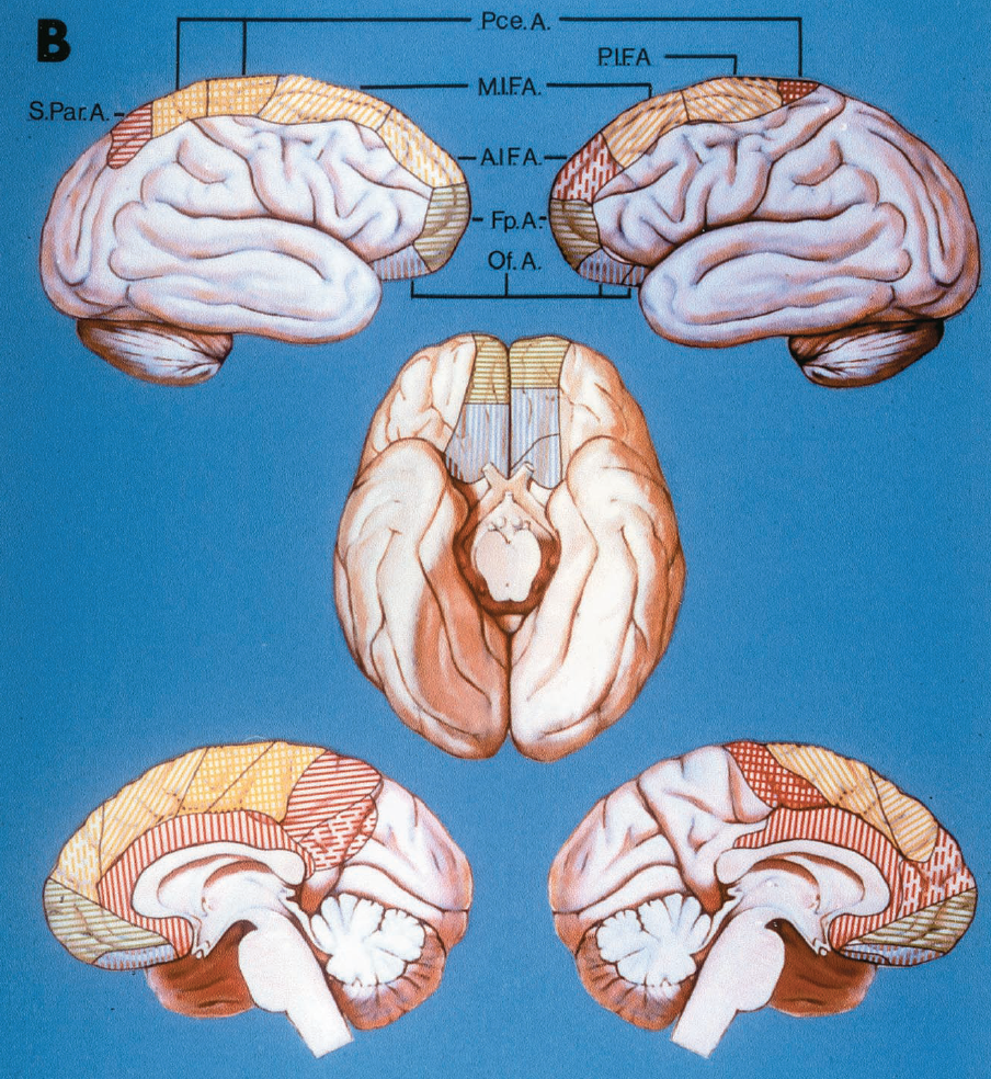
點擊這裏查看此圖像的交互模塊和相關內容。
圖2.27。B,右半球(左上、左下和基麵左半)顯示ACA異常大麵積供應,從頂枕裂延伸至楔形。額內後動脈缺失。胼胝體邊際動脈產生前、中、後額動脈和旁中心動脈(黃色區域)。黑線細分了中心旁動脈的交叉窩狀區域,顯示出由胼胝體周圍動脈發出的兩個獨立分支,供應中心旁動脈的區域。左半球(右上、右下和底圖右半部分)顯示ACA的供應麵積異常小。分支隻到達近中心區域。胼胝體邊際動脈起源於兩個皮層分支:額內動脈的中間和後部(黃色區域)。上、下壁動脈不可見。黑線分隔眼窩前額區(藍線),表示它是由胼胝體周圍動脈的兩個獨立分支提供的。
![圖2.27。C,右半球(左上、左下、基圖左半部分)顯示來自共同主幹的眶額動脈和額極動脈(均用綠色表示),來自胼胝體結節動脈的三個分支供應的相對後部區域(黃色區域),以及頂下動脈的缺失。左半球(右上、右下和基麵右半部分)顯示了兩個起源於胼胝體凹陷區(黃色)的皮層分支,頂上動脈不存在,頂下動脈存在。黑線細分了額後內動脈的區域,顯示出兩個獨立的分支從胼胝體周圍動脈發出,供應這一區域。一個,動脈;A.I.F.A,額前內動脈;《外交政策》。額極;我,自卑;M.I.F.A,額內中動脈; Of., orbitofrontal; Par., parietal; Pce., paracentral; P.I.F.A., posterior internal frontal artery; S., superior. (From, Perlmutter D, Rhoton AL Jr: Microsurgical anatomy of the distal anterior cerebral artery. J Neurosurg 49:204–228, 1978 [27].)](https://assets.neurosurgicalatlas.com/neuroanatomy/Rhoton_Book_-_Supratentorial/Figure_2.27_C.png)
點擊這裏查看此圖像的交互模塊和相關內容。
圖2.27。C,右半球(左上、左下、基圖左半部分)顯示來自共同主幹的眶額動脈和額極動脈(均用綠色表示),來自胼胝體結節動脈的三個分支供應的相對後部區域(黃色區域),以及頂下動脈的缺失。左半球(右上、右下和基麵右半部分)顯示了兩個起源於胼胝體凹陷區(黃色)的皮層分支,頂上動脈不存在,頂下動脈存在。黑線細分了額後內動脈的區域,顯示出兩個獨立的分支從胼胝體周圍動脈發出,供應這一區域。一個,動脈;A.I.F.A,額前內動脈;《外交政策》。額極;我,自卑;M.I.F.A,額內中動脈; Of., orbitofrontal; Par., parietal; Pce., paracentral; P.I.F.A., posterior internal frontal artery; S., superior. (From, Perlmutter D, Rhoton AL Jr: Microsurgical anatomy of the distal anterior cerebral artery. J Neurosurg 49:204–228, 1978 [27].)
ACA有大麵積的外側皮層分布,在那裏有很好的機會找到足夠直徑的血管,與顳淺動脈額支進行旁路吻合。通過在半球外周畫一條圓周線,從sylvian裂隙開始,繼續圍繞額極,並越過半球上緣向枕極,確定在外側表麵找到適當ACA分支的最佳機會區域。吻合所需的最小直徑通常被認為是0.8毫米(27)。在圓周線上2厘米處畫一條相同的線。ACA分支穿越這些線的最大百分比位於半球的前部,在圓周線上的5cm和15cm點之間。
ACA是供應胼胝體的主要動脈。胼胝體周圍動脈有分支,分別分布於骨前庭、膝、體和脾部,並常從脾部下方通過。終末胼胝體旁支由PCA的脾支向後連接。胼胝體最常見的是由穿孔分支供應,稱為短胼胝體動脈,因為它們起源於胼胝體周圍動脈,直接滲透到胼胝體。在一個半球中可發現多達20個短胼胝體分支(平均為7)(27)。這些分支不僅支配胼胝體,還通過胼胝體繼續支配透明隔、穹窿前柱和部分前連連。
在少數病例中,形成良好的較長的分支,稱為長胼胝體動脈,起源於胼胝體周圍動脈,並平行於胼胝體周圍動脈,在其與胼胝體表麵之間,形成胼胝體穿支(圖2.22)。除了向胼胝體輸送分支外,它們還可支配鄰近的皮層、隔核、透明隔和穹窿柱的上部(27)。胼胝體周圍動脈經常繼續圍繞胼胝體脾,遠端到最後一個皮質分支的起源,並在胼胝體下表麵向前,在少數情況下到達Monro孔。
胼胝體前動脈是一種罕見的A2或AComA分支,它像胼胝體周圍動脈和終板之間的一根長胼胝體動脈一樣向上延伸,分支進入前間腦,並向胼胝體的喙部和膝關節下部發出多個小分支。
在大約15%的大腦中發現遠端ACA異常,包括後交流段的三倍,遠端ACA配對失敗,以及雙半球分支(2)。雙半球分支是在AComA遠端分裂的分支,提供兩個半球的主要供應。在這種異常的情況下,阻塞AComA遠端的一個ACA可能會產生雙側腦損傷,類似於阻塞兩個ACA所產生的損傷。在近三分之二的大腦中,一側的遠端ACA向對側半球發送分支(圖2.28)。然而,大多數隻供應對側半球內側表麵的一小塊區域。一種罕見的異常是A1段遠端ACA未配對,而一個遠端ACA分裂供養兩個半球(26)。

點擊這裏查看此圖像的交互模塊和相關內容。
圖2.28。大腦半球的內側表麵。A,右半球;B,左半球。黑點表示對麵大腦前動脈分支到達大腦半球的位置。基於25個大腦的左右半球(來自,Perlmutter D, Rhoton AL Jr:遠端大腦前動脈的顯微外科解剖。中華神經外科雜誌(英文版)。
前穿支動脈是指通過前穿支物質進入大腦的一組動脈(圖2.29-2.31)。來自不同來源的前穿孔動脈與它們在大腦中央供應的重要血管和核之間的相互關係,使它們值得特別注意。這些動脈與頸內動脈、大腦中動脈和前動脈的動脈瘤以及大腦深處的腫瘤有著密切的關係(圖2.32)(31,35,36)。
前穿孔物是一個深埋於sylvian裂的菱形區域,前部為外側和內側嗅覺紋,後方為視神經束和顳葉,外側為島膜;向內側延伸至視交叉上方至半球間裂(圖2.29)。從下麵穿過並向前穿孔物質發送分支的動脈是ICA, MCA, ACA和AChA。每個母動脈的穿支動脈進入前穿支物質特定的中外側和前後區域。中外側方向的穿透部位與沿嗅束向後通過的一條線有關。這條線將前穿孔物質分為內側和外側區域,穿過前穿孔物質最大的前後尺寸附近,並在穿過腦梗時橫切視神經束。內側區域在視交叉上方延伸至半球間裂,外側區域延伸至sylvian裂至邊緣島。每組動脈的穿透位置在前後方向上也相對恒定,這是基於將前穿孔物質細分為前、中、後三個區域,從半球間裂延伸到腦島膜(圖2.29-2.31)。
進入前穿孔體的C4分支來自脈絡膜段(圖2.30,A和B)。脈絡膜段在幾乎100%的半球向前穿孔體發送分支(36)。這些分支傾向於起源更接近分叉而不是AChA的起源。在分叉處產生的分支往往比分叉下產生的分支更結實。通常情況下,這些C4分支沿後上路徑到達前穿孔物質的後部,靠近視神經束。大約一半的分支穿透前穿孔物的內側區域,一半穿透外側區域。大多數進入後區或中間區,很少進入前區。
AChA在90%的半球(13,33,36)向前部穿孔物質發送分支(圖2.30,C-F)。大多數分支沿後、上、內側通路,或直接沿後、上通路至前穿孔體。在AChA起源處出現的分支比遠端出現的分支略粗。這些分支進入前穿孔物質的後內側部分,靠近視神經束,並沿著嗅覺束將內側和外側區域分開。大約三分之二的分支進入內側,三分之一進入前部穿孔物質的外側區域。大多數進入前穿孔物質的後區或中間區相鄰部分。
前穿孔物質的分支,稱為透鏡狀紋狀動脈,起源於M1和M2和
偶爾從早期分支(圖2.30,G-J)。它們在每一種情況下都來自M1的分叉前部分,在一半的半球中來自M1段的分叉後部分。透鏡狀紋狀動脈分為內側、中間和外側三組。內側組出現在一半的腦半球,沿相對直接的路徑進入C4支外側的前穿孔體。90%的內側透鏡狀紋狀動脈進入前穿孔物質的外側區域,而隻有10%進入內側區域(36)。主要的模式是它們進入前穿孔物質的中間和後部區域。在透鏡狀紋狀動脈內側組缺失的半球中,透鏡狀紋狀動脈在前穿孔物質中的區域被C4、ACA、AChA和中間透鏡狀紋狀動脈的分支占據。
在90%以上的大腦半球中都有進入內側和外側紋狀透鏡體動脈之間的前穿孔物質的中間紋狀透鏡體動脈。它們在前穿孔物質的外側區域分布廣泛。近90%進入前穿孔物質的中間或後部區域,位於紋狀透鏡體內側和外側動脈之間,C4分支的外側,以及複發動脈分支的後部。
側透鏡狀紋狀動脈幾乎存在於所有腦半球,主要起源於M1的外側部分,但也可能起源於M1的早期分支或M2。它們沿s型路徑進入前穿孔物質的後外側。所有透鏡狀紋狀體外側動脈均進入前穿孔物質靠近島膜的外側區域,且幾乎全部進入前穿孔物質外側部分的後區。
大腦前動脈到前穿孔物質的分支有兩個來源。首先,A1產生直接通向前部穿孔物質的分支。其次,A1和A2的近端產生了再發動脈,其分支延伸到前部穿孔物質的很大範圍內(圖2.30,M-P)。幾乎所有的a1都向前部穿孔體發送分支。近90%起源於A1的近半部分,並直接向前部穿孔物質的後部和上部發展。起始點在內側,向外側到達前穿孔物質。大部分進入視交叉和視束附近前穿孔物質的內側區域,其餘進入外側區域。大多數進入前穿孔物質的中間和後部區域,主要是在複發動脈分支的後部,頸內動脈和脈絡膜前動脈分支的前內側,以及大腦中動脈分支的內側。
返動脈是通向前穿孔體的最大最長的分支。它存在於各個半球,向前部穿孔物質發送分支。複發分支進入前穿孔物的全部中外側範圍,但在前後維度有有限的表現。複發支所滲透的區域延伸到視交叉上方狹窄的內側區域,並延伸到外側區域,直到外膜島的內邊緣。與豐富的中外側表現相比,其前後分布有限,主要局限於前穿孔物質的前半部分。發源於A1外側的複發動脈分支比發源於A1和A2交界處的分支更傾向於進入中間和後部區域。由於其較長的中外側範圍,返動脈與所有進入前穿孔物質的其他組接壤。
總之,ICA和AChA分支進入前穿孔物質中央部分的後半部分。MCA進入前穿孔物質外側半部分的中間和後部。A1產生分支,進入視神經和交叉上方的前穿孔物質的內側。再發動脈的分支進入前穿孔物質全部中外側範圍的前三分之二。不同組之間在前穿孔物水平上有最小的吻合和有限的重疊,因此在該區域的手術入路中保護每一組都是最重要的。ICA、AChA、ACA和MCA的實質內和前穿孔物質區域之間存在互反關係,即一條動脈區域的大小以互反方式增加或減少另一條動脈的區域。
位於前部穿孔物質正上方的深層大腦結構是額角、尾狀核前部、殼核和內囊(23)。前穿支動脈穿過尾狀核、殼核和內囊的部分,直接位於前穿支物質上方,並向後方擴散,供應這些結構的較大部分以及白球和丘腦的鄰近區域(圖2.32)(39,pp 30-33)。穿透前穿孔物質的C4分支灌注內囊膝和蒼白球鄰近部分、內囊後肢和丘腦。AChA的分支進入前穿孔物質供應蒼白球的內側兩段,內囊後肢的下部,以及丘腦的前核和腹外側核。透鏡狀紋狀動脈外側組和中間組穿過殼核,向內側和後方拱起,幾乎供應內囊上部和尾狀核體及頭部的整個前後長度。內側紋狀透鏡體動脈灌溉外側和中間紋狀透鏡體動脈供給的內側及以下區域;該區域包括蒼白球外側部、內囊前肢上部、尾狀核頭前上部。
A1支提供前內側以下的區域,由透鏡狀紋狀動脈提供。該區域包括視交叉周圍區域、前連合、下丘腦前部、內囊膝和蒼白球前部。其供應區域可能很少延伸到內囊後肢的毗連部分和丘腦前部(26)。再循環動脈供應尾狀核和殼核頭部的前部和下部,以及內囊前肢的鄰近部分(26)。
進入前穿孔物質的動脈與頸內動脈、前交通動脈和大腦中動脈的動脈瘤有內在聯係,在手術中通常暴露。這些關係將在第三章中回顧。硬膜內暴露C4和所有向前穿孔物質發送分支的動脈可以使用以翼點為中心的小額顳皮瓣實現。所有這些與前穿支動脈相關的動脈瘤均可通過此入路沿同側蝶骨脊顯露,並打開側裂。選擇紋狀體動靜脈畸形,涉及進入前穿孔物質的動脈,已通過切開前穿孔物質和阻塞供血動脈而不產生缺損(圖2.16I)(41)。這些動靜脈畸形的手術治療通常隻考慮病變位於內囊膝前區域的前穿孔物質正上方,除非內囊膝和後肢已經受損。
![圖2.29。前穿孔物質的範圍和區域。前穿孔物質位於額葉和顳葉之間。前部由內側和外側嗅紋構成,後部由顳葉和視神經束構成。前穿孔物質被沿著嗅束向後畫的一條線分為內側和外側區域。內側區域在視神經和交叉上方延伸至半球間裂,外側區域在sylvian裂向外側延伸至邊緣島。前穿孔物也分為前、中、後三個橫帶,從半球間裂延伸到腦島邊緣,大致對應於前穿孔物的前、中、後三分之一。每條動脈穿過這些區域的位置都被記錄下來。內側和外側嗅紋與嗅束向前連續。螞蟻。, anterior; Interhem., interhemispheric; Fiss., fissure; Front., frontal; N., nerve; Olf., olfactory; Post., posterior; Temp., temporal; Tr., tract. (From, Rosner SS, Rhoton AL Jr, Ono M, Barry M: Microsurgical anatomy of the anterior perforating arteries. J Neurosurg 61:468–485, 1984 [36].)](https://assets.neurosurgicalatlas.com/neuroanatomy/Rhoton_-_Missed_Images/Supratentorial_2.29.jpg)
圖2.29。前穿孔物質的範圍和區域。前穿孔物質位於額葉和顳葉之間。前部由內側和外側嗅紋構成,後部由顳葉和視神經束構成。前穿孔物質被沿著嗅束向後畫的一條線分為內側和外側區域。內側區域在視神經和交叉上方延伸至半球間裂,外側區域在sylvian裂向外側延伸至邊緣島。前穿孔物也分為前、中、後三個橫帶,從半球間裂延伸到腦島邊緣,大致對應於前穿孔物的前、中、後三分之一。每條動脈穿過這些區域的位置都被記錄下來。內側和外側嗅紋與嗅束向前連續。螞蟻。, anterior; Interhem., interhemispheric; Fiss., fissure; Front., frontal; N., nerve; Olf., olfactory; Post., posterior; Temp., temporal; Tr., tract. (From, Rosner SS, Rhoton AL Jr, Ono M, Barry M: Microsurgical anatomy of the anterior perforating arteries. J Neurosurg 61:468–485, 1984 [36].)

點擊這裏查看此圖像的交互模塊和相關內容。
圖2.30 a-f .A,動脈進入前穿孔物質。頸內動脈、大腦前動脈和大腦中動脈、脈絡膜前動脈和返動脈向前部穿孔物發送分支。頸動脈分支位於脈絡膜前動脈起點的遠端,遠高於眼動脈和後交通動脈起點。大腦中支起源於M1和M2段。腦前分支,包括再回動脈,起源於A1和A2段。前穿孔物質在視交叉上方向內側延伸至半球間裂,向外側延伸至limen insulae,向前延伸至嗅覺紋,向後延伸至視神經束和顳葉。嗅道位於額葉的下表麵,位於直回和眶回的交界處。A和B是頸內動脈。A,從頸內動脈到前穿孔物的分支起於前脈絡膜動脈上方的後壁,並在頸動脈分叉後向上延伸。 Inset: lateral view of the carotid artery. Eighty-one percent of the branches to the anterior perforated substance arise from the posterior wall below the bifurcation and 18% arise from the posterosuperior surface of the wall, at or near the level of the bifurcation. B, internal carotid zone and territory in the anterior perforated substance. Most of the branches of the internal carotid artery enter the posterior and middle zones of the medial territory of the anterior perforated substance. C–F, anterior choroidal artery. Inferior views showing three patterns of origin (C–E). C, a branch to the anterior perforated substance arises in common with the origin of the anterior choroidal artery. D, the superior branch of the anterior choroidal artery gives rise to branches to the anterior perforated substance. E, the main trunk of the anterior choroidal artery gives off branches to the anterior perforated substance along its course to the choroid plexus in the temporal horn. F, the anterior choroidal branches enter the posterior and middle zones near the junction of the medial and lateral territories of the anterior perforated substance.

點擊這裏查看此圖像的交互模塊和相關內容。
圖2.30 G-L,大腦中動脈的透鏡狀紋狀分支。透鏡紋狀分支分為內側、中間和外側組。G和H,內側透鏡狀紋狀動脈。G,內側透鏡狀紋狀動脈起源於M1段的近端。插圖:這些動脈主要起源於動脈的後壁和上壁。H,它們進入前穿孔物質外側區域內側的中間和後部區域。I和J,中間透鏡狀紋狀動脈。I,這些動脈起源於M1節段,由於複雜的分支,在接近前穿孔物質時形成燭台狀外觀。插圖:它們主要出現在壁的後部、上部和後上部。J,動脈主要進入前穿孔物質外側區域中心部分的中間和後部。 K and L, lateral lenticulostriate arteries. K, these arteries arise in closer proximity to the bifurcation of the middle cerebral artery, from the M1 and M2 segments, and have an S-shaped course. First, they pass posterior, medial, and superior, then turn laterally, and finally complete an S-curve by turning medially just before entering the anterior perforated substance. Approximately half arise proximal and half arise distal to the bifurcation of the middle cerebral artery. Inset: site of origin. These arteries arise from the posterior, superior, or posterosuperior aspect of the parent trunk. L, the lateral lenticulostriate arteries enter predominantly the middle and posterior zones of the lateral territory of the anterior perforated substance.
![圖2.30 m-p。M和N,大腦前動脈A1段的穿支。M, A1分支起於前穿孔體內側下方,並向上延伸。A1段外側半段比內側半段有更豐富的穿孔分支。插圖:分支起源於A1節段的後表麵、上表麵或後上表麵。N, A1支進入視交叉上方的前穿孔物質狹窄帶。它們主要進入前穿孔物質內側區域的後區和中間區。O和P,再發動脈。O,多達4個複發動脈可能起源於大腦前動脈,或近端或近端AComA水平。它們在頸動脈分叉上方的外側穿過,並在前穿孔物質的整個中外側範圍內形成分支。 They may wander forward on the posterior part of the orbital surface of the frontal lobe. Inset (lower right): Site of origin of the recurrent arteries. Left inset: Recurrent artery origins near the junction of the A1 and A2 segments. The cross section of the artery at this level is oriented in a transverse plane. These branches arise predominantly from the lateral side of the vessel. Right inset: Site of origin of recurrent arteries arising from the A1 segments. The cross section of the artery at this level has an orientation in the sagittal plane. The branches arise predominantly from the superior or posterosuperior surface. P, the branches of the recurrent artery enter predominantly the anterior half of the anterior perforated substance along its full mediolateral extent, from the interhemispheric fissure to the limen insulae. A., arteries, artery; A.C.A., anterior cerebral artery; Ant., anterior; Car., carotid; Chor., choroidal; Comm., communicating; Fiss., fissure; Front., frontal; Gyr., gyrus; Inf., inferior; Int., intermediate; Interhem., interhemispheric; Lat., lateral; Len. Str., lenticulostriate; M.C.A., middle cerebral artery; Med., medial; N., nerve; Olf., olfactory; Ophth., ophthalmic; Orb., orbital; Perf., perforated, perforating; Post., posterior; Subst., substance; Sup., superior; Tr., tract, trunk; Temp., temporal. (From, Rosner SS, Rhoton AL Jr, Ono M, Barry M: Microsurgical anatomy of the anterior perforating arteries. J Neurosurg 61:468–485, 1984 [36].)](https://assets.neurosurgicalatlas.com/neuroanatomy/Rhoton_Book_-_Supratentorial/Figure_2.30_M-P.png)
點擊這裏查看此圖像的交互模塊和相關內容。
圖2.30 m-p。M和N,大腦前動脈A1段的穿支。M, A1分支起於前穿孔體內側下方,並向上延伸。A1段外側半段比內側半段有更豐富的穿孔分支。插圖:分支起源於A1節段的後表麵、上表麵或後上表麵。N, A1支進入視交叉上方的前穿孔物質狹窄帶。它們主要進入前穿孔物質內側區域的後區和中間區。O和P,再發動脈。O,多達4個複發動脈可能起源於大腦前動脈,或近端或近端AComA水平。它們在頸動脈分叉上方的外側穿過,並在前穿孔物質的整個中外側範圍內形成分支。 They may wander forward on the posterior part of the orbital surface of the frontal lobe. Inset (lower right): Site of origin of the recurrent arteries. Left inset: Recurrent artery origins near the junction of the A1 and A2 segments. The cross section of the artery at this level is oriented in a transverse plane. These branches arise predominantly from the lateral side of the vessel. Right inset: Site of origin of recurrent arteries arising from the A1 segments. The cross section of the artery at this level has an orientation in the sagittal plane. The branches arise predominantly from the superior or posterosuperior surface. P, the branches of the recurrent artery enter predominantly the anterior half of the anterior perforated substance along its full mediolateral extent, from the interhemispheric fissure to the limen insulae. A., arteries, artery; A.C.A., anterior cerebral artery; Ant., anterior; Car., carotid; Chor., choroidal; Comm., communicating; Fiss., fissure; Front., frontal; Gyr., gyrus; Inf., inferior; Int., intermediate; Interhem., interhemispheric; Lat., lateral; Len. Str., lenticulostriate; M.C.A., middle cerebral artery; Med., medial; N., nerve; Olf., olfactory; Ophth., ophthalmic; Orb., orbital; Perf., perforated, perforating; Post., posterior; Subst., substance; Sup., superior; Tr., tract, trunk; Temp., temporal. (From, Rosner SS, Rhoton AL Jr, Ono M, Barry M: Microsurgical anatomy of the anterior perforating arteries. J Neurosurg 61:468–485, 1984 [36].)
圖2.31。動脈進入前麵的穿孔物質。A,劣勢視角。左側的前穿孔物質向前延伸到內側和外側嗅紋,向後延伸到視神經束和顳葉,向外側延伸到外膜島,在視交叉上方的內側延伸到半球間裂。顳葉前部已被切除以暴露顳角。頸內動脈、脈絡膜前動脈、大腦前動脈和中動脈(M1和M2)形成了前穿孔物質的分支。PComA不為前部穿孔物質提供分支。大腦的中間分支,稱為紋狀透鏡體動脈,分為內側、中間和外側紋狀透鏡體組。外側透鏡狀紋狀動脈在M1段分叉附近外側出現。脈絡膜前支和頸動脈前支進入前穿孔物質的後部,靠近視神經束。 The branches from the anterior cerebral artery enter the narrow strip of the anterior perforated substance above the optic chiasm. The recurrent artery arises from the anterior cerebral artery, near the level of the AComA, and passes laterally above the carotid bifurcation to enter the anterior perforated substance anterior to the branches from the other sources. The M1 segment gives rise to an early branch. B, another specimen. The optic nerve and chiasm have been reflected inferiorly. The branches from the left A1 segment enter the narrow medial sector of the anterior perforated substance extending above the optic chiasm to the interhemispheric fissure. A perforating artery arises from an early branch of the M1. Some of the lateral lenticulostriate arteries arise near the M1 bifurcation. The intermediate lenticulostriate arteries have a candelabra appearance. The anterior choroidal artery sends branches to the posterior half of the anterior perforated substance. Two recurrent arteries arise near the anterior communicating artery. C, another specimen. The anterior choroidal branches to the anterior perforated substance arise near the origin of the anterior choroidal artery. The lateral lenticulostriate arteries arise near the M1 trifurcation and have a roughly S shaped course. The intermediate lenticulostriate arteries have a candelabra appearance. The medial lenticulostriate arteries pass near the perforating branches arising from the carotid artery and the medial half of the A1 segment. D, inferior view, right side. The intermediate lenticulostriate arteries have a candelabra appearance. The A1 branches enter the anterior perforated substance medial to those from the internal carotid, anterior choroidal, and middle cerebral arteries. The recurrent artery arises above the optic chiasm, passes laterally above the carotid bifurcation, and gives rise to branches that enter the anterior perforated substance in front of those from other sources. E, perforating branches of the anterior cerebral artery, anterior view. The recurrent artery arises above the optic chiasm near the level of the AComA. The A1 segment arises from the carotid artery and its perforating branches to enter the medial half of the anterior perforated substance in the narrow sector extending above the optic chiasm. A., arteries, artery; A.C.A., anterior cerebral artery; Ant., anterior; Bifurc., bifurcation; Car., carotid; Chor., choroid, choroidal; Comm., communicating; Br., branch; Fiss., fissure; Front., frontal; Gyr., gyrus; I.C.A., internal cerebral artery; Infund., infundibulum; Int., intermediate; Interhem., interhemispheric; Lat., lateral; Len. Str., lenticulostriate; Med., medial; N., nerve; Olf., olfactory; Orb., orbital; P.C.A., posterior cerebral artery; Ped., peduncle; Perf., perforated, perforating; Plex., plexus; Post., posterior; Rec., recurrent; Subst., substance, substantia; Temp., temporal; Tr., tract; Trifurc., trifurcation. (From, Rosner SS, Rhoton AL Jr, Ono M, Barry M: Microsurgical anatomy of the anterior perforating arteries. J Neurosurg 61:468–485, 1984 [36].)

點擊這裏查看此圖像的交互模塊和相關內容。
圖2.32 a-b .A,頸內動脈,前脈絡膜,大腦前動脈和中動脈的分支進入前穿孔物質的位置。前穿孔物質位於額葉和顳葉之間,前部與內側和外側嗅紋接壤,外側與島膜接壤,後方與視神經束和顳葉接壤,並在視神經和交叉上方向內側延伸至半球間裂。大腦前動脈A1段產生分支(藍色),這些分支進入視神經和交叉上方的前穿孔物質的內側。頸內動脈(紫色)和脈絡膜前動脈(紅色)形成分支,進入前穿孔物質中心部分的後部。大腦中動脈產生內側(棕色)、中間(橙色)和外側透鏡狀紋狀動脈(綠色),它們進入前穿孔物質外側半部分的中間和後部。再發動脈(黃色)分支進入前穿孔物質全部中外側範圍的前半部分。嗅覺束在直回和眶回之間劃分額葉。B,前穿支動脈與深部大腦結構的關係。右腦半球和左腦半球上部全部切除的上視圖。 The site of the anterior perforated substance is shown on both sides by dotted lines. The deep neural structures above the anterior perforated substance are shown on the left side. The transverse section of the left cerebrum extends through the caudate nucleus, thalamus, globus pallidus, putamen, the anterior limb, genu, and posterior limb of the internal capsule, and the frontal horn and atrium of the lateral ventricle. The right side shows the site of origin of the perforating branches to the anterior perforated substance. The branches to the anterior perforated substance pass through the deep structures directly above the anterior perforated substance and spread posteriorly to supply larger parts of the caudate nucleus, putamen, internal capsule, and the adjacent parts of the globus pallidus and thalamus. The C4 branches (purple) perfuse the genu of the internal capsule, and the adjacent part of the globus pallidus, posterior limb of the internal capsule, and thalamus. The anterior choroidal branches (red) supply the medial two segments of the globus pallidus, the inferior part of the posterior limb of the internal capsule, and the anterior and ventrolateral nuclei of the thalamus. The lateral (green) and intermediate groups (orange) of lenticulostriate arteries pass through the putamen and the adjacent part of the globus pallidus and arch medially and posteriorly (arrows) to supply almost the entire anterior-to-posterior length of the upper part of the internal capsule and the body and head of the caudate nucleus. The medial lenticulostriate arteries (brown) irrigate the lateral part of the globus pallidus, the superior part of the anterior limb of the internal capsule, and the anterosuperior part of the head of the caudate nucleus. The A1 branches (blue) supply the genu of the internal capsule and the anterior part of the globus pallidus, and may extend to the adjacent part of the posterior limb of the internal capsule and, less commonly, to the thalamus. The recurrent artery (yellow) supplies the most anterior and inferior part of the head of the caudate nucleus and putamen, and the adjacent part of the anterior limb of the internal capsule.
![圖2.32 C-D,前穿支動脈與沿蝶骨脊的腫瘤的關係。C,優越的視野。前穿支動脈橫跨蝶脊腦膜瘤的上表麵。D,翼點暴露。切口如圖所示。額葉和顳葉被收縮,露出被腫瘤包裹的頸動脈。前穿支動脈伸展於腫瘤的上表麵。最好的方法是在使用精細的顯微技術將腫瘤囊與穿支動脈分離之前先將這種類型的腫瘤減壓。A、動脈、動脈;螞蟻。, anterior; Cap., capsule; Car., carotid; Chor., choroidal; Fiss., fissure; Front., frontal; Gyr., gyrus; Int., intermediate, internal; Interhem., interhemispheric; Lat., lateral; Lent. Str., lenticulostriate; Med., medial; N., nerve; Nucl., nucleus; Olf., olfactory; Orb., orbital; Pall., pallidus; Perf., perforating; Post., posterior; Rec., recurrent; Temp., temporal; Tr., tract. (From, Rosner SS, Rhoton AL Jr, Ono M, Barry M: Microsurgical anatomy of the anterior perforating arteries. J Neurosurg 61:468–485, 1984 [36].)](https://assets.neurosurgicalatlas.com/neuroanatomy/Rhoton_Book_-_Supratentorial/Figure_2.32_C-D.png)
點擊這裏查看此圖像的交互模塊和相關內容。
圖2.32 C-D,前穿支動脈與沿蝶骨脊的腫瘤的關係。C,優越的視野。前穿支動脈橫跨蝶脊腦膜瘤的上表麵。D,翼點暴露。切口如圖所示。額葉和顳葉被收縮,露出被腫瘤包裹的頸動脈。前穿支動脈伸展於腫瘤的上表麵。在將腫瘤包膜與穿支動脈分離之前,最好先對這種類型的腫瘤進行減壓
顯微技術。A、動脈、動脈;螞蟻。前;帽,膠囊;的車。頸動脈;楚。脈絡膜的;費斯。, fissure; Front., frontal; Gyr., gyrus; Int., intermediate, internal; Interhem., interhemispheric; Lat., lateral; Lent. Str., lenticulostriate; Med., medial; N., nerve; Nucl., nucleus; Olf., olfactory; Orb., orbital; Pall., pallidus; Perf., perforating; Post., posterior; Rec., recurrent; Temp., temporal; Tr., tract. (From, Rosner SS, Rhoton AL Jr, Ono M, Barry M: Microsurgical anatomy of the anterior perforating arteries. J Neurosurg 61:468–485, 1984 [36].)
Willis圓的後部由近端PCA和PComA組成,它們在不同程度上共同為遠端PCA提供血流(圖2.8、2.33和2.34)。後圓是手術最難接近的部位之一,因為它位於第三腦室下方的中線,周圍有一係列複雜的穿孔血管,並由其產生,它與眼外神經和上腦幹有密切的關係。它的分支在基底尖、幕頂切跡、側腦室和第三腦室、顳下和內側頂枕區以及鬆果體區域的手術入路中暴露出來——所有這些都是相對難以接近的區域。
在大約一半的大腦中存在一個正常的後圓,其中近端pca直徑大於其pcoma且不發育不良(圖2.8和2.34)。另一半存在後圓異常,包括發育不良的PComA或胎兒形態,其中近端PCA較小,PComA為PCA提供主要供應,大於P1(24)。
動脈段發育不良是指直徑小於或等於1毫米的動脈段。在我們的研究中,單側發現PComA發育不全的比例為26%,雙側發現PComA發育不全的比例為6%,單側發現PCA主要起源於頸動脈的胎兒形態為20%,雙側發現PCA發育不全的胎兒形態為2%(37)。8%的人一邊有發育不良的通訊動脈,另一邊有胎兒複合體。通訊動脈或P1段缺失是非常罕見的。PComA已經在上麵描述過了。
威利斯環的後部向間腦和中腦輸送一係列穿通動脈,這些穿通動脈可能在鞍上腫瘤或後環動脈瘤周圍拉伸(圖2.33和2.34)。基底動脈上部的一些穿支與後圓的一些穿支重疊。在腫瘤或動脈瘤手術中阻塞這些重要的穿孔血管的風險包括視力喪失、體覺障礙、運動無力、記憶障礙、自主神經失衡、複視、意識改變、異常運動和內分泌紊亂。

點擊這裏查看此圖像的交互模塊和相關內容。
圖2.33 a-d .後脈絡膜動脈。A,大腦後動脈的下視圖。脈絡膜後內側動脈起於P1,環繞P2和P3內側的腦幹,沿其路線向腦幹發出小分支。P3由四叉神經池的分支組成。B、放大視圖。內側後脈絡膜動脈在腳側、周圍和四叉神經池內環繞腦幹,並在四叉神經池的鬆果體旁向前拐,到達第三腦室頂部。C,另一個標本中大腦後動脈的下視圖,第三腦室的底部被移除。內側後動脈環繞中腦,在四叉神經池內向前轉,到達第三腦室頂部。右側海馬旁回的內側部分被切除以暴露P2的分支。 D, enlarged view. The lower layer of tela in the roof of the third ventricle has been opened to expose the medial posterior choroidal arteries coursing in the velum interpositum with the branches of the internal cerebral vein. The choroid plexus in the body of the lateral ventricle is continuous at the posterior margin of the foramen of Monro with the choroid plexus in the roof of the third ventricle, which has been removed.

點擊這裏查看此圖像的交互模塊和相關內容。
圖2.33 e-h .;E,切除左海馬旁回的內側部分,暴露出來自P2的脈絡膜後外側動脈,穿過位於毛毛和丘腦之間的脈絡膜裂隙,到達顳角的脈絡膜叢。像丘腦膝狀動脈這樣的穿孔分支也起源於P2,並上升穿透丘腦膝狀體區域的下表麵。F,放大圖,切除海馬回下部,保留絨毛。P2向內側縮回,暴露出脈絡膜後外側動脈,它穿過位於陰毛和丘腦之間的脈絡膜裂隙,進入顳角的脈絡膜叢。脈絡膜前動脈也穿過裂隙。G,另一個樣本。M1、P1和P2產生了一係列進入大腦基麵的穿孔分支。P2被收縮暴露出脈絡膜後側分支通過脈絡膜裂到達顳角和心房的脈絡膜叢。海馬旁回已被切除。 The fimbria and thalamus border the choroidal fissure. The lateral geniculate body protrudes from the lower margin of the thalamus. H, enlarged view. The lateral posterior choroidal artery passes laterally through the choroidal fissure to reach the choroid plexus. The medial posterior choroidal encircles the brainstem. A., arteries, artery; A.Ch.A., anterior choroidal artery; Calc., calcarine; Car., carotid; Cer., cerebral; Chor., choroid, choroidal; Cist., cistern; CN, cranial nerve; Dent., dentate; Fiss., fissure; For., foramen; Gen., geniculate; Gyr., gyrus; Int., internal; Lat., lateral; Lent. Str., lenticulostriate; L.P.Ch.A., lateral posterior choroidal artery; M.C.A., middle cerebral artery; M.P.Ch.A., medial posterior choroidal artery; Parahippo., parahippocampal; P.Co.A., posterior communicating artery; Plex., plexus; Quad., quadrigeminal; Temp., temporal; V., vein; Vent., ventricle.
![圖2.34。基底動脈、小腦上動脈、P1、大腦後段、後交通動脈、頸內動脈和近端脈絡膜前動脈的上視圖。半色調顯示後穿孔體、乳頭體、視束、交叉和神經下方的動脈分支。第三和第四神經位於小腦上動脈和大腦後動脈之間。腦橋上部、後中腦、梗間窩、後穿孔物質、乳頭體、灰膜結節、視神經束和交叉的動脈分支來自基底動脈、P1、後交通動脈和頸內動脈。A、正常配置威利斯後半圓;p1均大於交通動脈,後者不發育不良(直徑大於1mm)。右小腦上動脈重複。最大的右P1分支產生丘腦切開動脈和後脈絡膜動脈。右側P1上隻有兩條穿通動脈。 The left posterior choroidal arises on P2. Both premamillary arteries (largest communicating trunk to premamillary area) arise from the middle third of the PComAs. AChAs arise as a single trunk. B, hypoplastic left communicating artery. Thalamoperforating artery arises on P1 medial to the posterior choroidal arteries on both sides. The left premamillary artery arises from the posterior and the right from the anterior portion of the PComA. The superior cerebellar arteries are duplicated on both sides. C, PComAs are hypoplastic bilaterally. The largest right P1 branch gives rise to both the thalamoperforating and the PChAs. The thalamoperforating artery arises medial to the posterior choroidal artery on the left P1. The premamillary artery arises from the anterior third of the right PComA and from the middle third on the left. The left anterior choroidal arises from the carotid as two trunks. D, fetal origin of the right posterior cerebral artery. The thalamoperforating artery on the right arises near the basilar bifurcation. The right posterior choroidal artery arises on P2. The left posterior choroidal artery arises medial to the thalamoperforating artery. The right premamillary artery arises from the anterior portion of the communicating artery. The left premamillary area is supplied by a group of nearly equal-sized arteries. The anterior choroidal artery bifurcates immediately after origin on the left. E, bilateral fetal origin of the posterior cerebral artery. The right posterior choroidal artery arises lateral to the thalamoperforating artery. The largest left P1 branch gives rise to the thalamoperforating and choroidal arteries. The right premamillary artery arises from the middle portion of the communicating artery. A premamillary arterial complex is present on the left. F, fetal type of right posterior cerebral origin and hypoplastic left communicating artery. The right posterior choroidal artery arises lateral to the well-developed thalamoperforating artery. No thalamoperforating branches are present on the left. The right premamillary artery arises from the anterior and the left from the posterior portion of the communicating artery. A., artery; A.C.A., anterior cerebral artery; A.Ch.A., anterior choroidal artery; B., body; B.A., basilar artery; C.A., carotid artery; Mam., mamillary; N., nerve; O., optic; P.C.A., posterior cerebral artery; P.Ch.A., posterior choroid artery; P.Co.A., posterior communicating artery; P.Perf.S., posterior perforated substance; Premam., premamillary; S.C.A., superior cerebellar artery; Th.Pe., thalamoperforating. (From, Saeki N, Rhoton AL Jr: Microsurgical anatomy of the upper basilar artery and the posterior circle of Willis. J Neurosurg 46:563–578, 1977 [37].)](https://assets.neurosurgicalatlas.com/neuroanatomy/Rhoton_Book_-_Supratentorial/Figure_2.34.png)
點擊這裏查看此圖像的交互模塊和相關內容。
圖2.34。基底動脈、小腦上動脈、P1、大腦後段、後交通動脈、頸內動脈和近端脈絡膜前動脈的上視圖。半色調顯示後穿孔體、乳頭體、視束、交叉和神經下方的動脈分支。第三和第四神經位於小腦上動脈和大腦後動脈之間。腦橋上部、後中腦、梗間窩、後穿孔物質、乳頭體、灰膜結節、視神經束和交叉的動脈分支來自基底動脈、P1、後交通動脈和頸內動脈。A、正常配置威利斯後半圓;p1均大於交通動脈,後者不發育不良(直徑大於1mm)。右小腦上動脈重複。最大的右P1分支產生丘腦切開動脈和後脈絡膜動脈。右側P1上隻有兩條穿通動脈。 The left posterior choroidal arises on P2. Both premamillary arteries (largest communicating trunk to premamillary area) arise from the middle third of the PComAs. AChAs arise as a single trunk. B, hypoplastic left communicating artery. Thalamoperforating artery arises on P1 medial to the posterior choroidal arteries on both sides. The left premamillary artery arises from the posterior and the right from the anterior portion of the PComA. The superior cerebellar arteries are duplicated on both sides. C, PComAs are hypoplastic bilaterally. The largest right P1 branch gives rise to both the thalamoperforating and the PChAs. The thalamoperforating artery arises medial to the posterior choroidal artery on the left P1. The premamillary artery arises from the anterior third of the right PComA and from the middle third on the left. The left anterior choroidal arises from the carotid as two trunks. D, fetal origin of the right posterior cerebral artery. The thalamoperforating artery on the right arises near the basilar bifurcation. The right posterior choroidal artery arises on P2. The left posterior choroidal artery arises medial to the thalamoperforating artery. The right premamillary artery arises from the anterior portion of the communicating artery. The left premamillary area is supplied by a group of nearly equal-sized arteries. The anterior choroidal artery bifurcates immediately after origin on the left. E, bilateral fetal origin of the posterior cerebral artery. The right posterior choroidal artery arises lateral to the thalamoperforating artery. The largest left P1 branch gives rise to the thalamoperforating and choroidal arteries. The right premamillary artery arises from the middle portion of the communicating artery. A premamillary arterial complex is present on the left. F, fetal type of right posterior cerebral origin and hypoplastic left communicating artery. The right posterior choroidal artery arises lateral to the well-developed thalamoperforating artery. No thalamoperforating branches are present on the left. The right premamillary artery arises from the anterior and the left from the posterior portion of the communicating artery. A., artery; A.C.A., anterior cerebral artery; A.Ch.A., anterior choroidal artery; B., body; B.A., basilar artery; C.A., carotid artery; Mam., mamillary; N., nerve; O., optic; P.C.A., posterior cerebral artery; P.Ch.A., posterior choroid artery; P.Co.A., posterior communicating artery; P.Perf.S., posterior perforated substance; Premam., premamillary; S.C.A., superior cerebellar artery; Th.Pe., thalamoperforating. (From, Saeki N, Rhoton AL Jr: Microsurgical anatomy of the upper basilar artery and the posterior circle of Willis. J Neurosurg 46:563–578, 1977 [37].)
PCA起於基底分叉,與位於腦梗間池側緣的PComA相連,環繞腦幹,通過腳側和周圍池到達四叉神經池,分布於腦半球的後部(圖2.1、2.3、2.7-2.9、2.12、2.13、2.33和2.34)。大腦後動脈顧名思義,不僅供應大腦半球的後部,還向丘腦、中腦和其他深層結構(包括脈絡膜叢和側腦室和第三腦室壁)發送關鍵分支。在胚胎學上,它是頸內動脈的分支,但在出生時,它最常見的起源是基底動脈。基底分叉,也就是PCA的起點,可以遠至腦橋交界處以下1.3 mm的尾側,遠至乳頭體和鄰近的第三腦室底,高分叉可以抬高第三腦室。該動脈通常分叉於花序梗間窩對麵,但有些分叉可能低至腦橋上部或高至擠壓乳頭體和第三腦室底部。基底分叉和乳頭體之間的平均距離為8.1 mm(範圍0-14 mm)。16%的病例基底動脈分叉處變寬,使基底動脈頂端和分叉呈眼鏡蛇狀(37,43)。
PCA分為四個段,P1到P4(圖2.12-2.14和2.33)(37,43)。
P1段,也稱為預溝通段,從基底分叉延伸到與PComA的交界處。以P1直徑小於PComA和PCA為主的胎兒形態
從頸動脈開始,發生在大約三分之一的半球。一個正常的結構,其中P1段大於PComA,在近三分之二的半球中被發現。一些半球的PComA和P1的直徑相同。胎兒形態可能出現在兩側。
P1的長度不同,如果有胎兒模式則更長。平均P1長度為3 - 14毫米,胎兒形態組的P1長度約為9.0毫米,而正常模式組的P1長度為7.0毫米(37)。如果形態正常,動眼神經在PComA下方和稍外側通過;但如果存在胎兒模式,P1較長,神經位於通訊動脈下方或內側。
P1產生的相對恒定的分支有:1)丘腦穿支動脈,根據定義,它通過後穿孔物質進入大腦;2)脈絡膜後內側動脈,通往第三腦室和側腦室的脈絡膜叢;3)分支至四邊形板塊;和4)支到腦梗和中腦被蓋。小腦上動脈(SCA)起源於基底動脈,位於P1起始點和7mm以下(平均2.5 mm)之間(37)。SCA可能很少與P1有共同的起源或產生於P1。起始段產生穿孔血管,其末端可與基底尖和P1產生的穿孔血管重疊。
P2段開始於PComA,位於腳池和周圍池內,並終止於中腦後緣外側。P2分為前半和後半部分,因為該節段的前半和後半部分的手術入路經常不同,因為它有助於識別來自P2的許多分支的起源。前部被稱為P2A或腳梗段,因為它在腳池中繞過腦梗。後部被稱為P2P或周圍或外側中腦段,因為它在周圍池的中腦外側。兩段大約25毫米長。P2A從PComA開始,在形成腳池內側壁的腦梗和鉤突之間,在穿過池頂的視神經束和基底靜脈下方,進入周圍池的近端。P2P開始於腦梗的後邊緣,在腳池和周圍池的交界處。它穿行於外側中腦和海馬旁回和齒狀回之間,形成周圍池的內側和外側壁,在視神經束、基底靜脈和膝狀體下方和池頂的枕骨下外側部分,以及滑車神經和幕邊的超內側。
P3或四叉神經段從中腦外側表麵和周圍池的後緣向後方延伸,到達四叉神經池的外側部分,結束於鈣質裂的前極限。在到達鈣質裂隙的前極限之前,PCA通常分為主要的終末分支,鈣質動脈和頂枕動脈。P3節段平均長度為2cm。兩邊的p3在丘後方相互靠近。每一側的pca最近的點被稱為丘點或四邊形點。分離平均8.9mm(範圍3.5-17 mm)(43)。形成丘點的動脈在大約一半的半球中是PCA幹,在另一半半球中,PCA在到達丘點之前分叉成其末端分支,由鈣質動脈形成。
P4節段包括分布於皮層表麵的分支。在後方,它開始於鈣質溝的前端。
PCA產生了三種類型的分支:1)通往間腦和中腦的中央穿孔分支;2)腦室分支至脈絡叢、側腦室和第三腦室壁及鄰近結構;3)通向大腦皮層和胼胝體脾的腦分支(圖2.33)。中心分支包括直穿動脈和旋穿動脈,包括丘腦穿動脈、椎梗穿動脈和丘腦膝狀動脈。腦室分支是後脈絡膜外側和內側動脈。腦支包括顳下支組,分為海馬支、顳前支、顳中支、顳後支和顳總支,再加上頂枕支、鈣支和脾支。
旋長動脈和旋短動脈和丘腦穿通動脈主要起源於P1,其他PCA分支最常起源於P2或P3。海馬動脈、前顳動脈、脈絡膜蒂穿支和內側後脈絡膜動脈最常發源於P2A。顳中動脈、後顳動脈、顳總動脈和後脈絡膜外側動脈最常發源於P2P。丘腦彎狀動脈在點對點出現的頻率僅略高於P2A。鈣質動脈和頂枕動脈最常發源於P3。
中心分支分為兩組:直接穿支和旋支(圖2.34和2.35)。直接穿支直接從主幹到腦幹。這一組包括丘腦手術
來自P1的動脈以及來自P2的丘腦膝狀動脈和花梗穿支動脈。彎曲的分支在進入間腦之前環繞腦幹一段可變的距離,而中腦則分為長組和短組,這取決於它們在腦幹周圍運動的距離。
平均有4個,但多達12個穿孔分支,其中最大的直徑可達1.5 mm,主要起源於P1的上表麵和後表麵,上表麵和後表麵,並分為許多分支,終止於梗間窩、後穿孔物、腦梗、乳頭體和後腦。穿孔分支很少出現在基底尖的前側,但在三分之一的p1中出現在前表麵,並終止於後穿孔體和乳頭體。P1最大的分支是丘腦穿孔動脈(占大腦半球的42%),後脈絡膜動脈(40%),或兩條動脈都起源於的大幹(18%)(37)。分支較大的p1具有較少的穿孔分支。隻有一個或兩個P1穿孔的P1往往具有較大的分支。如果最大的P1分支相對較小,則會有更多的P1分支。P1最大穿支的外側比內側有更多的穿支血管。
基底動脈上厘米的後表麵和外側表麵也是穿孔動脈的豐富來源,這些穿孔動脈與P1的穿孔動脈重疊。平均有8個(範圍3-18個)枝條從上麵的厘米長出來;大約一半來自後表麵,四分之一來自兩側(37)。更內側的分支,稱為正中或旁正中分支,進入中線附近的中腦和腦橋,而外側的分支,稱為橫向或周向分支,終止於外側腦橋、腦梗和後穿孔物。

點擊這裏查看此圖像的交互模塊和相關內容。
圖2.35 a-d .P1和PComA的穿孔分支。A,來自P1段的丘腦手術動脈的上視圖。左丘腦穿孔動脈比右動脈大。A1s的內側部分被切除,以提供基底尖的視圖。B,另一個基底分叉的下視圖。這兩個p1都是丘腦穿通動脈叢進入花序梗間窩的重要組成部分。右側AChA在PComA上方和外側,並在uncus上方外側轉彎。MPChA產生於右側P1。C,來自P1的thalamoperating動脈的上視圖。 The basilar artery, below the P1 origins, also send perforating branches in the same area. D, inferior view. The right P1 is much smaller than the left P1, but the right P1 gives rise to a tuft of thalamoperforating arteries that pass upward to enter the interpeduncular fossa. A nerve hook holds up a premamillary branch of the PComA.

點擊這裏查看此圖像的交互模塊和相關內容。
圖2.35 e-h .;E,這兩個p1都比pcoma小,但是這兩個p1都產生了thalamoperating動脈。較小的(左側)P1比較大的(右側)P1產生更多更大的穿孔動脈。F,解剖器支撐起兩個穿支,這兩個穿支起於小腦上動脈的起點,並通過與丘腦穿支動脈相同的區域進入大腦。G,左側PCA為PComA的胎兒起源。一係列穿通動脈起於PComA,並在乳頭體區域和第三腦室底部進入視神經束內側的間腦。P1沿著曲折的路線到達與P2.H的交界處,劣勢視圖。p1的外側部分形成丘腦穿通動脈。穿孔分支也產生於PComA。一個,動脈; A.Ch.A., anterior choroidal artery; Bas., basilar; Car., carotid; CN, cranial nerve; Dup., duplicate; M.P.Ch.A., medial posterior choroidal artery; P.Co.A., posterior communicating artery; Perf., perforating; Pit., pituitary; S.C.A., superior cerebellar artery; Thal. Perf., thalamoperforating; Tr., tract.
丘腦穿支動脈起於P1,通過後穿支體和腦梗內側進入大腦,位於腦梗間窩上部的乳狀體後方(圖2.35)。進入同一區域的PComA分支稱為乳頭前動脈。大部分thalamoperating動脈起源於P1的中間三分之一,作為最靠近基底分叉的P1分支,但它們也可能起源於內側或外側三分之一。如果第一分支不是丘腦穿支,則為旋支,終止於椎梗或後中腦區。在大多數情況下,丘腦穿孔動脈是P1最大的分支(37)。它們幾乎總是出現在P1的後側麵或上側麵,很少出現在前表麵。P1即使大小正常或較大,也可能很少不產生丘腦穿孔動脈,在這種情況下,對側有發育良好的丘腦穿孔分支,供應正常由缺失的丘腦穿孔動脈灌注的區域。它們供應後丘腦和下丘腦的前部和部分,下丘腦和中腦上部的內側部分,包括黑質、紅核、動眼核和滑車核、動眼神經、中腦網狀結構、前蓋、第四腦室的前內側底和內囊的後部(39,pp 96-99;43)。
與這些動脈缺失相關的缺陷包括:內側丘丘或丘腦的傳入通路受累引起的體覺障礙;由於內囊或椎梗的皮質脊髓束受累而引起的運動無力;由進出乳腺體的下丘腦通路所引起的記憶缺陷;前間腦和後間腦交感神經和副交感神經中樞紊亂引起的自主神經失衡;中腦的眼外神經或核核受累引起的複視;中腦網狀結構缺血引起的意識改變;神經紊亂:由於中腦和丘腦的小腦丘腦回路受到影響而引起的異常運動;丘腦開孔動脈的阻塞,取決於缺血區域的大小,可能會產生各種更多的局灶性綜合征,包括對側偏癱,小腦共濟失調,或與同側動眼神經麻痹相關的“紅”震顫(Nothnagel綜合征)。如果病變影響到下丘腦,可能會產生對側偏癱,隨著時間或治療,偏癱會減弱為舞蹈動作(43)。
腦梗穿支,通常為2 - 3支,最多可達6支,起於P2段,並直接從PCA進入腦梗。它們供應皮質脊髓和皮質球通路,以及黑質、紅核和被蓋的其他結構,並可能向動眼神經發送分支。
彎曲的動脈群起於P1和P2,並環繞中腦平行和內側的PCA。它們分為短旋組和長旋組。短的旋枝隻達到膝曲體。彎曲的長枝到達丘頂。短旋動脈在P2和後脈絡膜內側和長旋動脈的內側走行,並在其遠端終止時向腦梗發送分支,其範圍可從腦梗的後外側邊界到內側膝狀體。由P2產生的隻供應膝狀體和中腦被蓋。短旋動脈可將支束送到椎梗間窩和後穿孔物區域,主要由丘腦穿通動脈供應(37)。
長而彎曲的動脈,稱為四叉神經動脈,幾乎分布在大腦的所有半球,繞過腦幹到達四叉神經池,並為四叉神經體供血。它們環繞中腦主脊骨內側,並向腦梗和膝狀體發送小支,偶爾也向被蓋、枕部發送小支,並在四邊形板結束。通常發生於P1或P2A。長旋支的末端分支在丘上形成豐富的動脈網絡,在那裏它們與小腦上動脈的分支吻合。上丘由PCA的分支提供,下丘由小腦上動脈的分支提供。長旋動脈(四頭動脈)閉塞可導致後連接或Darkschewitsch或Cajal核(Parinaud’s綜合征)梗死引起的垂直規缺損(40)。
丘腦膝狀動脈直接起源於外側丘腦下方的P2,並穿透由膝狀體和周圍區域形成的周圍池的頂部部分。PCA最常見的起源為2或3條丘腦彎狀動脈,但也可能多達7條。它們出現在腳(P2A)和周圍(P2P)節段的交界處附近,每個節段產生的數量幾乎相等。
丘腦膝狀動脈供應外側丘腦的後半部分、內囊的後肢和視神經束(39,pp 96-99)。它們與靠近丘腦中部的P1的丘腦操作分支和前部側核內PComA的丘腦分支相遇。長、短旋和內側脈絡膜後動脈在環繞腦幹時也有分支到這一區域,但丘腦膝狀動脈這個術語是指那些起源於P2並穿過膝狀體和周圍池頂部相鄰部分的分支。
丘腦膝狀動脈供血區域的梗死可導致Dejerine和Roussy丘腦綜合征,包括對側淺表感覺喪失,特別是深部感覺喪失,患側出現強烈、難治性、過度疼痛,對輕度觸摸、疼痛和溫度刺激極度過敏,對側偏癱,通常是短暫性的,有時與癱瘓側的舞蹈運動或肌張力障礙運動有關。可能伴有同型偏盲(7,22)。通常有永久性的深層感覺障礙(體位感、重接觸和深壓),盡管皮膚刺激的閾值升高,但閾值刺激會引起不愉快的灼燒感、痛苦類型的疼痛反應,並可能有自發疼痛。四肢比臉部受影響更大。
在1906年報道的一例此類病例中,Dejerine和Roussy(7)發現丘腦外側核的後三分之一、內側核和中心核的部分、枕骨、內囊的後肢和慢狀核的後部都有梗死,但他們沒有發現任何PCA分支的閉塞。事實上,該區域不僅由多個丘腦膝狀動脈供應,而且還由PCA的回旋分支和脈絡膜分支供應,這使得單個丘腦膝狀動脈閉塞不太可能產生完整的綜合征。它更有可能是由所有這些分支的起源近端PCA閉塞引起的。動脈閉塞是典型丘腦綜合征的最常見原因,盡管丘腦血管畸形或腫瘤也可能是原因之一(43)。
脈絡膜後動脈是PCA的分支,進入側腦室和第三腦室,供給脈絡膜叢和腦室壁,根據供給的來源和區域,分為內側和外側兩組,稱為內側後動脈(MPChA)和外側後脈絡膜動脈(LPChA)(圖2.12、2.13和2.33)(13)。mpcha最常出現於PCA近半部分或其分支的後內側,環繞中腦PCA主幹的內側,在鬆果體外側前轉,進入丘腦之間的第三腦室頂部,最後通過脈絡膜裂和Monro孔,進入側腦室的脈絡膜叢。MPChAs沿其路線向花序梗、被蓋、膝狀體(內側和外側,但主要是前者)、丘、枕葉、鬆果體和內側丘腦發送分支。
大多數大腦半球隻有一個MPChA,但也有可能多達三個(43個)。大多數發生於P2,但也可能發生於P3或頂枕支和鈣質支。從頂枕動脈和鈣質動脈以及遠端PCA開始逆行進入第三腦室頂部的mpcha。
LPChAs起源於PCA或其分支,並向外側通過脈絡膜裂供應側腦室的脈絡膜叢。一個半球的lpcha數量從1到9(平均為4)(13)。它們通常直接來自P2P,但也可能來自P2A或P3,或來自一些PCA分支。最大的lpcha直接產生於周圍池的P2P,橫向通過脈絡膜裂隙到達顳角脈絡膜叢和心房脈絡膜叢血管球,並在AChA和MPChA分支內的脈絡膜叢上吻合。lpcha可向腦梗、後連連、穹窿腳部和主體的部分、外側膝狀體、枕葉核、背內側丘腦核和尾狀核主體發送分支(13,43)。
PCA的皮質分支包括顳下支、頂枕支、鈣質支和脾支(圖2.36和2.37)。

點擊這裏查看此圖像的交互模塊和相關內容。
圖2.36。大腦後動脈。A, P2分為P2A,穿過位於鉤突後段和腦梗之間的腳池,和P2P,穿過位於外側中腦和海馬旁回下方的周圍池。P3穿過四叉神經池,在那裏產生由皮層分支形成的P4,包括頂枕動脈和鈣質動脈,它們在頂枕溝和鈣質溝中運動,通常隱藏在溝唇之間。B,回縮頂枕溝和鈣質溝的唇部,露出沿溝延伸的頂枕溝和鈣質溝分支。MPChA環繞腦幹到達第三腦室頂。楔形形成上唇,舌形成鈣質溝的下唇。楔前葉形成上唇,楔前葉形成頂枕溝的下唇。C,另一個半球。PCA的末梢分支在頂枕溝和鈣質溝的後方通過。 The arrows are on branches that pass around the occipital pole to reach the adjacent lateral surface. D, the lips of the parieto-occipital and calcarine sulci have been retracted. The parieto-occipital artery courses within its sulcus. The calcarine artery courses just below the calcarine sulcus and gives rise to several small branches that course along the depths of the sulcus. E, posteroinferior view of occipital pole showing the branches (red arrow) of the PCA coursing around the occipital pole to reach the adjacent part of the lateral convexity. F, posterior view of both occipital lobes. The P4 branches course around the posterior and lower border of the occipital lobe to reach the lateral cortical surface. The P3s course on the quadrigeminal cistern. A., artery; A.C.A., anterior cerebral artery; Calc., calcarine; Car., carotid; Cist., cistern; M.P.Ch.A., medial posterior choroidal artery; P.Co.A., posterior communicating artery; Par. Occip., parieto-occipital; Quad., quadrigeminal; Sag., sagittal; Splen., splenial; Str., straight; V., vein.

點擊這裏查看此圖像的交互模塊和相關內容。
圖2.37 a-f .大腦的側麵、中間和基礎視圖,用顏色編碼的扇區代表特定的PCA皮層分支分布。每個PCA分支對應的顏色代碼如下:紅色,海馬動脈;黃色是顳動脈;綠色,鈣質動脈;藍色是頂枕動脈。顳動脈進一步細分:橫黃色條紋,顳前動脈;垂直的黃色條紋,顳總動脈;斜條紋,向上向右,顳前動脈;斜條紋向下向右,是顳後動脈。 The most common pattern (44% of hemispheres) is represented on the right cerebral hemisphere (A and D, and the left half of the basal view, C). This pattern includes hippocampal, anterior temporal, and posterior temporal arteries. The cortical distribution of the parieto-occipital artery is larger than that of the calcarine artery. The second most-common pattern (20% of hemispheres) is represented on the left cerebral hemisphere (B and E, and the right half of the basal view, C). This pattern includes anterior, middle, and posterior temporal, calcarine, and parieto-occipital arteries. In this pattern the anterior temporal artery supplies the region usually supplied by the hippocampal artery. The third most-common pattern (16% of hemispheres) is shown on the right hemisphere (F and I, and left half of basal view, H). In this pattern, there is a common temporal artery that supplies the entire inferior surface of the temporal lobe. The calcarine and parieto-occipital arteries are also present.

點擊這裏查看此圖像的交互模塊和相關內容。
圖2.37 g-l .;鈣質動脈和頂枕動脈也存在。第四種最常見的模式(10%的半球)描繪在左半球(G和J,以及基麵右半部分,H)。這種排列包括前、後顳動脈、鈣質動脈和頂枕動脈,但沒有PCA的海馬或顳中分支。鈣質動脈的區域被分成兩個扇區,以說明有兩個鈣質動脈起源於PCA,發生在10%的半球。
![圖2.37 m-o。第五種最常見的模式(占大腦半球的10%)顯示在大腦右半球(K和N,以及基本視圖的左半部分,M)。這種模式包括海馬、前、中和後顳、鈣質動脈和枕頂動脈。後顳動脈所提供的區域被分成兩部分,以表明兩個後顳動脈起源於PCA,在6%的大腦半球中出現。頂枕動脈支配內側表麵的大部分。最後一種模式顯示了一些顯著的變異(L和O,以及基礎視圖右半部分,M)。兩條海馬動脈起源於PCA,在12%的大腦半球中都有發現。顳前動脈在顳前表麵和顳外側表麵的供血量比平時少,其餘部分由大腦中動脈供血。鈣質動脈在內側表麵供應異常大的麵積。摘自,Zeal AA, Rhoton AL Jr:大腦後動脈顯微外科解剖。中華神經外科雜誌48:534-559,1978[43]。](https://assets.neurosurgicalatlas.com/neuroanatomy/Rhoton_Book_-_Supratentorial/Figure_2.37_M-O.png)
點擊這裏查看此圖像的交互模塊和相關內容。
圖2.37 m-o。第五種最常見的模式(占大腦半球的10%)顯示在大腦右半球(K和N,以及基本視圖的左半部分,M)。這種模式包括海馬、前、中和後顳、鈣質動脈和枕頂動脈。後顳動脈所提供的區域被分成兩部分,以表明兩個後顳動脈起源於PCA,在6%的大腦半球中出現。頂枕動脈支配內側表麵的大部分。最後一種模式顯示了一些顯著的變異(L和O,以及基礎視圖右半部分,M)。兩條海馬動脈起源於PCA,在12%的大腦半球中都有發現。顳前動脈在顳前表麵和顳外側表麵的供血量比平時少,其餘部分由大腦中動脈供血。鈣質動脈在內側表麵供應異常大的麵積。摘自,Zeal AA, Rhoton AL Jr:大腦後動脈顯微外科解剖。中華神經外科雜誌48:534-559,1978[43]。
顳下動脈來自PCA顳上動脈來自MCA。顳下動脈包括海馬和前、中、後和顳總動脈。這些動脈分布在顳葉的下部。顳下動脈的分支繞過大腦半球的下緣,到達大腦外側表麵,在42%的大腦半球中到達顳中回(43)。它們也會產生一些lpcha。顳下動脈根據存在的分支和它們所支配的區域分為五組:
海馬動脈,如果存在,出現在腳側或周圍池,是PCA的第一個皮質分支。它
供應鉤扣,前海馬旁回,海馬形成,和齒狀回。一個小分支可以延伸到顳葉的外側表麵,並向前延伸到顳尖。如果第一個皮層分支除海馬回外還支配顳下葉的很大一部分,則該分支被歸類為顳前動脈。由海馬動脈供血的顳內側區域的雙側血管閉塞可導致嚴重的記憶喪失和類似Korsakoff綜合征的缺陷(43)。
顳前動脈通常是PCA皮層的第二個分支。如果沒有海馬動脈,它就是第一個分支。它通常起於周圍池近端,供應顳葉前下表麵,偶爾到達部分顳極和顳中溝和回區域的腦外側表麵。
這條動脈起源於腳側和周圍池並支配顳葉的下表麵。它是最小的,經常沒有,並且有最少的分支的顳下動脈。
這條動脈幾乎存在於所有腦半球,起源於PCA的下側或外側,最常見的是在周圍池,但偶爾在腳池或四頭池,並斜向後外側向枕極延伸,供應顳下麵和枕麵,包括枕極和舌回。在所有顳動脈中,它的主幹直徑最大,分支數量最多,隻有一條普通的顳動脈是所有顳動脈分支的來源。後顳動脈閉塞後的缺陷包括語言障礙,通常是輕度和短暫性的,這是一種遺忘綜合征,通常是短暫性的,伴有同義性偏盲,但沒有偏癱或感覺喪失,無法將顏色與他們的名字匹配(21)。
顳總動脈在略少於20%的大腦半球中可見,起源於腳側或周圍池,作為單一的PCA分支,支配顳葉和枕葉下表麵的大部分。
頂枕動脈是PCA的兩個末端分支之一,幾乎分布在所有的半球。它始終作為一個分支出現,並在頂枕裂中運行,支配後矢狀旁區、楔形、楔前葉、枕外側回,很少支配中央前小葉和頂上小葉。它發生在周圍池或四叉神經池。起源較近的動脈往往較大,當它們在海馬裂內後方通過時,向中腦、丘腦、枕葉和外側膝狀體捐贈分支。這些近端動脈也有分支通過脈絡膜裂到達側腦室的脈絡膜叢。這條動脈偶爾在MPChA供血區域的第三腦室或胼胝體的脾部有分支。
鈣質動脈,終末分支,存在於所有半球。它在鈣裂內延伸至枕極,並有分支向舌回和下楔形肌扇形伸展。它通常直接起源於周圍或四叉神經池的PCA,但偶爾是頂枕動脈的分支。鈣質動脈供應視覺皮層,該血管閉塞的標誌是視場缺損,通常伴有黃斑保留。閉塞可能與同側眼疼痛有關。雙側枕葉梗死可導致保留瞳孔反射的失明或Anton綜合征,其中包括皮質盲、幻覺、否認失明和保留瞳孔對光的反應。結紮或封堵鈣質動脈後視野可恢複(19)。
PCA,或其分支,在各個半球產生分支,供應胼胝體的脾。它們可能起源於以下動脈:頂枕動脈、鈣質動脈、內側後脈絡膜、後顳動脈和外側後脈絡膜。如前所述,脾動脈與胼胝體周圍動脈的分支在脾後尖前幾厘米處吻合。這條動脈通過胼胝體周圍動脈逆行充盈提示主動脈在脾動脈起點近端閉塞。顯性枕極的梗死(產生偏盲)加上脾動脈分布中的胼胝體脾,中斷了完好的枕極和對側角回之間的纖維,導致無書寫困難的閱讀障礙綜合征(43)。
PCA的所有皮質分支均可向半球外側表麵發送分支,但在7條皮質動脈中,後顳動脈是外側皮質分支最常見的起發部位。其次是頂枕動脈。如果在顳淺動脈或枕動脈與PCA皮質支之間進行微血管吻合的血管重建手術,由顳後動脈提供的區域最有希望發現足夠口徑的血管作為受體,在該區域內發現足夠大小的血管的幾率高於75%(43)。這與枕前切跡正前方的區域相對應。PCA的大部分皮質分支直徑為0.4 - 0.6毫米,它們環繞邊緣到達大腦外側表麵。
動眼神經和滑車神經與PCA和SCA之間的關係是恒定的(圖2.1和2.3)(32)。動眼神經始終貫穿於主動脈和SCA之間的原點附近,滑車神經貫穿於兩者之間的腦幹側緣。即使小腦上起點重複,這種關係也不會改變。當SCA作為重複中繼出現時,神經在SCA的上級中繼和PCA之間傳遞。PCA始終在滑車上方運動。彎曲的SCA偶爾會在滑車神經上方循環。
PCA比任何其他顱內血管更能輔助視覺功能。它支持一長串的眼部功能,包括乳頭反射、眼運動、視覺記憶、視覺信息的半球內傳遞、雙眼和視覺空間整合,通過它對視神經束、膝狀體、丘、眼外神經及其核、膝狀突束、紋狀和周狀皮層的供應。由閉塞引起的功能障礙
單獨的PCA分支已經在與這些分支相關的小節中進行了審查。各種分支的阻塞也可能導致內側丘板或丘腦傳入通路受累引起的軀體障礙,內囊或椎梗皮質脊髓束受累引起的運動無力,進出乳頭體的下丘腦通路受累引起的記憶障礙,前間腦和後間腦交感神經和副交感神經通路受累引起的自主神經失衡。中腦網狀結構缺血引起的意識改變,中腦和丘腦小腦丘腦回路受累引起的異常運動,下丘腦垂體軸受累引起的內分泌紊亂。
垂體手術的血管並發症主要是頸動脈損傷和頸動脈閉塞後的循環障礙。在討論垂體手術並發症時,後圓穿孔支的阻塞通常被忽視。本研究中回顧的動脈分支在鞍上腫瘤邊緣被拉伸,當被阻塞時,有可能導致人格障礙、記憶障礙、眼外麻痹、視力喪失和意識狀態改變(12,34)。腦垂體腫瘤周圍的分支將在第8章進一步討論。
貢獻者:Albert L. Rhoton, Jr, MD
內容來自Rhoton AL, Jr.幕上顱空間:顯微外科解剖和手術入路。神經外科2002年,51:S1-iii-S1-vi。doi.org/10.1097/00006123 - 200210001 - 00001.經牛津大學出版社許可,代表神經外科醫生協會。©神經外科醫生協會。
神經外科188bet手机app圖集很榮幸能夠保持Albert L. Rhoton, Jr, MD的遺產。
請登錄發表評論。
請務必在社交媒體上關注我們,獲取令人興奮的內容,並保持更新生活科恩醫生的會議,關於手術技術的問題,以及更多!


您必須登錄才能查看此資料。
的188bet手机app幾乎完全取決於你的捐款。
如果沒有你們的大量捐贈我們無法繼續製作《地圖集》。
請承諾每年至少向Atlas捐贈250美元。如果沒有這種承諾,Atlas將很快需要付費訂閱,世界各地的許多外科醫生將無法使用它,他們的病人的護理依賴於它。
請立即捐款!
如果沒有你們的大量捐贈我們無法繼續製作《地圖集》。請承諾每年至少向Atlas捐贈250美元。
如果沒有這個承諾,Atlas將很快需要付費訂閱世界各地的許多外科醫生都無法使用它,他們的病人的護理都依賴於它。請立即捐款!